. 2000 Jun;156(6):2017-31.
doi: 10.1016/S0002-9440(10)65074-2.

Proliferation and differentiation of fetal liver epithelial progenitor cells after transplantation into adult rat liver

Affiliations

Proliferation and differentiation of fetal liver epithelial progenitor cells after transplantation into adult rat liver

M D Dabeva et al. Am J Pathol. 2000 Jun.

Abstract

To identify cells that have the ability to proliferate and differentiate into all epithelial components of the liver lobule, we isolated fetal liver epithelial cells (FLEC) from ED 14 Fischer (F) 344 rats and transplanted these cells in conjunction with two-thirds partial hepatectomy into the liver of normal and retrorsine (Rs) treated syngeneic dipeptidyl peptidase IV mutant (DPPIV(-)) F344 rats. Using dual label immunohistochemistry/in situ hybridization, three subpopulations of FLEC were identified: cells expressing both alpha-fetoprotein (AFP) and albumin, but not CK-19; cells expressing CK-19, but not AFP or albumin, and cells expressing AFP, albumin, and cytokeratins-19 (CK-19). Proliferation, differentiation, and expansion of transplanted FLEC differed significantly in the two models. In normal liver, 1 to 2 weeks after transplantation, mainly cells with a single phenotype, hepatocytic (expressing AFP and albumin) or bile ductular (expressing only CK-19), had proliferated. In Rs-treated rats, in which the proliferative capacity of endogenous hepatocytes is impaired, transplanted cells showed mainly a dual phenotype (expressing both AFP/albumin and CK-19). One month after transplantation, DPPIV(+) FLEC engrafted into the parenchyma exhibited an hepatocytic phenotype and generated new hepatic cord structures. FLEC, localized in the vicinity of bile ducts, exhibited a biliary epithelial phenotype and formed new bile duct structures or were incorporated into pre-existing bile ducts. In the absence of a proliferative stimulus, ED 14 FLEC did not proliferate or differentiate. Our results demonstrate that 14-day fetal liver contains lineage committed (unipotential) and uncommitted (bipotential) progenitor cells exerting different repopulating capacities, which are affected by the proliferative status of the recipient liver and the host site within the liver where the transplanted cells become engrafted. These findings have important implications in future studies directed toward liver repopulation and ex vivo gene therapy.


Figures

Figure 1.
Figure 1.
Phenotypic characteristics of ED 14–15 rat FLEC. Fetal liver cells were isolated as described in Materials and Methods, washed, and collected on slides. A: AFP mRNA expression (the fetal form), evaluated by ISH to 35S antisense riboprobe (cells exhibiting autoradiographic grains) in ED 14 FLEC. B: Albumin mRNA expression, evaluated by ISH to digoxigenin-labeled antisense riboprobe (cells exhibiting dark color) in ED 14 FLEC. C: γ-GT, histochemical staining (cells exhibiting dark color) in ED 15 FLEC. D: CK-19, immunohistochemical staining (cells exhibiting dark color) in ED 15 FLEC. Original magnification, ×200.
Figure 2.
Figure 2.
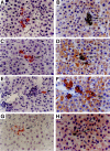
Dual phenotypic characteristics of ED 12 and ED 14 FLEC. Fetal liver cells were isolated as described in Materials and Methods, washed, and collected on slides. Dual ISH for AFP mRNA (brown color) and albumin mRNA (autoradiographic grains) of ED 12 (A) and ED 14 (B) FLEC. All cells expressing AFP mRNA are also positive for albumin mRNA. C and D: Immunohistochemistry (brown color) for CK-19 combined with ISH for AFP mRNA (autoradiographic grains). The majority of cells express only AFP mRNA (arrows in C and D). Some cells expressing AFP mRNA also express CK-19 (arrowhead in C), others express only CK-19 (small arrow in D). E and F: Immunohistochemistry (brown color) for CK-19 combined with ISH for albumin mRNA (autoradiographic grains). Most cells express only albumin mRNA (arrow in E). Some cells express only CK-19 (arrow in F) and others express both albumin mRNA and CK-19 (small arrow in F). G and H: Immunohistochemistry of ED 12 FLEC for CK-19 (brown color) combined with ISH for albumin mRNA (autoradiographic grains). Most cells express only albumin mRNA (arrows in G and H). Some cells express only CK-19 (small arrow in G) and others express both albumin mRNA and CK-19 (arrowheads in G and H). Original magnifications, ×400 (A-C, E, F, and H) and ×200 (D and G).
Figure 3.
Figure 3.
Differentiation of rat FLEC in normal adult regenerating liver. FLEC were isolated from the liver of ED 14 rat DPPIV+ fetuses and transplanted into the liver of mutant DPPIV female rats, as described in Materials and Methods. One, 2 and 4 weeks later, the livers were removed and frozen sections were stained for histochemical detection of DPPIV enzyme activity (red color). A: One week after transplantation, scattered cells, diffusely stained for DPPIV, were identified. B: Two weeks after transplantation, FLEC in the parenchyma (zones 2 and 3) both expanded in number and differentiated into mature hepatocytes. C: Two weeks after transplantation, some cells in the regions of bile ducts (zone 1) had differentiated into BDEC (diffuse staining for DPPIV). D: One month after transplantation, larger clusters of morphologically fully differentiated Hc were observed. Fully mature bile duct structures comprised of either transplanted cells or a mixture of transplanted and host cells were also present (data not shown). Original magnification, ×400.
Figure 4.
Figure 4.
Differentiation of rat FLEC in Rs-treated adult regenerating liver. FLEC were isolated from the liver of ED 14 rat DPPIV+ fetuses and transplanted into the liver of mutant DPPIV female rats treated with Rs, as described in Materials and Methods. One, 2 and 4 weeks later, the livers were removed and frozen sections were stained for histochemical detection of DPPIV enzyme activity (red color). A: One week after transplantation, small groups of cells diffusely stained for DPPIV, were found. B and C: Two weeks after transplantation, DPPIV+ cells formed larger clusters of hepatocytes that were beginning to show canalicular staining for DPPIV (B), or BDEC in the bile duct region, as evidenced by diffusely stained DPPIV+ small epithelial cells in bile duct-like structures (C). D: One month after transplantation, numerous clusters of morphologically fully differentiated Hc were observed. Original magnifications, ×200 (A-C) and ×100 (D).
Figure 5.
Figure 5.
Transplanted cells lose markers of undifferentiated FLEC. Fetal liver cells were isolated from ED 14 rat DPPIV+ fetuses and transplanted in conjunction with PH into the liver of mutant DPPIV female rats treated with Rs, as described in Materials and Methods. Serial sections were processed for DPPIV histochemical staining, shown by transplanted cells exhibiting dark staining in a membranous (bile canalicular) distribution in A, C, and E and for CK-19 by immunohistochemical staining, shown as darkly colored cells in B; γ-GT by histochemical staining, shown as darkly color cells in D and AFP mRNA by ISH, shown as autoradiographic grains, which are negative, in F. The clusters of transplanted cells are surrounded by arrows. As shown in these serial sections taken 1 month after cell transplantation, DPPIV+ hepatocytes are negative for γ-GT, CK-19, and AFP mRNA. Original magnifications, ×40 (A–D) and ×200 (E, F).
Figure 6.
Figure 6.
Transplanted cells acquire phenotypic markers of differentiated hepatocytes. FLEC were isolated from the liver of ED 14 rat DPPIV+ fetuses and transplanted in conjunction with PH into the liver of mutant DPPIV female rats treated with Rs, as described in Materials and Methods. One month after cell transplantation, livers were removed and serial sections were processed for DPPIV histochemical staining, (cells with dark staining in a membranous distribution and highlighted by arrows) in A, C, and E. B: ISH for albumin mRNA (autoradioactive grains) in the same region as the DPPIV+ Hc in A. This region shows a cluster of transplanted cells with high albumin mRNA expression (circumscribed by arrows). D: Histochemical staining for G-6P (dark color) expressed in Hc originating from transplanted cells in the same large cluster, which fills the microscopic field. F: Immunohistochemical staining for OV-6 (dark color) in epithelial cells within mature bile ducts, some of which are also positive for DPPIV (examples of dual positive cells are highlighted by arrows in E and F). Original magnifications, ×200 (A, B, E, and F) and ×100 (C and D).
Figure 7.
Figure 7.
Differentiation of rat FLEC in adult Rs-treated liver stimulated with T3. FLEC were isolated from the liver of ED 14 rat DPPIV+ fetuses and transplanted into the liver of Rs-treated mutant DPPIV female rats and stimulated with T3, as described in Materials and Methods. Four weeks later, the livers were removed and frozen sections were stained for histochemical detection of DPPIV. A: Cluster of transplanted cells differentiated morphologically into mature hepatocytes (area surrounded by arrowheads). B: Small bile duct (denoted by large arrow), originating from transplanted cells. Original magnification, ×200.
Figure 8.
Figure 8.
Detection and amplification of the rat sry gene located on the Y chromosome in transplanted cells. DNA was isolated from the liver of female rats 2 weeks after FLEC transplantation. A 104-bp fragment of the sry gene located on the Y chromosome was amplified, as described in Materials and Methods, and resolved on a 2% agarose gel. Lanes 1–3: Amplified fragments from 50, 5, and 0.5 ng, respectively, of recipient DNA, isolated 2 weeks after transplantation. Lanes 4–6: Amplified fragments from 50, 5 and 0.5 ng, respectively, of recipient DNA, isolated 2 weeks after transplantation from animals subjected to PH. Lane 7: Amplified fragment from 50 ng of control female DNA. Lanes 8–12: Amplified fragments from 50, 5, 0.5, 0.05, and 0.005 ng DNA from male F344 rats diluted into control female DNA. Lane 13: control tube with no DNA. Lane 14: Molecular weight markers.
Figure 9.
Figure 9.
Differentiation of lineage committed FLEC in regenerating liver of normal adult F344 rats. FLEC were isolated from the liver of ED 14 rat DPPIV+ fetuses and transplanted in conjunction with PH into the liver of mutant DPPIV female rats not treated with Rs. One week (A-D) and 2 weeks (E-H) after transplantation, livers were removed and serial sections prepared. Histochemical detection of DPPIV (red color) is shown in A, C, E, and G and dual immunohistochemical detection of CK-19 (brown color) and ISH for AFP mRNA (autoradiographic grains) is presented in serial sections in B, D, F, and H. B: The cluster of AFP mRNA+ cells does not express CK-19. D: AFP mRNA expressing cells do express CK-19. F: None of the CK-19+ cells forming bile duct structures (arrow) express AFP mRNA. G and H: Decreased expression of AFP mRNA and absent expression of CK-19 in transplanted cells that differentiated into hepatocytes (arrow). Original magnification, ×400.
Figure 10.
Figure 10.
Differentiation of lineage uncommitted FLEC in regenerating liver of Rs-treated adult F344 rats. FLEC were isolated from the liver of ED 14 rat DPPIV+ fetuses and transplanted in conjunction with PH into the liver of mutant DPPIV female rats treated with Rs. One week (A-D) and 2 weeks (E-H) after transplantation, livers were removed and serial sections processed. Histochemical detection of DPPIV (red color) is shown in A, C, E, and G and dual immunohistochemical detection of CK-19 (brown color) and ISH for AFP mRNA (autoradiographic grains) is shown in B, D, F, and H. B: Transplanted cells, shown in A, formed a large cluster of AFP mRNA+ and CK-19+ cells, shown in B. The expression of CK-19 is lower than in endogenous small epithelial cells (arrowhead in B). D: A mixed population of transplanted cells, expressing AFP mRNA. Those with lower expression of AFP mRNA also express CK-19 (arrows). The cells with higher expression of AFP mRNA do not express CK-19 (arrowhead). F: Transplanted cells, forming bile duct structures, express both CK-19 and AFP mRNA (arrows). Expression of AFP mRNA is generally lower in CK-19+ duct cells than that in Hc. Also note that CK-19+/AFP duct cells of host origin are also present (arrowheads). H: Expression of AFP mRNA in CK-19+ Hc (arrow) decreased faster than that in Hc not expressing CK-19 (arrowhead). Original magnifications, ×400 (A-F) and ×200 (G and H).

Similar articles

Cited by

References

    1. DuBois AM: The embryonic liver. The Liver. Edited by Rouiller CH. New York, Academic Press, 1963
    1. Wilson JW, Groat CS, Leduc EH: Histogenesis of the liver. Ann NY Acad Sci 1963, 111:8-22 - PubMed
    1. Rugh R: The Mouse: Its Reproduction and Development. 1968. Oxford University Press, New York
    1. Le Douarin NM: An experimental analysis of liver development. Med Biol 1975, 53:427-455 - PubMed
    1. Houssaint E: Differentiation of mouse hepatic primordium: an analysis of tissue interactions in hepatocyte differentiation. Cell Growth Differ 1980, 9:269-279 - PubMed

Publication types

MeSH terms