. 2000 Jun 15;19(12):2946-57.
doi: 10.1093/emboj/19.12.2946.

Phosphorylation status of the SCR homeodomain determines its functional activity: essential role for protein phosphatase 2A,B'

Affiliations

Phosphorylation status of the SCR homeodomain determines its functional activity: essential role for protein phosphatase 2A,B'

M Berry et al. EMBO J. .

Abstract

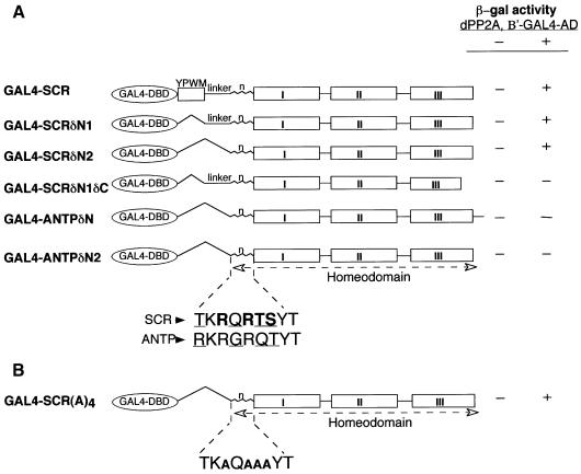
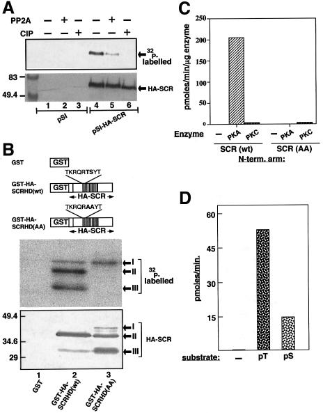
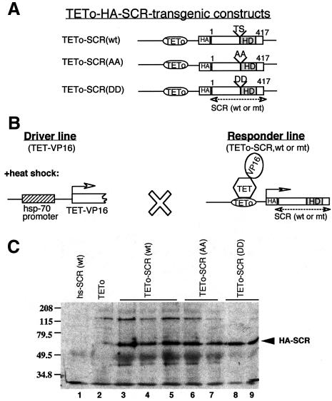
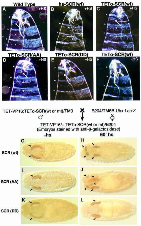
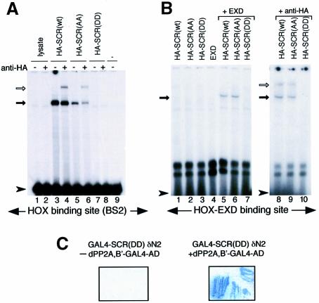
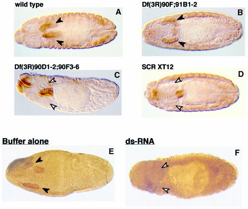
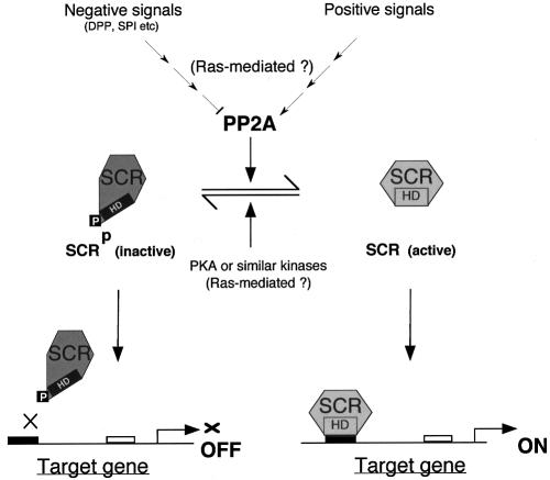
Sex combs reduced (SCR) is a Drosophila Hox protein that determines the identity of the labial and prothoracic segments. In search of factors that might associate with SCR to control its activity and/or specificity, we performed a yeast two-hybrid screen. A Drosophila homologue of the regulatory subunit (B'/PR61) of serine-threonine protein phosphatase 2A (dPP2A,B') specifically interacted with the SCR homeodomain. The N-terminal arm within the SCR homeodomain was shown to be a target of phosphorylation/dephosphorylation by cAMP-dependent protein kinase A and protein phosphatase 2A, respectively. In vivo analyses revealed that mutant forms of SCR mimicking constitutively dephosphorylated or phosphorylated states of the homeodomain were active or inactive, respectively. Inactivity of the phosphorylated mimic form was attributed to impaired DNA binding. Specific ablation of dPP2A,B' gene activity by double-stranded RNA-mediated genetic interference resulted in embryos without salivary glands, an SCR null phenotype. Our data demonstrate an essential role for Drosophila PP2A,B' in positively modulating SCR function.


Figures

None
Fig. 1. (A) dPP2A,B′ interacts specifically with the SCR homeodomain in a yeast two-hybrid screen. Different GAL4 fusion constructs containing the GAL4 DNA-binding domain (amino acids 1–147) fused to distinct regions of SCR and ANTP are shown in the scheme. The amino acid co-ordinates of the regions of SCR and ANTP in the constructs are provided in Materials and methods. The GAL4–HOX protein chimeras were co-transformed into yeast, with either GAL4 AD alone (–) or the GAL4 AD fused to amino acids 245–670 of dPP2A,B′ (+). The differences in the amino acids within the N-terminal arm of SCR and ANTP homeodomains are underlined. Two consensus phosphorylation sites in the SCR homeodomain are depicted in bold. (B) Conservation of the phosphorylation sites within the N-terminal arm of the SCR homeodomain is not essential for the interaction with dPP2A,B′. The four amino acids (RRTS) of the consensus phosphorylation sites in the SCR homeodomain are substituted by alanines in GAL4–SCR(A)4.
None
Fig. 2. Full-length nucleotide and deduced amino acid sequence of dPP2A,B′ cDNA. The 5′- and 3′-untranslated sequences are presented in lower case. The open reading frame is shown in upper case. The numbers corresponding to the nucleotide and the amino acid sequence are indicated on either side of the respective sequences.
None
Fig. 3. In vivo and in vitro phosphorylation and dephosphorylation of SCR. (A32P-labelled extracts of empty vector (pSI) or pSI-HA-SCR-transfected COS-1 cells were immunoprecipitated and subjected to dephosphorylation by the PP2A catalytic subunit (lanes 2 and 5) and by calf intestine alkaline phosphatase (CIP, lanes 3 and 6). Western blot was revealed by autoradiography (upper panel) and by alkaline phosphatase immunostaining to normalize the SCR content in each lane (lower panel). The position of HA-SCR is indicated with arrows. Molecular size markers in kDa are shown on the left. (B) Schematic representation of GST fusion constructs utilized in the present study. The depicted scale is arbitrary. The GST fusion constructs encode the GST protein alone (GST), GST–HA-SCRHD(wt) and GST–HA-SCRHD(AA). The homeodomain is represented by the shaded box. The wild-type and AA mutant sequences of the N-terminal arm in the homeodomain are also indicated (upper panel). The GST–SCRs were detected on western blot by alkaline phosphatase staining, to monitor the relative levels of protein products in each lane (lower panel), and subsequently by autoradiography (middle panel). Note that the GST–SCRs are visible in three different sizes corresponding to the full-length (band I) and its C-terminal truncations (bands II and III), which are indicated by arrows. (C) Comparison of phosphorylation of the wild-type and the AA mutant versions of the N-terminal arm of the SCR homeodomain in the absence and presence of kinases. Biotinylated peptides (200 µM) SCR(wt): TKRQRTSYT and SCR(AA): TKRQRAAYT were used as substrates. The specific activity of the [γ-32P]ATP per reaction was typically 2000 c.p.m./pmol. A 5 ng aliquot of PKA, PKC or no enzyme (–) was included in the assays. The enzyme reactions were carried out at 30°C for 2–5 min. The incorporation of [γ-32P]ATP into the peptides is depicted on the y-axis in pmol/min/µg enzyme. The values are an average of six and three independent experiments for PKA and PKC, respectively. (D) Dephosphorylation of the N-terminal arm of the SCR homeo-domain by PP2A: no peptide (–) or 200 µM of phosphopeptides pT: TKRQR(pT)SYT and pS: TKRQRT(pS)YT (x-axis) were used as substrates for dephosphorylation by PP2A. The assays were performed at 30°C for 15 min using a serine-threonine phosphatase (non-radioactive) assay kit (Promega) in the presence of 50 ng of the catalytic subunit of PP2A. The activity was monitored spectrophoto-metrically by measuring the release of pmoles of free phosphate per minute (y-axis). The depicted values are an average of four individual experiments.
None
Fig. 4. Expression of SCR transgenes in Drosophila embryos. (A) Schematic representation of the constructs used to generate transgenic flies. (B) Schematic representation of the tetracycline repressor-VP16 (TET-VP16)-mediated ectopic induction of SCR transgenes. TET-VP16 is produced upon heat shock. (C) Western blot showing the expression of HA-SCR(wt), HA-SCR(AA) and HA-SCR(DD) proteins in fly embryos. Lane 1, hs-SCR(wt); lane 2, TETo alone; lanes 3–5, HA-SCR(wt); lanes 6 and 7, and 8 and 9, HA-SCR(AA) and HA-SCR(DD), respectively, expressed from different transgenic lines for each construct. The arrow indicates the position of the distinct HA-SCR transgene products. The positions of protein size markers (in kDa) are indicated on the left.
None
Fig. 5. SCR(wt) and SCR(AA) but not SCR(DD) transgenes transform T2 and T3 towards T1 identity and cause head involution defects (A–F). Darkfield photomicrographs of the anterior ends of cuticles of (A) a wild-type embryo, (B) an embryo expressing SCR(wt) from a direct hs-SCR transgene (see Gibson et al., 1990), heat-shocked embryos from a cross between TET-VP16 and (C) TETo-SCR(wt), (D) TETo-SCR(AA) and (E) TETo-SCR(DD), and (F) an embryo of a cross between TET-VP16 and TETo-SCR(wt) without a heat shock. Arrowheads indicate segmental borders of the anterior region of larval cuticles. Arrows show the normal (A–F) and ectopic T1 beards (B–D). Note the head involution defects observed in embryos in (B), (C) and (D) compared with the normal head involution of embryos in (A), (E) and (F). Both SCR(wt) and SCR(AA) induce additional salivary glands in transgenic embryos (G–L). A scheme of the cross with the indicated genotypes is given above (G–L). B204 is a dCREB-A-lacZ enhancer trap line. Embryos were stained with anti-β-galactosidase to locate salivary glands. Embryos expressing dCREB-A-lacZ and SCR(wt) (G and H), SCR(AA) (I and J) or SCR(DD) transgenes (K and L) are shown. Normal and extra salivary glands are indicated by arrows and arrowheads, respectively.
None
Fig. 6. SCR(DD) does not bind DNA either on its own or in the presence of EXD. (A) Band-shifts performed with BS2 as a probe using lysate alone (lanes 1 and 2), HA-SCR(wt) (lanes 3 and 4), HA-SCR(AA) (lanes 5 and 6) and HA-SCR(DD) (lanes 7 and 8), or in the absence of any lysate (lane 9). (B) Band-shifts with a consensus HOX–EXD-binding site. HA-SCR(wt) (lane 1), HA-SCR(AA) (lane 2), HA-SCR(DD) (lane 3) or EXD alone (lane 4). In the presence of EXD, HA-SCR(wt) (lane 5) and HA-SCR(AA) (lane 6) specifically bind the HOX–EXD motif. Solid arrows indicate the position of specific complexes. Supershifts of the specific complexes are indicated with unfilled arrows (lanes 4, 6, 8 and 9). The position of the unbound probes is marked with an arrowhead. Note that HA-SCR(DD) does not bind to DNA at all (lanes 7 and 8 in A and lanes 3, 7 and 10 in B). (C) The SCR(DD) homeodomain interacts with dPP2A,B′ in a yeast two-hybrid system: co-transformation of GAL4–SCR(DD)δN2 (323–378;TS→DD) and dPP2A,B′–GAL4 AD induces the expression of β-galactosidase detected as blue colour of the colonies (right panel). No β-galactosidase is induced when GAL4–SCR(DD)δN2 is present by itself (left panel).
None
Fig. 7. dPP2A,B′ is essential for SCR activity in developing fly embryos. Normal salivary glands appear in wild-type embryos (A) and in the embryos with a deficiency that does not remove the dPP2A,B′ gene (B). In contrast, salivary glands are missing in the deficient embryos that lack the dPP2A,B′ gene (C) and in the SCR null embryos (D). Wild-type embryos (0–60 min) were injected either with buffer (E) or with dPP2A,B′ dsRNA (F). Normal salivary glands are shown by filled arrowheads. The positions where salivary glands are expected to form, but fail to appear, are indicated by unfilled arrowheads.
None
Fig. 8. Proposed model for the regulation of SCR function.

References

    1. Andrew D.J., Baig,A., Bhanot,P., Smolik,S.M. and Henderson,K.D. (1997) The Drosophila dCREB-A gene is required for dorsal/ventral patterning of the larval cuticle. Development, 124, 181–193. - PubMed
    1. Bartel P.L. and Fields,S. (1995) Analyzing protein–protein interactions using two-hybrid system. Methods Enzymol., 254, 241–263. - PubMed
    1. Bello B., Resendez-Perez,D. and Gehring,W.J. (1998) Spatial and temporal targeting of gene expression in Drosophila by means of a tetracycline-dependent transactivator system. Development, 125, 2193–2202. - PubMed
    1. Boube M., Benassayag,C., Seroude,L. and Cribbs,D.L. (1997) Ras1-mediated modulation of Drosophila homeotic function in cell and segment identity. Genetics, 146, 619–628. - PMC - PubMed
    1. Bourbon H.M., Martin-Blanco,E., Rosen,D. and Kornberg,T.B. (1995) Phosphorylation of the Drosophila engrailed protein at a site outside its homeodomain enhances DNA binding. J. Biol. Chem., 270, 11130–11139. - PubMed

Publication types

LinkOut - more resources