. 2000 Jun 15;19(12):3080-9.
doi: 10.1093/emboj/19.12.3080.

The Epstein-Barr virus lytic program is controlled by the co-operative functions of two transactivators

Affiliations

The Epstein-Barr virus lytic program is controlled by the co-operative functions of two transactivators

R Feederle et al. EMBO J. .

Abstract

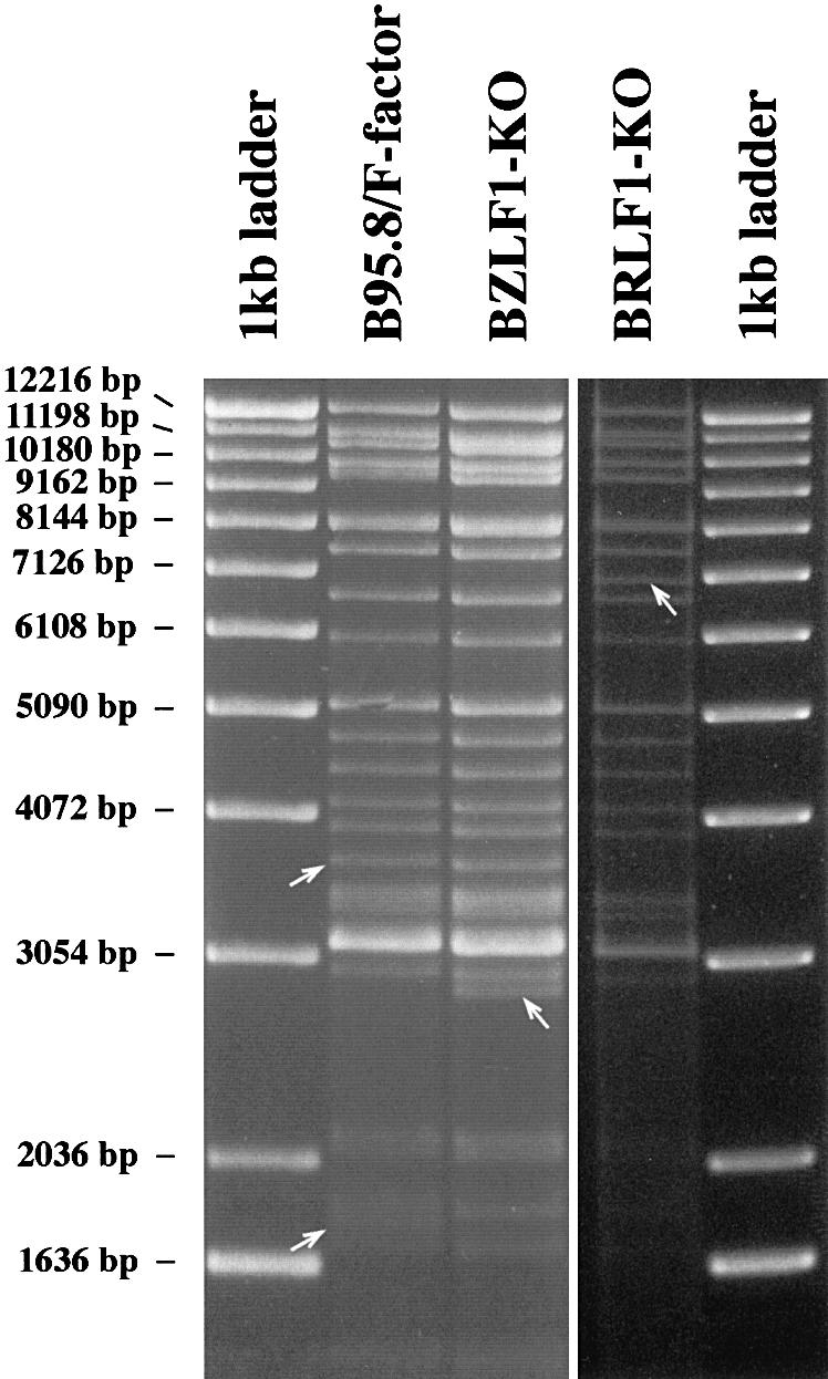
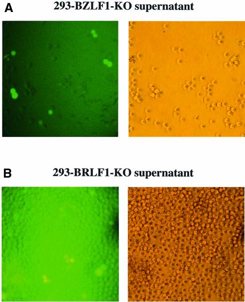
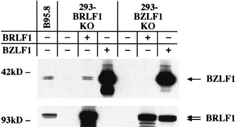
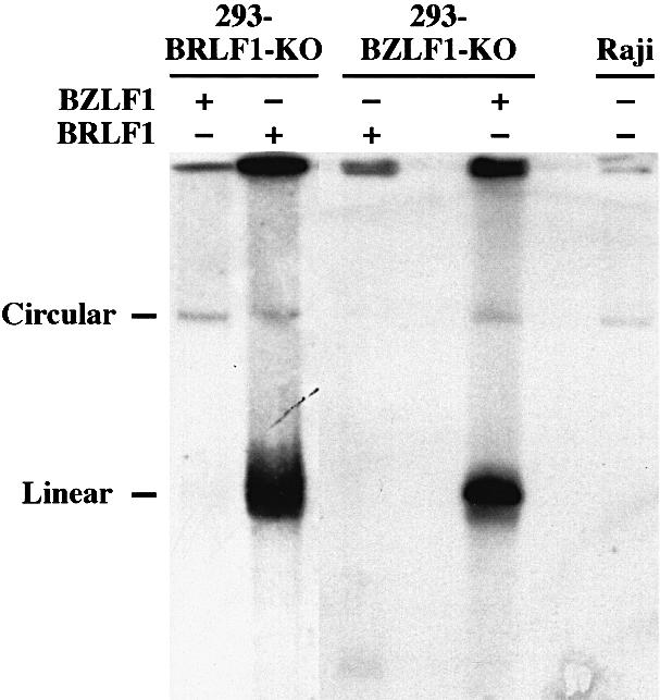
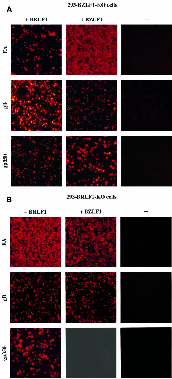
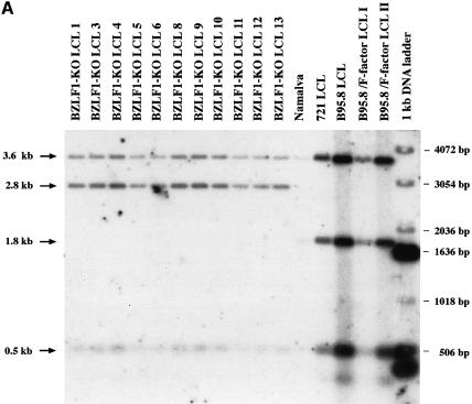
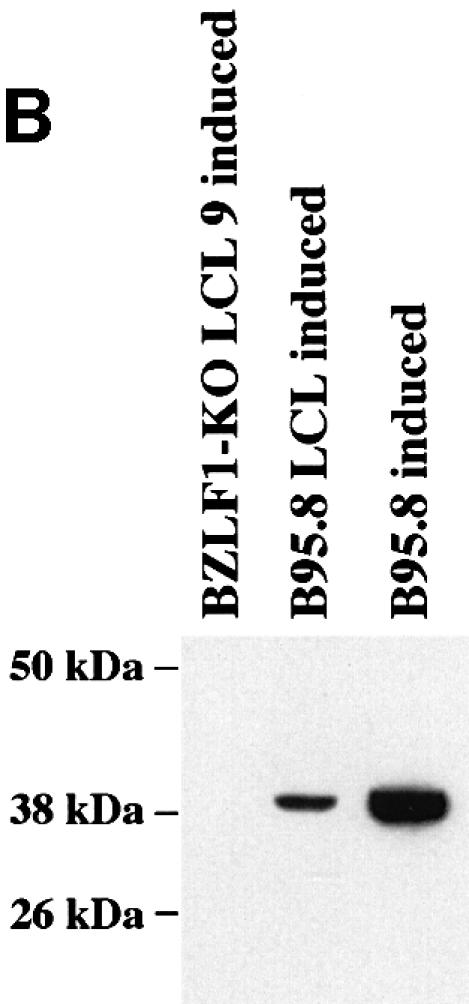
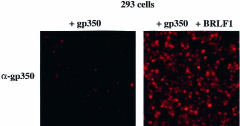
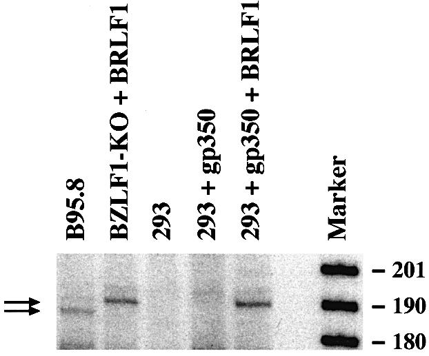
The propagation of herpesviruses has long been viewed as a temporally regulated sequential process that results from the consecutive expression of specific viral transactivators. As a key step in this process, lytic viral DNA replication is considered as a checkpoint that controls the expression of the late structural viral genes. In a novel genetic approach, we show that both hypotheses do not hold true for the Epstein-Barr virus (EBV). The study of viral mutants of EBV in which the early genes BZLF1 and BRLF1 are deleted allowed a precise assignment of the function of these proteins. Both transactivators were absolutely essential for viral DNA replication. Both BZLF1 and BRLF1 were required for full expression of the EBV proteins expressed during the lytic program, although the respective influence of these molecules on the expression of various viral target genes varied greatly. In replication-defective viral mutants, neither early gene expression nor DNA replication was a prerequisite for late gene expression. This work shows that BRLF1 and BZLF1 harbor distinct but complementary functions that influence all stages of viral production.


Figures

None
Fig. 1. Restriction fragment analysis of both BZLF1-KO and BRLF1-KO mutant DNAs in comparison with the wild-type B95.8/F-factor DNA. The BZLF1 gene was exchanged against the kanamycin resistance gene via homologous recombination. DNA was purified from a chloramphenicol- and kanamycin-resistant E.coli clone and digested with the BamHI restriction enzyme. The restriction pattern of the mutant BZLF1-KO DNA was compared with that obtained with wild-type B95.8/F-factor DNA. Both restriction fragment patterns were identical, with two exceptions. A new fragment was generated by replacement of the BamHI fragment that carries the BZLF1 gene (disappearance of the 1.8 kb fragment in B95.8/F-factor) with the one that carries the kanamycin resistance gene (appearance of a 2.8 kb fragment in BZLF1-KO DNA) (see also Figure 6A). Both rearranged fragments are indicated by an arrow. The same procedure was applied to the BRLF1 gene. In this case, the 3.6 kb BamHI fragment that carries the BRLF1 gene was exchanged with a 6.9 kb fragment that carries the tetracycline resistance gene.
None
Fig. 2. Complementation of the BRLF1 and BZLF1 mutants. GFP expression in Raji cells incubated with supernatants from 293-BZLF1-KO cells transfected with an expression plasmid encoding BZLF1 (A) or from 293-BRLF1-KO cells transfected with BRLF1 (B). A 1 ml aliquot of supernatant was mixed with 104 Raji cells. GFP fluorescence was investigated 48 h after infection (left panel); corresponding phase contrast light microscopy (right panels) (magnification ×200). After incubation with supernatants from 293-BZLF1-KO cells, ∼10% of the Raji cells were GFP positive, indicating a virus titer of at least 103 infectious viruses/ml. In the case of 293-BRLF1-KO cells, the virus titer could also be estimated as being at least 103 infectious viruses/ml.
None
Fig. 3. Reciprocal induction of BZLF1 and BRLF1. Western blot analysis of 293-BZLF1-KO or 293-BRLF1-KO cells transfected with an expression vector encoding either BRLF1 or BZLF1. Upper panel: BZLF1 expression pattern. Lower panel: BRLF1 expression pattern. The B95.8 cell line that spontaneously produces EBV virions was used as a positive control. Complementation of 293-BRLF1-KO with a BRLF1 expression plasmid or of 293-BZLF1-KO with a BZLF1 expression plasmid leads to the expression of both transactivators, showing that BZLF1 induces the expression of BRLF1, and vice versa. In 293-BZLF1-KO cells transfected with a BRLF1 expression plasmid or in 293-BRLF1-KO cells transfected with a BZLF1 expression plasmid, the expression pattern was limited to the expression of the transfected gene. Untransfected 293-BZLF1-KO or 293-BRLF1-KO cells provided additional negative controls.
None
Fig. 4. Analysis of DNA replication in BZLF1-KO and BRLF1-KO viral mutants after transfection of expression plasmids that carry the BZLF1 or BRLF1 genes. In cells latently infected with EBV, the viral DNA is present as an episome. In contrast, during lytic replication, newly synthesized viral DNA is linear. Both circular and linear forms can be distinguished readily after separation on a Gardella agarose gel electrophoresis, followed by Southern blot hybridization with an EBV-specific probe. Lytic replication was clearly identified after transfection of BZLF1 into the 293-BZLF1-KO cell line or of BRLF1 into the 293-BRLF1-KO cell line. In contrast, neither transfection of BRLF1 into the 293-BZLF1-KO cell line nor transfection of BZLF1 into the 293-BRLF1-KO cell line could give rise to any detectable DNA replication. The cell line Raji is defective for the lytic program and was used as a negative control.
None
Fig. 5. Production of the EBV lytic proteins EA-D, gB and gp350 after transfection of a BRLF1 or BZLF1 expression plasmid into 293-BZLF1-KO cells (A) or 293-BRLF1-KO cells (B). In each panel, a negative control consisting of untransfected cells is included. Fixed cells were incubated with a specific monoclonal antibody and a second anti-mouse antibody coupled to the Cy5 fluorochrome. Stained cells were visualized under UV light (magnification ×100).
None
Fig. 6. Immortalized B cells generated by infection of primary lymphoid cells with the BZLF1-KO EBV mutant do not carry the BZLF1 gene. (A) Southern blot analysis of cell lines infected either with the BZLF1-KO mutant or with wild-type EBV. Blots were hybridized with the construct used to generate the BZLF1-KO mutant that consists of the kanamycin resistance gene flanked by the first two-thirds of the first BZLF1 exon and sequences 5′ and 3′ from this gene. Whereas the wild-type EBVs carry the intact BZLF1 gene (1.8 kb fragment), only recombined fragments could be detected in the cell population infected by the viral mutant (2.8 kb fragment) (see also Figure 1). (B) Western blot analysis of cell lines infected either with the BZLF1-KO mutant or with wild-type EBV. After TPA and butyrate induction, cells that carry the wild-type EBV DNA (B95.8 and B95.8 LCL), but not those that carry the EBV viral mutant (BZLF1-KO LCL9), show clear production of the BZLF1 protein. Proteins extracted from the above-described lymphoid cell lines (20 µg for the BZLF1-KO mutant, 4 µg for the control cells) were separated by SDS–PAGE, blotted onto a nitrocellulose membrane and incubated with an antibody specific to the BZLF1 protein (Z130).
None
Fig. 7. The BRLF1 immediate early protein induces the expression of gp350 in EBV-negative cells. Expression of the EBV late protein gp350 after transfection of the gp350 gene that codes for gp350 (left panel, magnification ×100) and after co-transfection of the gp350 gene and BRLF1 into 293 cells (right panel, magnification ×100).
None
Fig. 8. The BRLF1 protein activates the expression of the gp350 gene from its established promoter. The 5′ end of the gp350 mRNA from 293 or 293-BZLF1-KO cells transfected with an expression plasmid encoding BRLF1 was determined using primer extension. B95.8 cells and untransfected 293 cells provided the positive and negative controls, respectively. Introduction of a BRLF1 expression plasmid into 293 or 293-BZLF1-KO cells gave rise to the synthesis of a gp350-specific mRNA, similar in size to the gp350 mRNA found in the B95.8 cells. The BRLF1-mediated activation of the gp350 mRNA therefore reflects the situation during the EBV lytic program in B95.8 cells. However, an additional gp350 mRNA transcript, slightly shorter in size, could also be detected in B95.8. At this stage, it is not clear whether this reflects the fact that the B95.8 cell line is derived from marmoset cells, or the contribution of additional viral factors apart from BRLF1 during viral lytic replication.

References

    1. Baer R. et al. (1984) DNA sequence and expression of the B95-8 Epstein–Barr virus genome. Nature, 310, 207–211. - PubMed
    1. Beisel C., Tanner,J., Matsuo,T., Thorley-Lawson,D., Kezdy,F. and Kieff,E. (1985) Two major outer envelope glycoproteins of Epstein–Barr virus are encoded by the same gene. J. Virol., 54, 665–674. - PMC - PubMed
    1. Campbell B.A. and Villarreal,L.P. (1988) Functional analysis of the individual enhancer core sequences of polyomavirus: cell-specific uncoupling of DNA replication from transcription. Mol. Cell. Biol., 8, 1993–2004. - PMC - PubMed
    1. Cherepanov P.P. and Wackernagel,W. (1995) Gene disruption in Escherichia coli: TcR and KmR cassettes with the option of Flp-catalyzed excision of the antibiotic-resistance determinant. Gene, 158, 9–14. - PubMed
    1. Conley A.J., Knipe,D.M., Jones,P.C. and Roizman,B. (1981) Molecular genetics of herpes simplex virus. VII. Characterization of a temperature-sensitive mutant produced by in vitro mutagenesis and defective in DNA synthesis and accumulation of γ polypeptides. J. Virol., 37, 191–206. - PMC - PubMed

Publication types

MeSH terms