Comparative Study
. 2000 Jun 15;19(12):3142-56.
doi: 10.1093/emboj/19.12.3142.

A novel shuttling protein, 4E-T, mediates the nuclear import of the mRNA 5' cap-binding protein, eIF4E

Affiliations
Comparative Study

A novel shuttling protein, 4E-T, mediates the nuclear import of the mRNA 5' cap-binding protein, eIF4E

J Dostie et al. EMBO J. .

Abstract

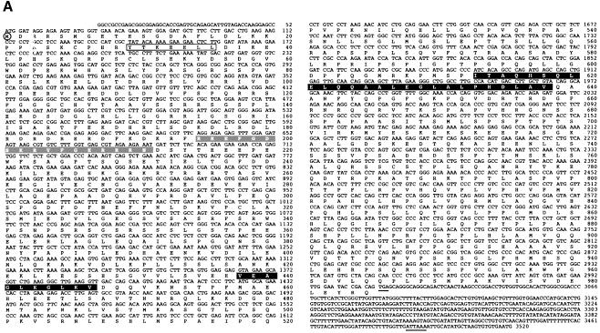
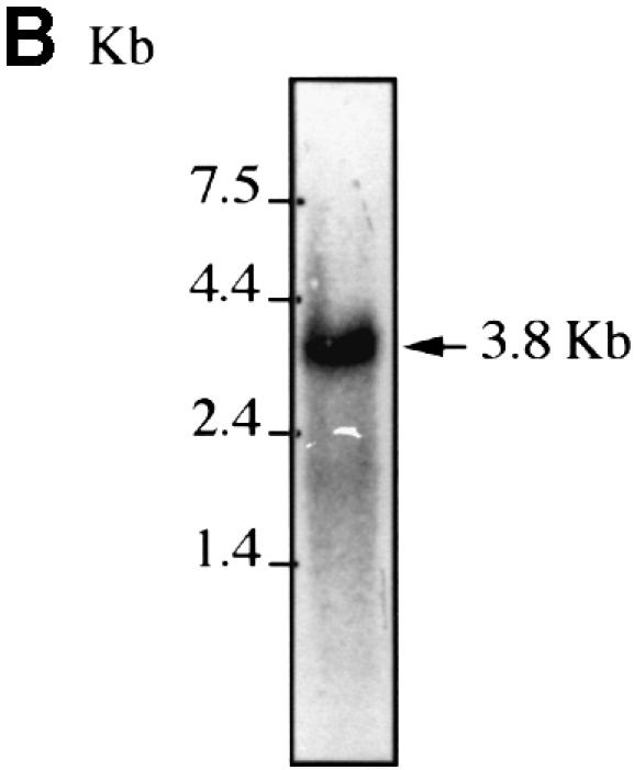
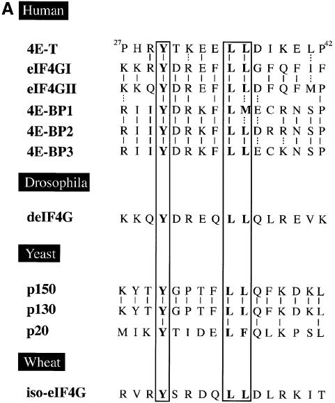
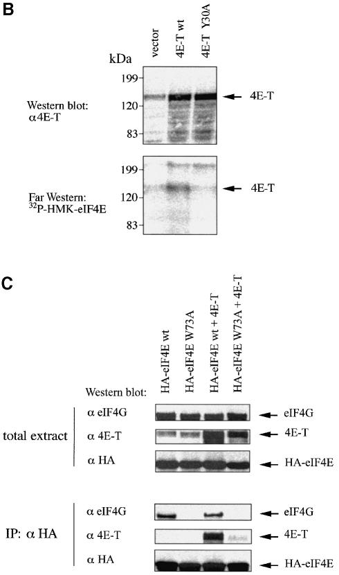
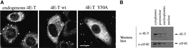
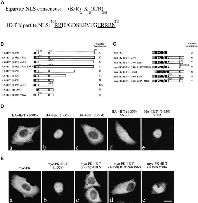
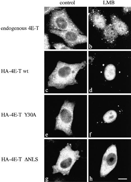
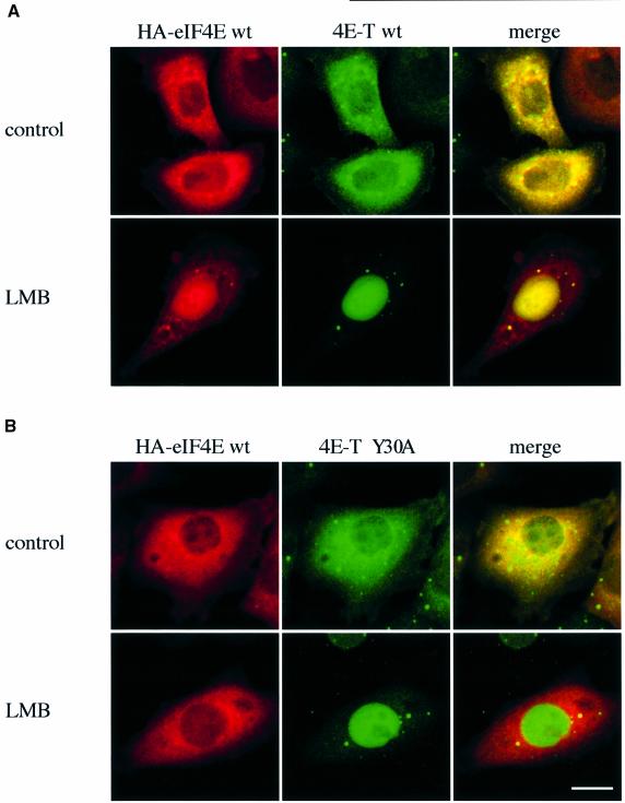
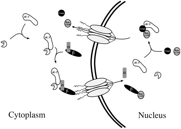
The eukaryotic translation initiation factor 4E (eIF4E) plays an important role in the control of cell growth. eIF4E binds to the mRNA 5' cap structure m(7)GpppN (where N is any nucleotide), and promotes ribosome binding to the mRNA in the cytoplasm. However, a fraction of eIF4E localizes to the nucleus. Here we describe the cloning and functional characterization of a new eIF4E-binding protein, referred to as 4E-T (eIF4E-Transporter). We demonstrate that 4E-T is a nucleocytoplasmic shuttling protein that contains an eIF4E-binding site, one bipartite nuclear localization signal and two leucine-rich nuclear export signals. eIF4E forms a complex with the importin alphabeta heterodimer only in the presence of 4E-T. Overexpression of wild-type 4E-T, but not of a mutant defective for eIF4E binding, causes the nuclear accumulation of HA-eIF4E in cells treated with leptomycin B. Taken together, these results demonstrate that the novel nucleocytoplasmic shuttling protein 4E-T mediates the nuclear import of eIF4E via the importin alphabeta pathway by a piggy-back mechanism.


Figures

None
Fig. 1. 4E-T cDNA. (A) Nucleotide and amino acid sequence of human 4E-T. The first methionine is circled. The stop codon and polyadenylation signal are underlined and the eIF4E-binding site is boxed. The nuclear localization signal and nuclear export signals are highlighted with gray and black boxes, respectively. The 4E-T sequence has been deposited in the DDBJ/EMBL/GenBank database (accession No. AF240775). (B) Northern blotting analysis of 4E-T. HeLa poly(A)+ RNA (5 µg) was probed with a 4E-T 5′ end probe as described in Materials and methods.
None
Fig. 2. Interaction of 4E-T with eIF4E. (A) eIF4E interacts with 4E-T as determined by far-western. HeLa cell extract (100 µg) was resolved by SDS–PAGE and stained with Coomassie Blue R-250 (left), or transferred to nitrocellulose and probed by far-western with a [32P]HMK-eIF4E probe (middle) or by western blotting with anti-4E-T antibody (right). (B) eIF4E specifically co-immunoprecipitates with 4E-T. HeLa cell extract (1 mg) was incubated with either pre-immune sera or anti-4E-T antibody as described in Materials and methods. Total extract [5% of input (50 µg)] and total immunoprecipitates from 1 mg of extract were resolved by SDS–PAGE, and analyzed by far-western with a [32P]HMK-eIF4E probe or by immunoblotting with anti-actin or anti-eIF4E monoclonal antibodies. (C) 4E-T interacts with eIF4E on a cap affinity resin. HeLa cell extract (1 mg) was incubated with resin or m7GDP resin, and processed as described in Materials and methods. Total extract [5% of input (50 µg)], resin, m7GDP resin-bound material from 1 mg of extract and flow through (5%) were resolved by SDS–PAGE, and analyzed by far-western with a [32P]HMK-eIF4E probe or by immunoblotting with anti-actin or anti-eIF4E monoclonal antibodies.
None
Fig. 3. 4E-T and eIF4E interact through conserved binding sites. (A) Amino acid sequence alignment of the eIF4E-binding site of 4E-T with those found in human eIF4GI (AF012088), human eIF4GII (AF012072), human 4E-BP1 (L36055), human 4E-BP2 (L36056), human 4E-BP3 (AF038869), Drosophila eIF4G (AF030155), S.cerevisiae eIF4GI (p39935), S.cerevisiae eIF4GII (p39936), S.cerevisiae p20 (X15731) and wheat iso-eIF4G (M95747). Residues which are critical for eIF4E binding are boxed. Solid and dashed lines represent identical and conserved residues, respectively. (B) 4E-T associates with eIF4E through a conserved motif. Untagged 4E-T wt and Y30A mutant were overexpressed in HeLa cells using the vaccinia virus system. Top: immunoblotting with anti-4E-T antibody. Bottom: far-western with [32P]HMK-eIF4E. (C) eIF4E binds to 4E-T and eIF4G through a shared sequence. HeLa cells were infected with vTF7-3 and transiently transfected with the plasmids indicated. Total extract [25% of input (50 µg)] was analyzed by western blotting with the indicated antibodies. Immunoprecipitation (from 200 µg of extract) of HA-eIF4E with anti-HA 12CA5 was performed as described in Materials and methods, and analyzed by western blotting with the antibodies indicated.
None
Fig. 4. 4E-T localizes predominantly to the cytoplasm. (A) Indirect immunofluorescence analysis with anti-4E-T. Localization of endogenous 4E-T in HeLa cells (left). HeLa cells were transiently transfected with either 4E-T wt (middle) or Y30A (right). At 36 h post-transfection, cells were fixed and immunostained with anti-4E-T antibody as described in Materials and methods. Bar, 10 µm. (B) Cell fractionation. HeLa cells were fractionated as described in Materials and methods, and an equal cell volume from each fraction was resolved by SDS–PAGE. Fractions were analyzed by western blotting with anti-4E-T (top) or anti-eIF4E monoclonal antibody (bottom).
None
Fig. 5. 4E-T contains a functional bipartite NLS. (A) Putative bipartite NLS of 4E-T. Invariant residues are underlined. (B) and (C) Schematic maps of HA-4E-T and myc-PK-4E-T expression constructs used for NLS mapping. HeLa cells were transfected with either HA-4E-T or myc-PK-4E-T expression constructs. Cells were fixed at 36 h post-transfection and immunostained with either anti-HA 12CA5 (D) or anti-myc 9E10 antibody (E). The transfected construct is indicated above each panel. Bar, 10 µm.
None
Fig. 6. 4E-T is a shuttling protein. HeLa cells were incubated with medium in the absence (A) or the presence of leptomycin B (LMB) (B) for 5 h prior to fixation and immunostaining with anti-4E-T as described in Materials and methods. (CH) HeLa cells were transfected with either HA-4E-T wt, Y30A or ΔNLS expression constructs. At 36 h post-transfection, the medium was replaced with medium either lacking (control) or containing LMB. Cells were processed as described above, except that anti-HA 12CA5 antibody was used for immunoblotting. Bar, 10 µm.
None
Fig. 7. 4E-T contains two functional NESs. (A) Putative NESs of 4E-T. Invariant residues are boxed. HeLa cells were transfected with either HA-4E-T (B) or myc-NPc-4E-T expression constructs (C). Cells were fixed at 36 h post-transfection and immunostained with either anti-HA 12CA5 (B) or anti-myc 9E10 antibody (C). Transfected construct is indicated above each panel. (D) and (E) Schematic maps of HA-4E-T and myc-NPc-4E-T expression constructs used for NES mapping. Bar, 10 µm.
None
Fig. 8. eIF4E forms a complex with importin α and β only in the presence of 4E-T. The purified proteins indicated were incubated with glutathione–Sepharose beads as described in Materials and methods. Precipitates were analyzed by western blotting with anti-4E-T, anti-GST, S-protein (Novagen) and anti-eIF4E (No. 5853) as indicated. Input represents 25% of total protein used for pull-down; flow through represents 10% of unbound material.
None
Fig. 9. 4E-T imports HA-eIF4E into the nucleus. HeLa cells were co-transfected with either HA-eIF4E and 4E-T wt (A), or HA-eIF4E and 4E-T Y30A (B). At 36 h post-transfection, the medium was replaced with fresh medium (control) or medium containing leptomycin B (LMB), and cells were incubated for 5 h prior to fixation. The localization of HA-eIF4E was determined by indirect immunofluorescence with anti-HA 12CA5 and Texas red-conjugated anti-mouse IgG (left), and that of 4E-T with anti-4E-T and fluorescein-conjugated anti-rabbit IgG (middle). The co-localization of HA-eIF4E and 4E-T appears yellow (right). Bar, 10 µm.
None
Fig. 10. 4E-T mediates the nuclear import of eIF4E. See the text for details.

References

    1. Adam E.J. and Adam,S.A. (1994) Identification of cytosolic factors required for nuclear location sequence-mediated binding to the nuclear envelope. J. Cell Biol., 125, 547–555. - PMC - PubMed
    1. Blanar M.A. and Rutter,W.J. (1992) Interaction cloning: identification of a helix–loop–helix zipper protein that interacts with c-Fos. Science, 256, 1014–1018. - PubMed
    1. Chi N.C., Adam,E.J., Visser,G.D. and Adam,S.A. (1996) RanBP1 stabilizes the interaction of Ran with p97 nuclear protein import. J. Cell Biol., 135, 559–569. - PMC - PubMed
    1. Dostie J., Lejbkowicz,F. and Sonenberg,N. (2000) Nuclear eukaryotic initiation factor 4E (eIF4E) colocalizes with splicing factors in speckles. J. Cell Biol., 148, 239–247. - PMC - PubMed
    1. Duncan R., Milburn,S.C. and Hershey,J.W. (1987) Regulated phosphorylation and low abundance of HeLa cell initiation factor eIF-4F suggest a role in translational control. Heat shock effects on eIF-4F. J. Biol. Chem., 262, 380–388. - PubMed

Publication types

MeSH terms

Substances

Associated data