Comparative Study
. 2000 Sep 8;275(36):28017-27.
doi: 10.1074/jbc.M004009200.

Two-dimensional NMR spectroscopy and structures of six lipid A species from Rhizobium etli CE3. Detection of an acyloxyacyl residue in each component and origin of the aminogluconate moiety

Affiliations
Comparative Study

Two-dimensional NMR spectroscopy and structures of six lipid A species from Rhizobium etli CE3. Detection of an acyloxyacyl residue in each component and origin of the aminogluconate moiety

N L Que et al. J Biol Chem. .

Abstract

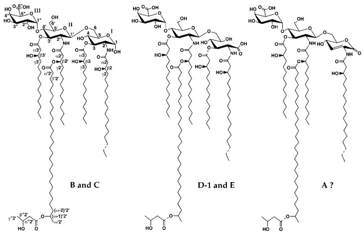
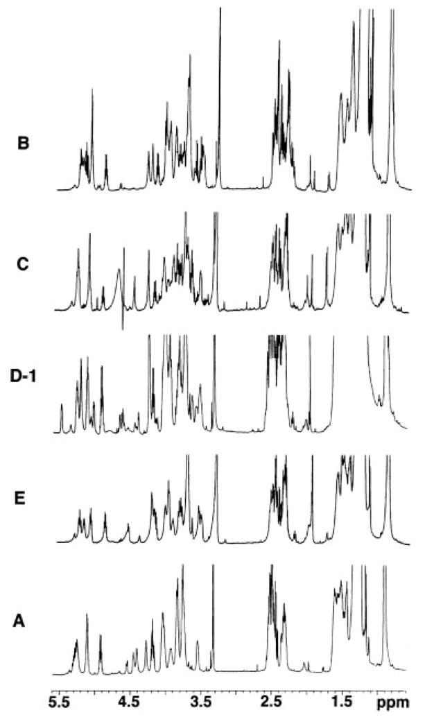
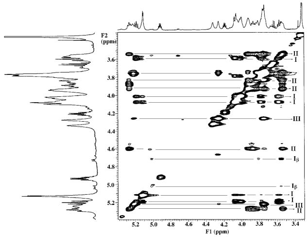
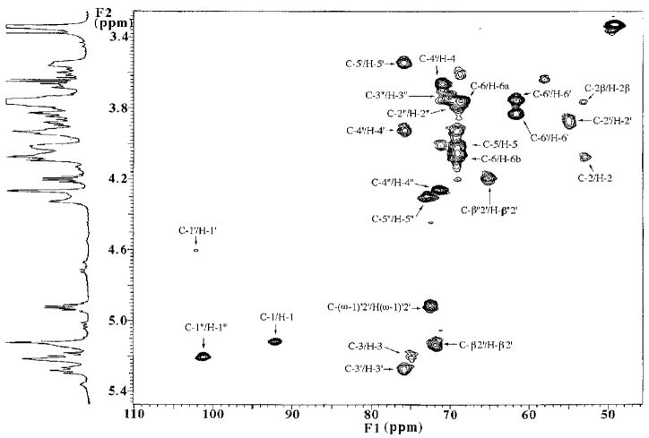
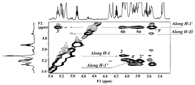
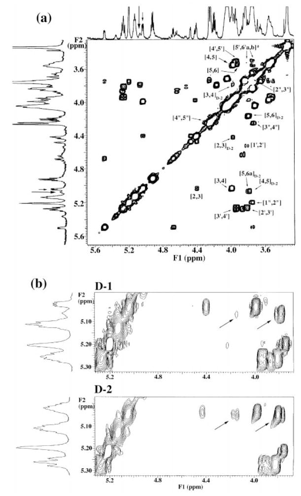
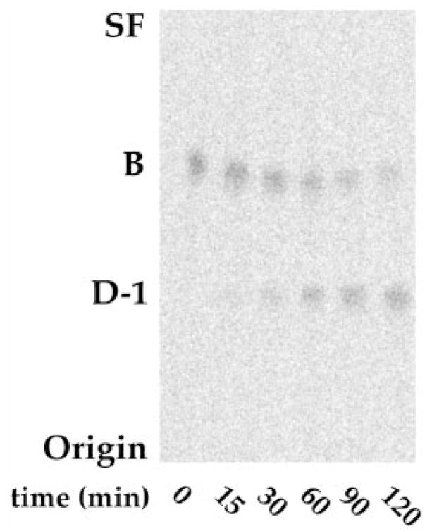
The chemical structures of six lipid A species (A, B, C, D-1, D-2, and E) purified from Rhizobium etli CE3 were investigated by one- and two-dimensional NMR spectroscopy. The R. etli lipid A subtypes each contain an unusual acyloxyacyl residue at position 2' as part of a conserved distal glucosamine moiety but differ in their proximal units. All R. etli lipid A species lack phosphate groups. However, they are derivatized with an alpha-linked galacturonic acid group at position 4', as shown by nuclear Overhauser effect spectroscopy. Component B, which had been not been reported in previous studies, features a beta, 1'-6 linked disaccharide of glucosamine acylated at positions 2, 3, 2', and 3' in a pattern that is typical of lipid A found in other Gram-negative bacteria. D-1 contains an acylated aminogluconate unit in place of the proximal glucosamine residue of B. C and E lack ester-linked beta-hydroxyacyl chains at position 3, as judged by their H-3 chemical shifts, and may be synthesized from B and D-1, respectively, by the R. etli 3-O-deacylase. D-2 is an isomer of D-1 that forms nonenzymatically by acyl chain migration. A may be an elimination product derived from D-1 during hydrolysis at 100 degrees C (pH 4.5), a step needed to release lipid A from lipopolysaccharide. Based on these findings, we propose a biosynthetic scheme for R. etli lipid A in which B is generated first by a variation of the E. coli pathway. The aminogluconate unit of D-1 could then be made from B by enzymatic oxidation of the proximal glucosamine. As predicted by our hypothesis, enzyme(s) can be demonstrated in extracts of R. etli that convert (14)C-labeled B to D-1.


Figures

Fig. 1
Fig. 1. Proposed structures of species B, C, D-1, E, and A isolated from R. etli CE3
Species B and C contain a glucosamine disaccharide unit typical of lipid A molecules found in most other Gram-negative bacteria, including E. coli. D-1 and E feature an aminogluconate unit in place of the proximal glucosamine. All lipid A species of R. etli contain a galacturonic acid substituent at position 4′ and an unusual C28 chain that is further substituted at C27, the position labeled as (ω-1)′2′. Dashed bonds show microheterogeneity with respect to acyl chain lengths or the presence of the β-hydroxybutyrate substituent. The uniform numbering scheme used to label the key sugar and lipid positions for the NMR analysis (Table I) is indicated in the structure of species B. Roman numerals indicate spin systems. Component A appears to be a chemical degradation product of D-1, and it may contain an aminoglucono-lactone residue (A. Ribeiro, C. Raetz, and N. Que, manuscript in preparation). D-2 (not shown) is an isomer of D-1 resulting from nonenzymatic acyl chain migration of the ester-linked chain at position 3 to position 5 of the aminogluconate moiety.
Fig. 2
Fig. 2. High resolution 1H NMR spectra at 600 MHz of species B, C, D-1, E, and A purified from R. etli
The NMR spectra were recorded at 25 °C in the solvent, CDCl3/CD3OD/D2O (2:3:1 v/v/v). Sample sizes were typically 1–4 mg in 0.6 ml.
Fig. 3
Fig. 3. A 600 MHz two-dimensional 1H-1H COSY analysis of species B highlighting the key acyl chain connectivities
The presence of an unusual acyloxyacyl chain is directly indicated by the cross-peaks to the downfield β-oxyme-thine designated α2′/β2′ and γ2′/β2′, compared with the remaining α/β and γ/β cross-peaks. The cross-peak pairs designated α22″ and γ22″ arise from the couplings within the β-hydroxybu-tyrate residue. The cross-peaks designated with thick and thin arrows indicate the coupling from the C27 oxymethine to the C26 methylene and C28 methyl groups of the C28 fatty acyl chain in the presence or absence of the β-hydroxybu-tyrate substituent, respectively.
Fig. 4
Fig. 4. Partial 600 MHz two-dimensional 1H-1H COSY analysis of species B showing sugar ring connectivities
The labeled cross-peaks indicate the assignments for the two glucosamine and the single galacturonic acid residue. The proximal glucosamine is not phosphoryl-ated, and both α and β anomeric forms are detected. Integration gave estimates of 82% α and 18% β anomeric forms.
Fig. 5
Fig. 5. Partial 600 MHz two-dimensional 1H-1H TOCSY of species B showing sugar ring connectivities
Spin systems I and Iβ correspond to the major α-anomeric and minor β-anomeric forms of the proximal glucosamine unit. Spin system II represents the distal β, 1–6 linked distal glucosamine. Spin system III arises from the galacturonic acid moiety attached to the 4′ position on the distal glucosamine unit.
Fig. 6
Fig. 6. Partial two-dimensional HMQC spectrum of species B, showing direct bond 1H-13C correlations within the sugar ring region
The assigned cross-peaks are labeled. Three anomeric cross-peaks are observed. The intensity of the C-1′/H-1′ cross-peak is reduced because of the presaturation sequence to suppress the solvent resonances.
Fig. 7
Fig. 7. Partial 600 MHz 1H-1H two-dimensional NOESY analysis of species B
With the diagonal phased negatively, the two-dimensional NOE cross-peaks phased negatively. Diagnostic NOEs are detected from H-1″ (5.23 ppm) to H-2″ (3.77 ppm) within the galacturonic acid moiety and to H-4′ (3.92 ppm) in the distal glucosamine unit (interglycosidic). In addition, cross-peaks are seen between H-1′ (4.53 ppm) and H-3′ (axial), and H-5′ (axial) within the distal glucosamine sugar, and to H-6a and H-6b in the proximal glucosamine unit (interglycosidic). A NOE from H-1 to H-2 is also detected within the proximal glucosamine. The mixing time was 500 ms.
Fig. 8
Fig. 8. Partial two-dimensional HMQC spectrum of species D-1, showing direct bond 1H-13C correlations within the sugar ring region
The assigned cross-peaks are labeled. Comparison with Fig. 6 reveals that the C-1′/H-1′, C-1″/H-1″, and C-2′/H-2′ cross-peaks in D-1 resonate at almost identical shifts as those in B. However, the C-2/H-2 cross-peak at 53.0/4.08 ppm in B is shifted to 57.0/4.42 ppm in D-1, and the C-1/H-1 cross-peak is not seen in D-1, consistent with the presence of a proximal aminogluconate unit in D-1.
Fig. 9
Fig. 9. Partial 600 MHz two-dimensional 1H-1H COSY analyses of freshly isolated D-1 and D-2 samples, showing sugar ring connectivities
a, the COSY cross-peaks arising from the galacturonic acid and the distal glucosamine unit in D-1 are similar to those observed for B (Fig. 6) and for D-2 (full spectrum not shown). However, even in a fresh sample of D-1, the proximal aminogluconate unit is revealed by NMR spectroscopy to be a mixture of a major 3-O-acylated form (authentic D-1) and a minor 5-O-acylated aminogluconate species (likely to be D-2, as labeled), which arises by a slow intramolecular trans-esterification. b, expansion of the acylated aminogluconate resonances of fresh D-1 and D-2 samples. The expanded D-1 spectrum reveals a prominent double-doublet at 5.03 ppm (H-3), which correlates to H-2 and H-4 in the COSY, providing evidence that this represents a 3-O-acylated species. A less prominent multiplet at 5.07 ppm (assigned to H-5 of the contaminating D-2 in the D-1 sample) correlates to H-6a, H-6b, and H-4 of the D-2 form in the COSY (indicated by the diagonal arrows). Integration of the D-1 spectrum yields estimates of 70% 3-O-acylated (D-1) and 30% 5-O-acylated (D-2) species in the fresh D-1 sample. Expansion of the acylated aminogluconate resonances in a fresh D-2 sample reveals a prominent multiplet at 5.04 ppm, which overlaps and obscures a less intense double-doublet (because of contaminating D-1). COSY reveals the prominent multiplet to correlate to H-6a, H-6b, and H-4 (indicated by the diagonal arrows) in D-2, thus indicating that the 5-O-acylated species is dominant in this sample. However, the COSY analysis also reveals “weaker” cross-peaks to H-2 and H-4, suggesting that the minor species present as an impurity in D-2 is in fact the 3-O-acylated isomer (contaminating D-1).
Fig. 10
Fig. 10. TLC analysis of the nonenzymatic interconversion of D-1 and D-2 after overnight incubation
Following incubation overnight in 50 mM aqueous MES, pH 6.5, and 0.1% Triton X-100, D-2 gradually appears in freshly isolated sample of D-1 (lane 1), whereas D-1 is generated in a sample of D-2 (lane 2). The 3-O-deacylated species E remains unchanged under these conditions (lane 3). The interconversion of D-1 and D-2 presumably occurs by a slow intramolecular acyl chain migration.
Fig. 11
Fig. 11. Rapid enzymatic conversion of B to D-1 by R. etli membranes
Upon incubation with R. etli or R. leguminosarum membranes, 14C- labeled B is converted to a more polar 14C- labeled species that migrates with purified standard of D-1.
Fig. 12
Fig. 12. A hypothetical pathway for the biosynthesis of the aminoglu-conate moiety of R. elti lipid A species D-1
Following the formation of component B, by the indicated pathway, a novel oxidation of the proximal glucosamine residue is proposed. If the reaction proceeds through a lactone intermediate (not shown), an additional lactonase would be needed to generate D-1. All the enzymes needed to generate component B have been detected (36, 37, 44), with the exception of the reactions that incorporate the galacturonic acid and the β-hydroxybutyrate residues.

References

    1. Long SR, Staskawicz BJ. Cell. 1993;73:921–935. - PubMed
    1. Spaink HP, Kondorosi A, Hooykaas PJJ. The Rhizobiaceae: Molecular Biology of Model Plant-associated Bacteria. Kluwer; Dordrecht, The Netherlands: 1998.
    1. Kannenberg EL, Brewin NJ. Trends Microbiol. 1994;2:277–283. - PubMed
    1. Carlson RW, Bhat UR, Reuhs B. In: Plant Bio/Technology and Development. Gresshoff PM, editor. CRC Press; Boca Raton, FL: 1992. pp. 33–44.
    1. Noel KD, Vandenbosch KA, Kulpaca B. J Bacteriol. 1986;168:1392–1401. - PMC - PubMed

Publication types

MeSH terms