. 2000 Jul;68(7):4064-74.
doi: 10.1128/IAI.68.7.4064-4074.2000.

Full capacity of recombinant Escherichia coli heat-stable enterotoxin fusion proteins for extracellular secretion, antigenicity, disulfide bond formation, and activity

Affiliations

Full capacity of recombinant Escherichia coli heat-stable enterotoxin fusion proteins for extracellular secretion, antigenicity, disulfide bond formation, and activity

I Batisson et al. Infect Immun. 2000 Jul.

Abstract

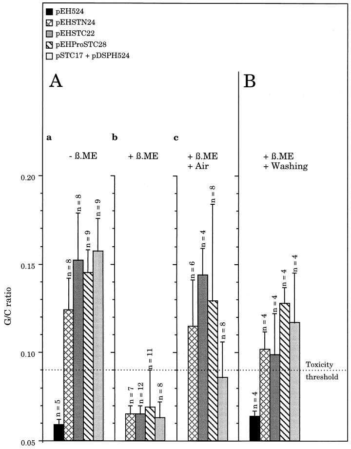
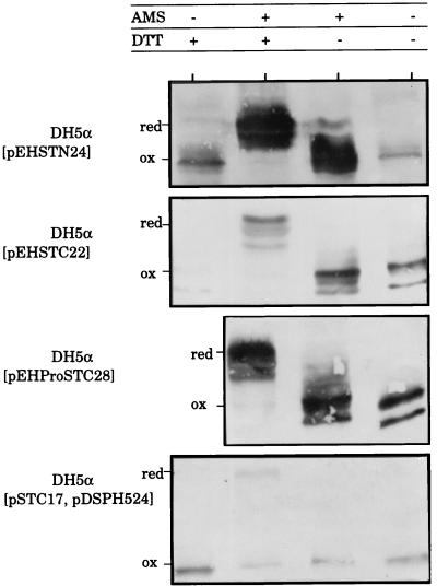
We have successfully used the major subunit ClpG of Escherichia coli CS31A fimbriae as an antigenic and immunogenic exposure-delivery vector for various heterologous peptides varying in nature and length. However, the ability of ClpG as a carrier to maintain in vitro and in vivo the native biological properties of passenger peptide has not yet been reported. To address this possibility, we genetically fused peptides containing all or part of the E. coli human heat-stable enterotoxin (STh) sequence to the amino or carboxyl ends of ClpG. Using antibodies to the ClpG and STh portions for detecting the hybrids; AMS (4-acetamido-4'-maleimidylstilbene-2, 2'-disulfonate), a potent free thiol-trapping reagent, for determining the redox state of STh in the fusion; and the suckling mouse assay for enterotoxicity, we demonstrated that all ClpG-STh proteins were secreted in vitro and in vivo outside the E. coli cells in a heat-stable active oxidized (disulfide-bonded) form. Indeed, in contrast to many earlier studies, blocking the natural NH(2) or COOH extremities of STh had, in all cases, no drastic effect on cell release and toxin activity. Only antigenicity of STh C-terminally extended with ClpG was strongly affected in a conformation-dependent manner. These results suggest that the STh activity was not altered by the chimeric structure, and therefore that, like the natural toxin, STh in the fusion had a spatial structure flexible enough to be compatible with secretion and enterotoxicity (folding and STh receptor recognition). Our study also indicates that disulfide bonds were essential for enterotoxicity but not for release, that spontaneous oxidation by molecular oxygen occurred in vitro in the medium, and that the E. coli cell-bound toxin activity in vivo resulted from an effective export processing of hybrids and not cell lysis. None of the ClpG-STh subunits formed hybrid CS31A-STh fimbriae at the cell surface of E. coli, and a strong decrease in the toxin activity was observed in the absence of CS31A helper proteins. In fact, chimeras translocated across the outer membrane as a free folded monomer once they were guided into the periplasm by the ClpG leader peptide through the CS31A-dependent secretory pathway. In summary, ClpG appears highly attractive as a carrier reporter protein for basic and applied research through the engineering of E. coli for culture supernatant delivery of an active cysteine-containing protein, such as the heat-stable enterotoxin.


Figures

FIG. 1
FIG. 1
Structure of fusion proteins. (A) The STh enterotoxin structure. The STh (STa3) sequence is from the work of Guzman-Verduzco and Kupersztoch (19). Only amino acid residues 46 to 72 of the Pre-Pro-STh precursor are shown in boldface. The six cysteines involved in the three disulfide bonds are indicated. (B) ClpG-STh fusion proteins. An additional valine at the C terminus of ClpG expressed by pHPCO838 did not affect the formation of CS31A fimbriae at the cell surface. Plasmids pEHSTN24 and pSTN24 carry the same fusion, as do pEHProSTC28 and pProSTC28, and pEHSTC22 and pSTC22 (see Materials and Methods). The numbers in lightface above the boxes are the positions of the amino acid residues relative to the signal peptide cleavage site −1/+1 of the ClpG precursor (B), and those in boldface are the positions of the amino acid residues relative to the STh precursor (A and B). Indicated amino acids represent either residues composing the sequence of the linker at the ClpG-STh junction or residues introduced in ClpG by site-directed mutagenesis (marked by an asterisk).
FIG. 2
FIG. 2
Detection of fusion proteins. (A) Culture supernatants were assayed for the presence of STh in the hybrid by competitive ELISA, consisting of the addition of various dilutions of the supernatant and of STa-monospecific HRP-conjugated antibodies to plates coated with synthetic STa. A positive sample was identified by binding inhibition of STa-specific HRP-conjugated monoclonal antibody to the immobilized STa as monitored by a decrease in A490. A sample giving an A490 of ≥1.2 contained no STh. (B) Double-antibody sandwich ELISA consisted of the addition of serially twofold-diluted supernatant sample to plates coated with the STa-monospecific 11C antibody followed by binding of rabbit anti-ClpG antibodies. Antibody binding was detected by the measurement of A405 using HRP-labeled goat anti-rabbit IgG. The double-sandwich ELISA was performed to ensure probing of the entire hybrid proteins, thus overcoming the risk of detection of intact STh free of ClpG.
FIG. 3
FIG. 3
Immunoblot analysis of fusion proteins produced by E. coli DH5α harboring the indicated plasmid. Lanes: 1, pEHSTN24; 2, pEHSTC22; 3, pEHProSTC28; 4, pSTC17+pDSPH524. Recombinants were cultured at 37°C in LB liquid (A) or LB agar medium (B to D). (A) Supernatants were from broth cultures containing 1 × 10⁹ to 2 × 10⁹ CFU/ml. (B) Supernatants were from plate-gown cultures containing 5 × 10¹⁰ to 2 × 10¹¹ CFU/ml. Supernatant samples were boiled in Laemmli buffer with β-ME and loaded onto 0.1% SDS–15% polyacrylamide gels. After electrophoresis, proteins were electrotransferred to nitrocellulose membranes and incubated either with ClpG-specific antiserum (A and B) or with the STa-specific monoclonal antibody 11C (C) or 20C1 (D). HRP-labeled goat anti-rabbit IgG (A and B) or biotin-labeled goat anti-mouse IgG in concert with HRP-conjugated streptavidin (C and D) was used as secondary antibody. The arrows at left point to the position of ClpG as assessed by the simultaneous migration of ClpG-containing solid culture supernatant from E. coli DH5α(pEH524) (data not shown).
FIG. 4
FIG. 4
Determination of minimal effective dose of the whole-cell (A) and culture supernatant (B) fractions. Only data obtained under solid culture conditions are presented as an example for minimal effective dose calculation. After centrifugation of overnight cultures, supernatants were separated from the cell pellets which were washed and suspended with PBS buffer (2 ml). Before centrifugation and after suspension in PBS, bacteria were enumerated on MacConkey lactose agar medium. Various dilutions of the supernatant and whole-cell preparations were tested in the suckling mouse assay. The enterotoxin titer was expressed as the highest dilution that gave a G/C ratio of 0.090 corresponding to an enterotoxin activity of 1 MU. Each datum point is the average of G/C ratios, which were plotted semilogarithmically versus bacterial densities (A) or supernatant dilution values (B), and the point at which the curve crossed the line equal to a G/C ratio of 0.090 (dotted line) was defined as the density (A) or the dilution (B) giving an enterotoxin activity of 1 MU. The points of the data with whole cells are 108.60, 108.82, 109.20, and 109.96 CFU, for DH5α(pEHProSTC28), DH5α(pEHSTN24), DH5α(pEHSTC22), and DH5α(pSTC17+pDSPH524), respectively.
FIG. 5
FIG. 5
Effect of reducing culture conditions on the suckling mouse activity of the supernatant (A) and whole-cell (B) fractions. LB broth-grown cells (2 ml) were poured onto LB agar medium containing (A, b and c, and B) or not (A, a) 5 mM (β-ME) and spread over the agar surface before overnight incubation at 37°C in a humid atmosphere with the agar surface facing up. Bacteria were then harvested by being scraped from the agar surface. After centrifugation, all of the supernatants were separated from cells (A), and each of them was then halved. One half was tested as it was (a and b), and the other was kept overnight at room temperature in contact with air before use (c). The corresponding β-ME-treated cell pellets were washed and resuspended in PBS, thus constituting the β-ME-cleared whole-cell fractions (B). Bars are the means ± standard errors of 2 determinations using 4 to 12 mice per sample as indicated above the bars. The dotted line equal to a G/C ratio of 0.090 represents the toxicity threshold above which the samples are considered positive.
FIG. 6
FIG. 6
Redox state of extracellular hybrids. LB broth-gown cells (2 ml) were poured onto LB agar medium and spread over the agar surface before overnight incubation at 37°C in a humid atmosphere with the agar surface facing up. Bacteria were then harvested by being scraped from the agar surface, separated from supernatants by centrifugation, and discarded. Supernatants were first reduced (+) or not (−) with 100 mM DTT at 37°C for 10 min, precipitated with TCA, washed with acetone, and then diluted in 1% SDS–1 mM EDTA–50 mM Tris-HCl (pH 8.0), containing (+) or not (−) 10 mM AMS. Samples were subjected to nonreducing SDS–12% PAGE, electrotransfer, and blotting with anti-ClpG antiserum. AMS is a potent thiol-blocking reagent highly soluble in aqueous solutions that blocks irreversibly free cysteines by producing thioesters (21). The reduced and oxidized forms can be separated by the charge difference due to AMS which increases the apparent molecular mass by 490 Da. Oxidized forms are resistant to reaction with AMS and migrate as a lower-molecular-weight protein band than do reduced forms. The position labeled “red” refers to reduced STh bound to AMS, while the position labeled “ox” designates either oxidized STh treated or not with AMS or unoxidized STh uncoupled to AMS.
FIG. 7
FIG. 7
STh in the fusion is disulfide bonded. The oxidation state of the fusion protein present in the overnight solid culture supernatant of DH5α(pEHProSTC28) was determined as described in the legend to Fig. 6, except that Western blots were additionally probed with anti-STa antibodies consisting of a mixture of the monoclonal antibodies 11C and 20C1.

References

    1. Acheson D W K. Enterotoxins in acute infective diarrhoea. J Infect. 1992;24:225–245. - PubMed
    1. Aitken R, Hirst T R. Development of an immunoassay using recombinant maltose-binding protein-STa fusions for quantitating antibody responses against STa, the heat-stable enterotoxin of Escherichia coli. J Clin Microbiol. 1992;30:732–734. - PMC - PubMed
    1. Aitken R, Hirst T R. Recombinant enterotoxins as vaccines against Escherichia coli-mediated diarrhoea. Vaccine. 1993;11:227–233. - PubMed
    1. Bakker D, Vader C E M, Roosendaal B, Mooi F R, Oudega B, de Graaf F K. Structure and function of periplasmic chaperone-like proteins involved in the biosynthesis of K88 and K99 fimbriae in enterotoxigenic Escherichia coli. Mol Microbiol. 1991;5:875–886. - PubMed
    1. Bertin Y, Girardeau J P, Der Vartanian M, Martin C. The ClpE protein involved in biogenesis of the CS31A capsule-like antigen is a member of a periplasmic chaperone family in Gram-negative bacteria. FEMS Microbiol Lett. 1993;108:59–68. - PubMed

Publication types

MeSH terms

LinkOut - more resources