Comparative Study
. 2000 Jul;68(7):4155-68.
doi: 10.1128/IAI.68.7.4155-4168.2000.

Comparative genomics of Helicobacter pylori: analysis of the outer membrane protein families

Affiliations
Comparative Study

Comparative genomics of Helicobacter pylori: analysis of the outer membrane protein families

R A Alm et al. Infect Immun. 2000 Jul.

Abstract

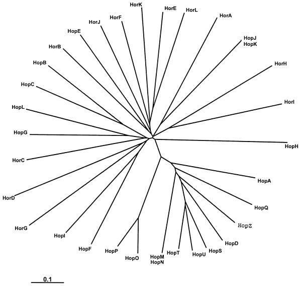
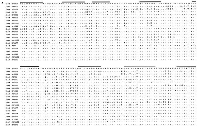
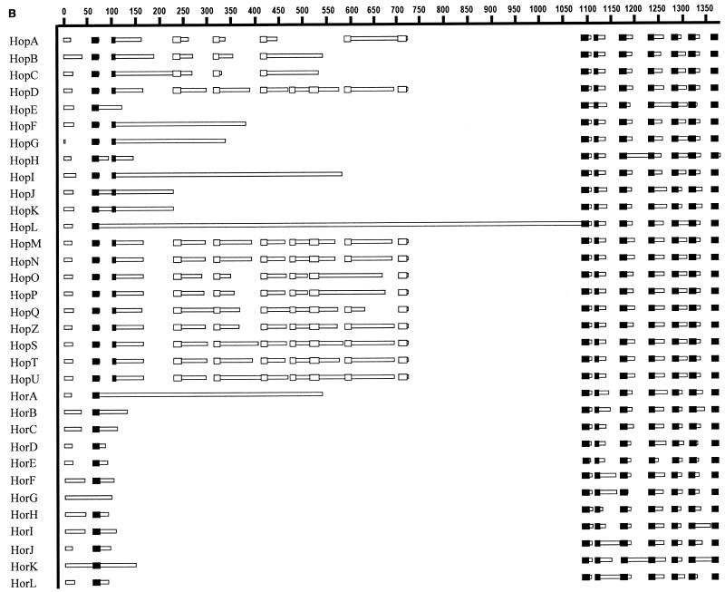
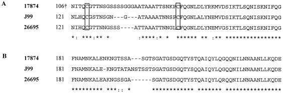
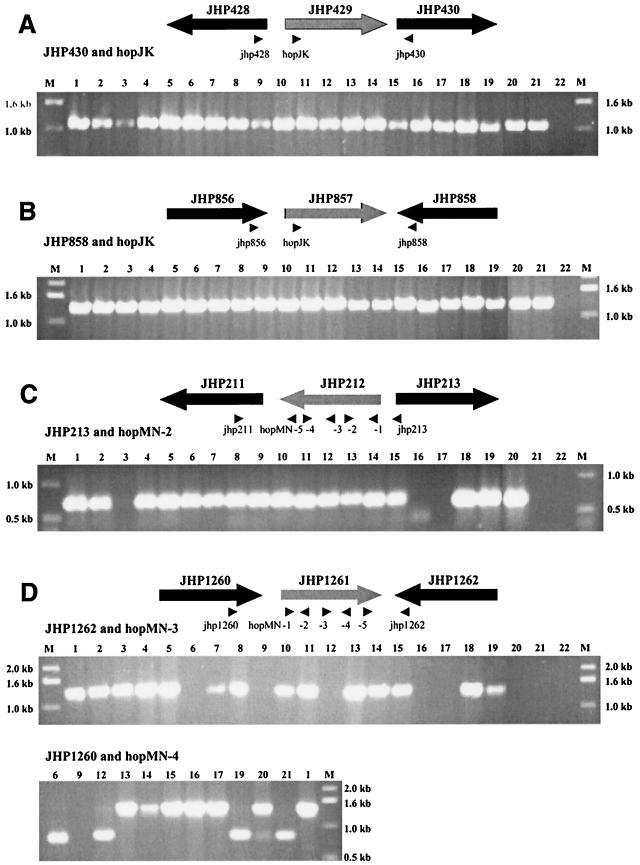
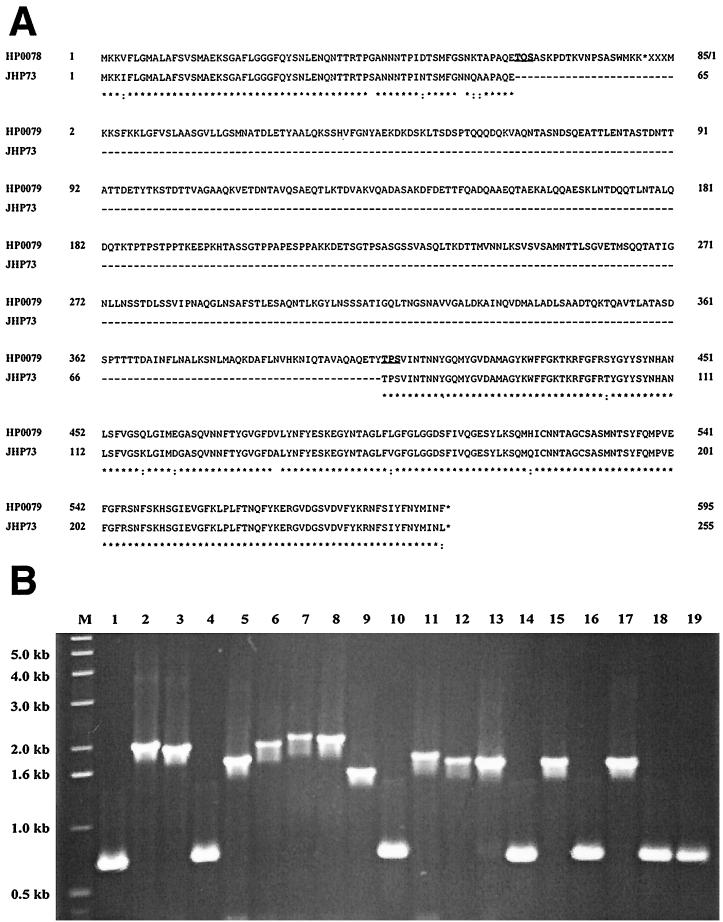
The two complete genomic sequences of Helicobacter pylori J99 and 26695 were used to compare the paralogous families (related genes within one genome, likely to have related function) of genes predicted to encode outer membrane proteins which were present in each strain. We identified five paralogous gene families ranging in size from 3 to 33 members; two of these families contained members specific for either H. pylori J99 or H. pylori 26695. Most orthologous protein pairs (equivalent genes between two genomes, same function) shared considerable identity between the two strains. The unusual set of outer membrane proteins and the specialized outer membrane may be a reflection of the adaptation of H. pylori to the unique gastric environment where it is found. One subfamily of proteins, which contains both channel-forming and adhesin molecules, is extremely highly related at the sequence level and has likely arisen due to ancestral gene duplication. In addition, the largest paralogous family contained two essentially identical pairs of genes in both strains. The presence and genomic organization of these two pairs of duplicated genes were analyzed in a panel of independent H. pylori isolates. While one pair was present in every strain examined, one allele of the other pair appeared partially deleted in several isolates.


Figures

FIG. 1
FIG. 1
Phylogenic tree of the large Hop and Hor outer membrane protein family. Protein sequences were analyzed using the PHYLIP program. The two pairs of duplicated Hop proteins (HopJ/K and HopM/N) were not differentiated and are each visualized as one line.
FIG. 2
FIG. 2
(A) Alignment of the C-terminal domains of the Y-Hop proteins from H. pylori J99 and 26695. The alignment is based on the sequence of HP0317, the strain-specific member from H. pylori 26695. The proteins are listed as orthologous pairs from the two strains. Identical residues are indicated by colon; the eight predicted transmembrane sequences are indicated above the sequence. (B) BLOCKS alignment of the Hop and Hor proteins. BLOCKS is a method used to demonstrate similarity among a group of proteins that contain repeated sections of high similarity across the family (filled boxes) or a subset of the family (unfilled boxes) flanked by regions of lesser similarity (empty bars) and variable size (blank regions representing sequence missing from a given protein).
FIG. 2
FIG. 2
(A) Alignment of the C-terminal domains of the Y-Hop proteins from H. pylori J99 and 26695. The alignment is based on the sequence of HP0317, the strain-specific member from H. pylori 26695. The proteins are listed as orthologous pairs from the two strains. Identical residues are indicated by colon; the eight predicted transmembrane sequences are indicated above the sequence. (B) BLOCKS alignment of the Hop and Hor proteins. BLOCKS is a method used to demonstrate similarity among a group of proteins that contain repeated sections of high similarity across the family (filled boxes) or a subset of the family (unfilled boxes) flanked by regions of lesser similarity (empty bars) and variable size (blank regions representing sequence missing from a given protein).
FIG. 3
FIG. 3
Alignment of the variable domains of HopB (A) and HopC (B). The H. pylori 17874 proteins are found in GenBank (accession number Z82988) and are called AlpB and AlpA, respectively. Positions of the proteins included in the alignment are indicated with numbers; † indicates that the difference in position within the HopB protein represents a difference in the prediction of the initiation codon. The conserved cysteine residues in the HopB proteins are boxed. Identical (∗) and conserved (:) residues are indicated.
FIG. 4
FIG. 4
Examination of H. pylori isolates for the duplication of hop genes. The genomic organization and primer binding location sites for JHP429 (hopJ) (A), JHP857 (hopK) (B), JHP212 (hopM) (C), and JHP1261 (hopN) (D) are shown. Representative PCRs are also shown in each panel, with the primer combinations used indicated. The loading order for each panel is as follows: marker (lane M), J99 (lane 1), 26695 (lane 2), ARHp64 (lane 3), SS1 (lane 4), UA861 (lane 5), ARHp12 (lane 6), ARHp18 (lane 7), ARHp25 (lane 8), ARHp210 (lane 9), ARHp65 (lane 10), ARHp55 (lane 11), ARHp124 (lane 12), ARHp54 (lane 13), CCUG17874 (lane 14), ARHp221 (lane 15), ARHp246 (lane 16), ARHp245 (lane 17), AH244 (lane 18), ARHp241 (lane 19), ARHp243 (lane 20), ARHp244 (lane 21), and no-DNA control (lane 22). The strains shown in the second gel in panel D (primers JHP1260 and hopMN-4) are indicated with the same numbering system. The sizes of the molecular weight markers are indicated.
FIG. 5
FIG. 5
(A) Alignment of the JHP73 and the HP0078/HP0079 proteins. Amino acid positions of the proteins are indicated by numbers. Identical (∗) and conserved (:) residues are indicated. As predicted by Tomb et al. (61), the short HP0078 protein ends after 85 residues and the HP0079 protein begins 11 nt later. (B) PCR analysis of multiple H. pylori isolates for the presence of a JHP73 ortholog, using the primer combination jhp73F/jhp73R. Molecular weight markers are shown in lane M, with the sizes indicated on the left. The strains analyzed are J99 (lane 1), 26695 (lane 2), ARHp64 (lane 3), SS1 (lane 4), UA861 (lane 5), ARHp12 (lane 6), ARHp18 (lane 7), ARHp25 (lane 8), ARHp210 (lane 9), ARHp65 (lane 10), ARHp55 (lane 11), ARHp124 (lane 12), ARHp54 (lane 13), CCUG17874 (lane 14), ARHp221 (lane 15), ARHp245 (lane 16), AH244 (lane 17), ARHp243 (lane 18), and ARHp244 (lane 19).

References

    1. Akopyants N S, Jiang Q, Taylor D E, Berg D E. Corrected identity of isolates of Helicobacter pylori reference strain NCTC11637. Helicobacter. 1997;2:48–52. - PubMed
    1. Akopyanz N, Bukanov N O, Westblom T U, Berg D E. PCR-based RFLP analysis of DNA sequence diversity in the gastric pathogen Helicobacter pylori. Nucleic Acids Res. 1992;20:6221–6225. - PMC - PubMed
    1. Akopyanz N, Bukanov N O, Westblom T U, Kresovich S, Berg D E. DNA diversity among clinical isolates of Helicobacter pylori detected by PCR-based RAPD fingerprinting. Nucleic Acids Res. 1992;20:5137–5142. - PMC - PubMed
    1. Alm R A, Ling L L, Moir D T, King B L, Brown E D, Doig P C, Smith D R, Noonan B, Guild B C, deJonge B L, Carmel G, Tummino P J, Caruso A, Uria-Nickelsen M, Mills D M, Ives C, Gibson R, Merberg D, Mills S D, Jiang Q, Taylor D E, Vovis G F, Trust T J. Genomic sequence comparison of two unrelated isolates of the human gastric pathogen Helicobacter pylori. Nature. 1999;397:186–190. - PubMed
    1. Appelmelk B J, Martin S L, Monteiro M A, Clayton C A, McColm A A, Zheng P, Verboom T, Maaskant J J, van den Eijnden D H, Hokke C H, Perry M B, Vandenbroucke-Grauls C M J E, Kusters J G. Phase variation in Helicobacter pylori lipopolysaccharide due to changes in the lengths of poly(C) tracts in α3-fucosyltransferase genes. Infect Immun. 1999;67:5361–5366. - PMC - PubMed

Publication types

MeSH terms

LinkOut - more resources