. 2000 Jul;68(7):4207-16.
doi: 10.1128/IAI.68.7.4207-4216.2000.

Effects of estradiol and progesterone on susceptibility and early immune responses to Chlamydia trachomatis infection in the female reproductive tract

Affiliations

Effects of estradiol and progesterone on susceptibility and early immune responses to Chlamydia trachomatis infection in the female reproductive tract

C Kaushic et al. Infect Immun. 2000 Jul.

Abstract

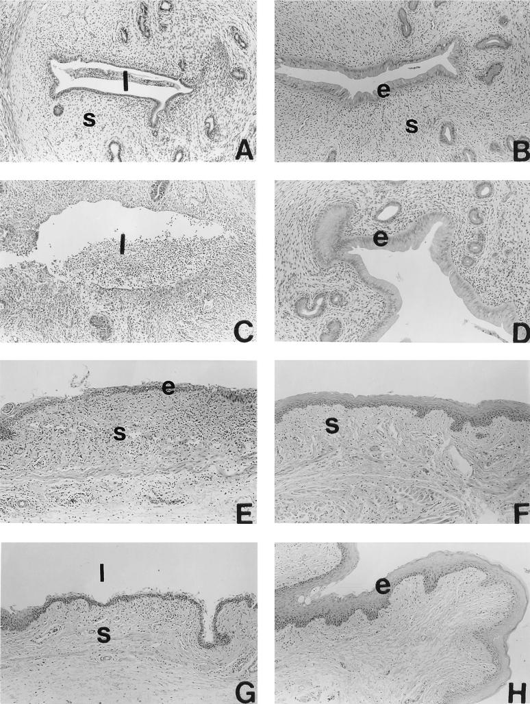
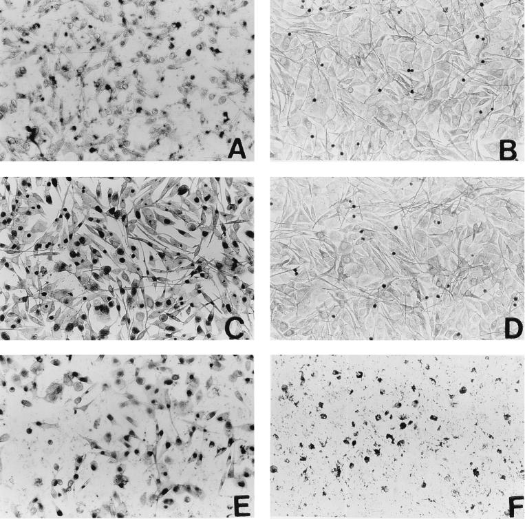
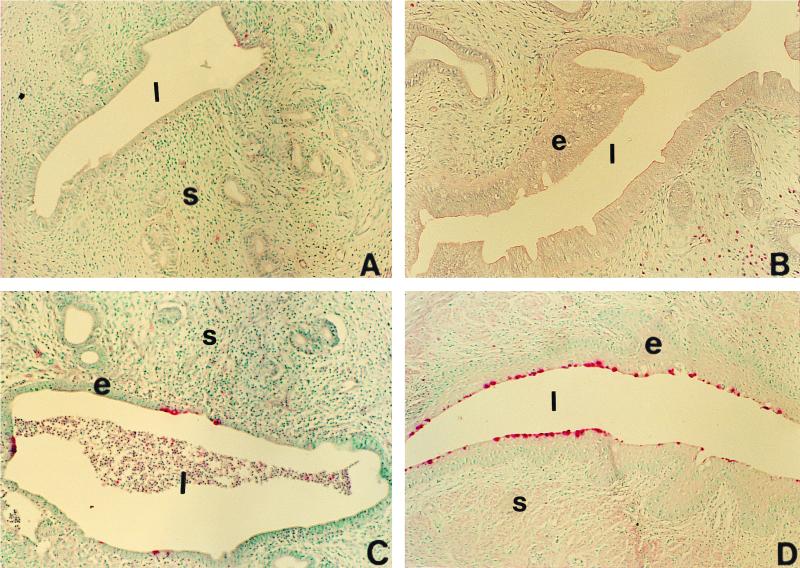
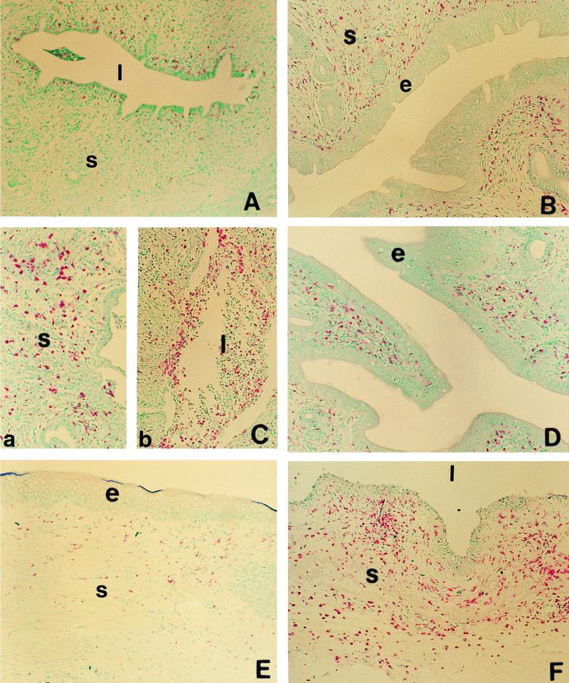
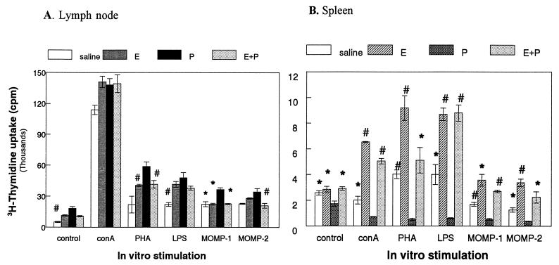
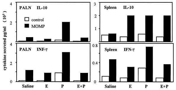
We have used a previously described rodent model to examine the influence of hormonal environment on susceptibility and immune responses to genital Chlamydia infection. Ovariectomized rats were administered estradiol, progesterone, or a combination of both, infected with Chlamydia trachomatis via the intrauterine route, and sacrificed 5 days later. Histopathological examination showed severe inflammation in the uteri and vaginae of progesterone-treated animals, whereas animals receiving estradiol or a combination of both hormones showed no inflammation. Large numbers of chlamydiae were found in vaginal secretions of progesterone-treated and combination-treated animals, while estradiol-treated animals had none. Tissue localization showed that numerous chlamydial inclusions were present in the uterine epithelium of the progesterone group and the cervicovaginal epithelium of the combination group. Examination of the acute immune responses of the infected animals showed that maximum activation was present in the draining lymph node cells from the progesterone-treated group, and these cells were producing large amounts of interleukin-10 and gamma interferon compared to other hormone-treated groups. In contrast, spleen cell proliferation was suppressed in progesterone-treated animals compared to other hormone-treated groups. We conclude that progesterone increases and estradiol decreases susceptibility to intrauterine chlamydial infection in this rat model. Our data demonstrate that hormone environment, at the time of infection, has a profound effect on the outcome of microbial infection in the female reproductive tract.


Figures

FIG. 1
FIG. 1
Histopathology of uteri (A to D) and vaginae (E to F) of rats infected with C. trachomatis following intrauterine treatment with saline (A and E), estradiol (B and F), progesterone (C and G), or estradiol and progesterone (D and H). Animals were sacrificed 5 days postinfection; reproductive tract tissues were removed, fixed, processed for histology, and stained with hematoxylin and eosin. Representative tissue sections from each treatment groups are shown. Estrogenic effects on epithelial cells of uteri and vaginae of rats receiving estradiol can be observed (B, D, F, and H). Note the acute inflammation in the uterus of a progesterone-treated rat, characterized by large number of infiltrating leukocytes (C). s, stroma; l, lumen; e, epithelium. Original magnification, ×100.
FIG. 2
FIG. 2
In vitro infectivity assay to assess bacterial shedding in vaginal washes of hormone-treated, infected rats. Vaginal washes from day 5 postinfection were layered on monolayers of McCoy cells and stained with an anti-Chlamydia antibody as described in Materials and Methods. Micrographs show stained monolayers from cultures layered with washings (all at same dilution) from animals infected with Chlamydia after treatment with saline (C), estradiol (D), progesterone (E), and estradiol plus progesterone (F). Positive (previously titered stock; A) and negative (mock infection; B) controls are also shown. Darkly stained inclusion bodies can be clearly distinguished in infected monolayers. Note that panels E and F are partially destroyed monolayers, indicating presence of too many IFU. These and similar samples were diluted further and retitered to obtain final titers. Results from estradiol-treated animals (D) were found to be similar to negative control results in all samples. The dark staining seen in panels B and D was due to staining of dead cells and was subtracted as background when titers were calculated.
FIG. 3
FIG. 3
Localization of infection in the reproductive tracts of hormone-treated, infected rats by immunohistochemical staining of chlamydial antigens. Polyclonal antibody was used to detect Chlamydia-specific staining as described in Materials and Methods. Representative tissue sections showing typical staining for each group are shown in the micrographs. Positive (pink) staining can be seen in the uterine epithelium of progesterone-treated animals (C) and cervicovaginal epithelium of estradiol-progesterone-treated animals (D). Very little positive staining was seen in saline-treated animals (A). Uteri and vaginae of estradiol-treated animals were negative in all sections examined. s, stroma; l, lumen; e, epithelium. Original magnification, ×100.
FIG. 4
FIG. 4
Localization of ED1-positive cells (macrophages and blood granulocytes) in the uteri (A to D) and vaginae (E and F) of hormone-treated, infected rats. Positively stained cells (pink) can be seen in uteri of animals treated with saline (A), estradiol (B), progesterone (C), or estradiol and progesterone (D). Micrographs of representative tissue sections from vaginae of estradiol-treated (E) and progesterone-treated (F) animals stained for ED1-positive cells are also shown. Note the accumulation of large number of positively stained cells in the subepithelial stroma (C, a) and the leukocytic infiltration in the lumen (C, b) of uteri of progesterone-treated animals. s, stroma; l, lumen; e, epithelium. Original magnification, ×100.
FIG. 5
FIG. 5
Lymph node (A) and spleen (B) cell proliferation assay in response to mitogens and MOMP in hormone-treated, infected animals. Four animals were used for each treatment group. Results shown are representative of two separate experiments. Spleen or lymph node cells were isolated as described in Materials and Methods and incubated in the presence of medium alone (control), ConA (1 μg/ml), PHA (5 μg/ml), LPS (10 μg/ml), MOMP-1 (1 μg/ml), and MOMP-2 (5 μg/ml). ∗, P < 0.01; #, P < 0.05 compared to progesterone-treated group. E, estradiol; P, progesterone.
FIG. 6
FIG. 6
IFN-γ and IL-10 measurements in supernatants from PALN and spleen cells of hormone-treated, infected animals. Both IFN-γ and IL-10 were in measured in PALN and spleen cell cultures from day 5 postinfection, incubated with or without MOMP (1 μg/ml). E, estradiol; P, progesterone.

References

    1. Baker D A, Plotkin S A. Enhancement of vaginal infection in mice by herpes simplex virus type II with progesterone. Proc Soc Exp Biol Med. 1978;158:131–134. - PubMed
    1. Barron A, Pasley J N, Rank R G, White H J, Mrak R E. Chlamydial salpingitis in female guinea pigs receiving oral contraceptives. Sex Transm Dis. 1988;15:169–173. - PubMed
    1. Igietseme J U, Rank R G. Susceptibility to reinfection after a primary chlamydial genital infection is associated with a decrease of antigen-specific T cells in the genital tract. Infect Immun. 1991;59:1346–1351. - PMC - PubMed
    1. Ito J J I, Harrison R, Alexander E R, Billings L J. Establishment of genital tract infection in the CF-1 mouse by intravaginal inoculation of a human oculogenital isolate of chlamydia trachomatis. J Infect Dis. 1984;150:577–582. - PubMed
    1. Kaushic C, Frauendorf E, Rossoll R M, Richardson J M, Wira C R. Influence of estrous cycle on the presence and distribution of immune cells in the rat reproductive tract. Am J Reprod Immunol. 1998;39:209–216. - PubMed

Publication types

MeSH terms