. 2000 Jul;68(7):4217-24.
doi: 10.1128/IAI.68.7.4217-4224.2000.

The most abundant glycoprotein of amebic cyst walls (Jacob) is a lectin with five Cys-rich, chitin-binding domains

Affiliations

The most abundant glycoprotein of amebic cyst walls (Jacob) is a lectin with five Cys-rich, chitin-binding domains

M Frisardi et al. Infect Immun. 2000 Jul.

Abstract

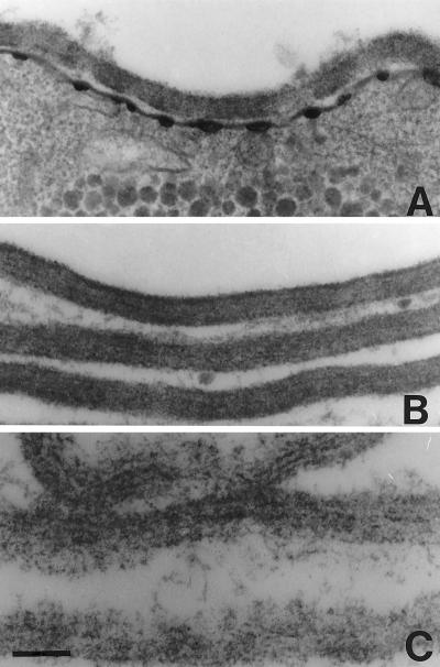
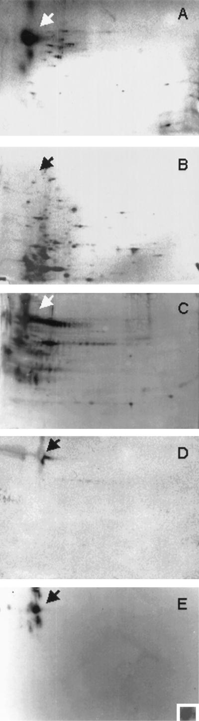
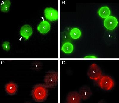
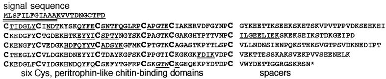
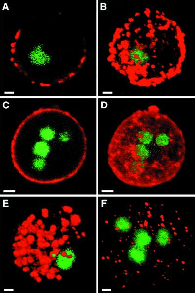
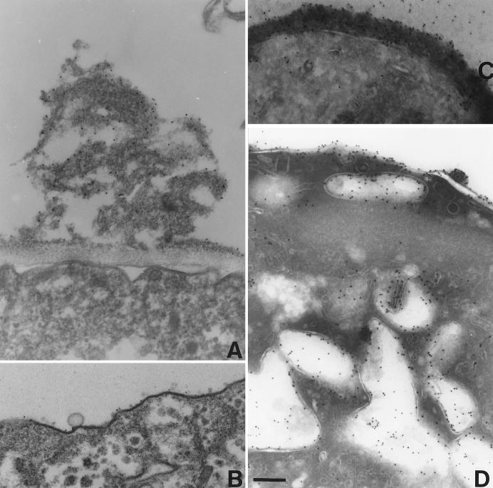
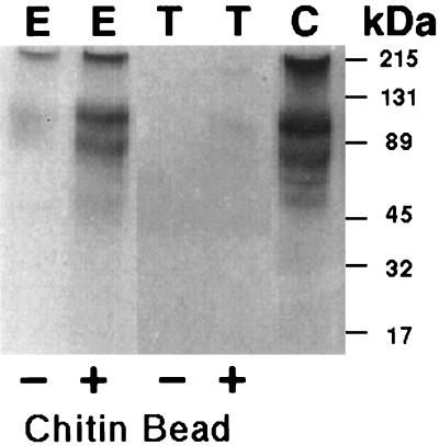
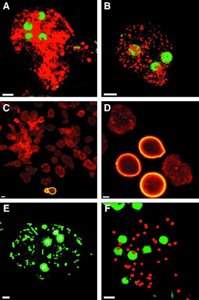
The infectious stage of amebae is the chitin-walled cyst, which is resistant to stomach acids. In this study an extraordinarily abundant, encystation-specific glycoprotein (Jacob) was identified on two-dimensional protein gels of cyst walls purified from Entamoeba invadens. Jacob, which was acidic and had an apparent molecular mass of approximately 100 kDa, contained sugars that bound to concanavalin A and ricin. The jacob gene encoded a 45-kDa protein with a ladder-like series of five Cys-rich domains. These Cys-rich domains were reminiscent of but not homologous to the Cys-rich chitin-binding domains of insect chitinases and peritrophic matrix proteins that surround the food bolus in the insect gut. Jacob bound purified chitin and chitin remaining in sodium dodecyl sulfate-treated cyst walls. Conversely, the E. histolytica plasma membrane Gal/GalNAc lectin bound sugars of intact cyst walls and purified Jacob. In the presence of galactose, E. invadens formed wall-less cysts, which were quadranucleate and contained Jacob and chitinase (another encystation-specific protein) in secretory vesicles. A galactose lectin was found to be present on the surface of wall-less cysts, which phagocytosed bacteria and mucin-coated beads. These results suggest that the E. invadens cyst wall forms when the plasma membrane galactose lectin binds sugars on Jacob, which in turn binds chitin via its five chitin-binding domains.


Figures

FIG. 1
FIG. 1
Transmission electron micrographs of E. invadens cyst walls. (A) Intact E. invadens cysts have an electron-dense wall, which overlies secretory vacuoles and electron densities along the plasma membrane. (B) Cyst walls purified by two sucrose gradients contain electron-dense material between chitin fibrils. (C) Chitin fibrils and little other electron-dense material remain in cyst walls after boiling in SDS. Bar, 200 nm. Magnification, ×4,500.
FIG. 2
FIG. 2
Two-dimensional gels of E. invadens cyst wall proteins. (A and B) Silver stains show that Jacob, which is by far the most abundant cyst wall protein (arrow in panel A), is absent from trophozoites (B). (C and D) Western blots show that ConA (C) and ricin (D), visualized with acid phosphatase, bind to Jacob and numerous other cyst wall glycoproteins. (E) In contrast, anti-Jacob antibodies, visualized by chemiluminescence, bind to Jacob and larger and smaller proteins with the same charge. Purified E. histolytica Gal/GalNAc lectin, which was detected with anti-lectin antibodies, binds to Jacob excised from a Ponceau-stained two-dimensional gel (inset in panel E). A negative control, in which the Gal/GalNAc lectin was omitted, did not bind the anti-Gal/GalNAc antibodies (data not shown).
FIG. 3
FIG. 3
Fluorescence micrographs of E. invadens cysts and trophozoites. (A and B) FITC-ConA (arrowheads in panel A) and FITC-ricin (B) stained the surface of cysts (c) much more intensely than the surface of trophozoites (t). (C) Similarly, anti-Jacob antibodies, made to the native Jacob protein, bound to the surface of cysts but not to trophozoites. (D) The E. histolytica Gal/GalNAc lectin, which was detected with anti-lectin antibodies, also bound to E. invadens cyst walls but not to the surface of trophozoites.
FIG. 4
FIG. 4
Primary structure of E. invadens Jacob, in single-letter code. An asterisk marks the stop codon. The signal sequence, proven by N-terminal sequencing of intact Jacob, is underlined twice, while the N-terminal sequences of Jacob tryptic peptides are each underlined once. PCR primers for cloning the E. invadens jacob gene were made to QYFECSNT and HDFQYYV. A possible site of Asn-linked glycosylation (NDT) is marked with a wavy underline. Conserved Cys residues in putative chitin-binding domains, which are aligned, are marked in bold.
FIG. 5
FIG. 5
Confocal micrographs of encysting E. invadens stained with anti-Jacob antibodies (red in panels A through E), anti-chitinase antibodies (red in panel F) and Sytox green (nuclear stain in panels A through F). Jacob was present in multiple places on the surface of encysting parasites with one nucleus (in section [A] and three-dimensional composite [B]) and became more dense on parasites with four nuclei (in section [C] and composite [D]). Jacob was present in numerous relatively large secretory vesicles (in composite [E]) that surround the nuclei of encysting parasites, which were permeabilized before labeling. Chitinase (in composite [F]) was present within hundreds of smaller secretory vesicles of an encysting parasite. Bars, 5 μm.
FIG. 6
FIG. 6
Immuno-EM of anti-Jacob antibodies binding to cysts and wall-less cysts. (A) Anti-Jacob antibodies, visualized with gold particles, were present over electron-dense material on the surface of encysting parasites, which were stained prior to fixation. (B) In contrast, wall-less cysts, which were made in the presence of galactose, lacked chitin and had a thin layer of electron-dense material that bound few anti-Jacob antibodies. (C and D) When encysting parasites were fixed and sectioned prior to staining, anti-Jacob antibodies bound to cyst walls and to numerous large secretory vesicles. Bars, 200 nm. Magnification, ×4,500.
FIG. 7
FIG. 7
SDS-PAGE and Western blots of Jacob binding to chitin beads. Western blots with anti-Jacob antibodies to trophozoite proteins (T) before (−) and after (+) binding to chitin beads, total proteins from encysting parasites (E) before (−) and after (+) binding to chitin beads, and cyst wall proteins (C) (positive control) are shown. Two bands labeled with anti-Jacob antibodies correspond to 100-kDa and high-molecular-mass forms of Jacob identified on two-dimensional protein gels (Fig. 2). Molecular mass standards are marked on the right.
FIG. 8
FIG. 8
Confocal micrographs of wall-less cysts, which were made by encysting E. invadens parasites for 2 days in the presence of 50 mM galactose. Wall-less cysts, which were permeabilized before labeling, had four nuclei (stained with Sytox green in panels A, B, E, and F) and contained numerous secretory vesicles stained with anti-Jacob antibodies (red in panel A) and anti-chitinase antibodies (red in panel B). The vast majority of wall-less cysts, which were labeled with anti-Jacob antibodies (red in panel C), lacked chitin on their surface, which was detected with WGA (yellow in panel C). In contrast, many control parasites encysting in the absence of galactose were spherical and bound WGA to their cyst walls (yellow in panel D). The galactose lectin was present on the surface of wall-less cysts, which phagocytosed GFP-labeled bacteria (stained green in panel E) or mucin-coated beads (stained red in panel F) when excess galactose was removed. Bars, 5 μm.

References

    1. Altschul S F, Madden T L, Schaffer A A, Zhang J, Zhang Z, Miller W, Lipman D J. Gapped BLAST and PSI-BLAST: a new generation of protein database search programs. Nucleic Acids Res. 1997;25:3389–3402. - PMC - PubMed
    1. Arroyo-Begovich A, Carbez-Trejo A. Location of chitin in the cyst wall of Entamoeba invadens with the colloidal gold tracers. J Parasitol. 1982;68:253–258. - PubMed
    1. Avron B, Deutsch R M, Mirelman D. Chitin synthesis inhibitors prevent cyst formation by trophozoites. Biochem Biophys Res Commun. 1982;108:815–821. - PubMed
    1. Beintema J J. Structural features of plant chitinases and chitin-binding proteins. FEBS Lett. 1994;350:159–163. - PubMed
    1. Bulawa C E. Genetics and molecular biology of chitin synthesis in fungi. Annu Rev Microbiol. 1993;43:505–534. - PubMed

Publication types

MeSH terms