. 2000 Jul;68(7):4225-37.
doi: 10.1128/IAI.68.7.4225-4237.2000.

Cryptococcus neoformans is a facultative intracellular pathogen in murine pulmonary infection

Affiliations

Cryptococcus neoformans is a facultative intracellular pathogen in murine pulmonary infection

M Feldmesser et al. Infect Immun. 2000 Jul.

Abstract

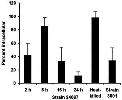
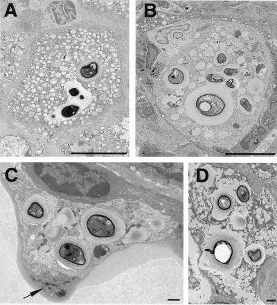
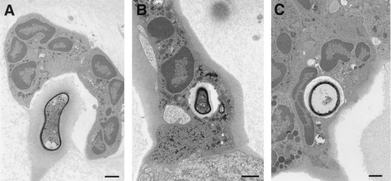
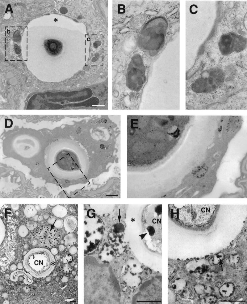
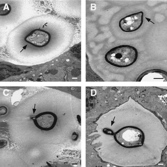
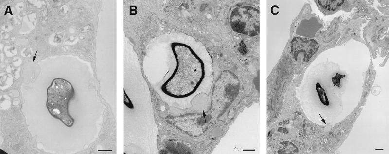
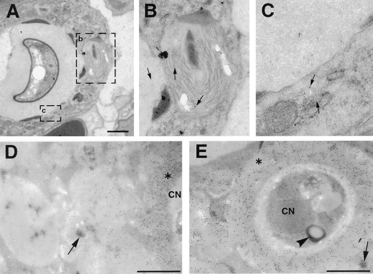
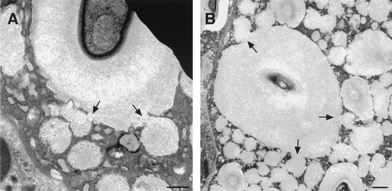
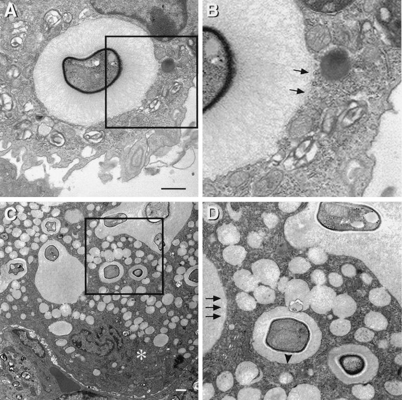
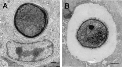
To produce chronic infection, microbial pathogens must escape host immune defenses. Infection with the human pathogenic fungus Cryptococcus neoformans is typically chronic. To understand the mechanism by which C. neoformans survives in tissue after the infection of immunocompetent hosts, we systematically studied the course of pulmonary infection in mice by electron microscopy. The macrophage was the primary phagocytic cell at all times of infection, but neutrophils also ingested yeast. Alveolar macrophages rapidly internalized yeast cells after intratracheal infection, and intracellular yeast cells were noted at all times of infection from 2 h through 28 days. However, the proportion of yeast cells in the intracellular and extracellular spaces varied with the time of infection. Early in infection, yeast cells were found predominantly in the intracellular compartment. A shift toward extracellular predominance occurred by 24 h that was accompanied by macrophage cytotoxicity and disruption. Later in infection, intracellular persistence in vivo was associated with replication, residence in a membrane-bound phagosome, polysaccharide accumulation inside cells, and cytotoxicity to macrophages, despite phagolysosomal fusion. Many phagocytic vacuoles with intracellular yeast had discontinuous membranes. Macrophage infection resulted in cells with a distinctive appearance characterized by large numbers of vacuoles filled with polysaccharide antigen. Similar results were observed in vitro using a macrophage-like cell line. Our results show that C. neoformans is a facultative intracellular pathogen in vivo. Furthermore, our observations suggest that C. neoformans occupies a unique niche among the intracellular pathogens whereby survival in phagocytic cells is accompanied by intracellular polysaccharide production.


Figures

FIG. 1
FIG. 1
Location of C. neoformans. The percentage of intracellular yeast for each group was determined by counting all yeast cells on 4 or 5 noncontiguous grids for each of two mice (8 to 10 grids per group). Heat-killed yeast of strain 24067 and live yeast of strain 3501 were studied 24 h after infection. Bars represent means; error bars denote standard deviations. All pairwise comparisons between tissues infected with strain 24067 were statistically significantly different as determined by Student's t test with the Bonferroni correction except for comparison between the tissues obtained at 2 and 16 h and between the tissue obtained at 8 h and tissue from mice infected with heat-killed yeast.
FIG. 2
FIG. 2
Intracellular replication and cytopathic effects of C. neoformans. (A and B) Phagosomes containing multiple yeast cells, demonstrating heterogeneity of size within the phagosome and budding forms. Bars, 10 μm. Time after infection: A, 28 days; B, 7 days. (C) Macrophage containing several intracellular C. neoformans cells at 7 days shows cytoplasmic disruption. The arrow points to membrane-bound cellular debris seen in proximity to this cell, suggesting that cellular destruction is a consequence of infection. (D) Multinucleated giant cell at 28 days demonstrating numerous intracellular yeast cells and abundant cytoplasmic vacuolation. Bars, 1 μm.
FIG. 3
FIG. 3
Neutrophils in close contact with C. neoformans in tissue. (A) A collection of neutrophils abuts an extracellular yeast in the alveolar space 48 h after infection. (B and C) At 7 days after infection, neutrophil phagocytosis of C. neoformans is seen, suggesting a role for neutrophils in host defense in vivo. Bars, 1 μm.
FIG. 4
FIG. 4
Phagolysosomal fusion. (A) Intracellular cryptococcus 2 h after infection. Macrophage lysosomes have moved toward the yeast and appear to fuse with the phagosome. Asterisk denotes artifactual contraction of the cryptococcal capsule from the wall of the phagosome. (B and C) Higher magnification of areas contained within rectangles in panel A demonstrates the fusion of lysosomes with the phagosome. Electron-dense material at the periphery of the phagosome likely represents entry of lysosomal components into phagosome. (D and E) Acid phosphatase cytochemistry 2 h after infection demonstrating lysosomal fusion with the phagosome, with entry of the black reaction product into the phagosome. Panel E is a higher-magnification view of the rectangular region shown in panel D. (F to H) Vacuoles containing black deposits representing acid phosphatase activity (arrows) are fused with phagosomes containing C. neoformans on day 14. Many small cytoplasmic vacuoles without yeast have acid phosphatase activity, suggesting that they are lysosomes. Some of these vacuoles are in direct continuity with the phagosome (E), while others are discontinuous. (F) Acid phosphatase reaction product is also visible at the periphery of the phagosome. Bars, 1 μm.
FIG. 5
FIG. 5
Geometry of extracellular budding. Extracellular organisms at various stages of budding. Photos were obtained in 129/SvEv mice 13 days after infection. Bars, 1 μm. Arrows point to buds. At all stages, the buds are encased in structures that label with MAb to CNPS. This phenomenon was seen in all three mouse strains and with all three encapsulated cryptococcal strains in C57BL/6 mice.
FIG. 6
FIG. 6
Cytoplasmic blebs in macrophages with internalized C. neoformans. Adjacent to phagosomes containing yeast, the macrophage cytoplasm developed hypolucent areas (blebs) with few organelles (arrows) at regions of contact with extracellular cryptococci. These may represent a precursor for the increasing cytoplasmic disruption seen with increasing time after infection. Bars, 1 μm. Times after infection: A, 48 h; B, 8 h; C, 24 h.
FIG. 7
FIG. 7
Immunoelectron microscopy for CNPS. (A) Two hours after infection, lysosomes fuse with the phagosome containing C. neoformans. (B and C) Higher magnification of the areas within rectangles seen in panel A. Gold particles inside lysosomes indicate that the CNPS has entered the lysosome (arrows). (D and E) Colocalization of gold particles demonstrating the presence of CNPS, with acid phosphatase activity in cytoplasmic vacuoles 14 days after infection. Note the continuity between spaces containing CNPS and lysosomes. Sections were not counterstained with uranyl or lead. CN, C. neoformans; Asterisks indicate cryptococcal capsules. Arrows point to representative substrate deposition indicative of lysosomal acid phosphatase activity. Arrowhead denotes acid phosphatase activity inside C. neoformans. Bars, 1 μm.
FIG. 8
FIG. 8
Vacuole formation. (A and B) Pulmonary macrophages from two C57BL/6 mice infected for 14 days demonstrating the network of cytoplasmic vacuoles. Direct continuity is seen between the contents of cytoplasmic vacuoles and phagosomes (arrows). Bars, 1 μm.
FIG. 9
FIG. 9
Phagosomal membrane disruption. Macrophage phagosomes containing C. neoformans in a C57BL/6 mouse 48 h after infection (A and B) and in an A/JCr mouse 14 days after infection (C and D) demonstrate areas in which the phagosome membrane is discontinuous (arrows), despite the appearance of well-defined membranes in cytoplasmic organelles. Panels B and D are enlargements of areas contained in the rectangles in panels A and C, respectively. The asterisk in panel C demonstrates well-preserved membranes in the Golgi apparatus. The arrowheads in panels C and D demonstrate sharply defined phagosomal membrane coexisting in a cell in which a phagosomal membrane is disrupted. Bars, 1 μm.
FIG. 10
FIG. 10
The distance between the phagosome membrane and the C. neoformans cell wall is dependent on the size of the capsule. (A) Phagosome containing a nonencapsulated Cap 67 mutant has the phagosomal membrane in close apposition to the fungal cell wall. (B) In a phagosome containing the encapsulated strain 3501, the polysaccharide capsule is interposed between the phagosomal membrane and the cell wall. Bars, 1 μm.
FIG. 11
FIG. 11
In vitro phagocytosis assay with J774 cells and C. neoformans strains 3501 (encapsulated, black bars) and Cap 67 (acapsular, white bars). Values represent means; error bars denote standard deviations. (A) Percentage of J774 cells with number of yeast cells (n = 1, 2, 3, 4, or >4) per J774 cell after various times (t) of incubation. Statistical analysis compared the percentage of J774 cells with >4 yeast cells per cell. Asterisks denote P values of <0.05 compared to values at 3 h. (B) Percentage of J774 cells with intracellular C. neoformans after various times of incubation. An asterisk denotes a P value of <0.05 compared to the value at 3 h of incubation with strain 3501. A double asterisk denotes a P value of <0.05 compared to the value at 3 h of incubation with strain Cap 67. (C) Percentage of dead J774 cells that contained intracellular C. neoformans. An asterisk denotes a P value of <0.05 compared to the value at 18 h of incubation with strain 3501. The percentage of dead cells may be underestimated because dead cells become unattached and are washed off during the staining process. The loss of dead cells likely explains the decrease in the percentage of J774 cells with intracellular yeast at 66 h of incubation with strain 3501 seen in panel B. (D) Immunohistochemistry for CNPS using MAb 2H1. Top left, single cell of strain 3501 inside an intact J774 cell after 3 h of incubation; top right, J774 cell after 18 h of incubation with collections of CNPS and single intracellular yeast demonstrating that, in some cases, yeast cells appeared to be destroyed; bottom, J774 cells after 66 h of incubation with strain 3501 show cells with large vacuoles containing multiple cryptococci with budding forms (left) and cells with multiple phagosomes containing yeast cells heterogeneous in size, consistent with active replication (right). All panels were photographed at a magnification of ×1,000.

References

    1. Adams D O. The granulomatous inflammatory response. Am J Pathol. 1976;84:164–191. - PMC - PubMed
    1. Antoine J-C, Prina E, Jouanne C, Bongrand P. Parasitophorous vacuoles of Leishmania amazonensis-infected macrophages maintain an acidic pH. Infect Immun. 1990;58:779–787. - PMC - PubMed
    1. Casadevall A, Cleare W, Feldmesser M, Glatman-Freedman A, Goldman D L, Kozel T R, Lendvai N, Mukherjee J, Pirofski L-A, Rivera J, Rosas A L, Scharff M D, Valadon P, Westin K, Zhong Z. Characterization of a murine monoclonal antibody to Cryptococcus neoformans polysaccharide that is a candidate for human therapeutic studies. Antimicrob Agents Chemother. 1998;42:1437–1446. - PMC - PubMed
    1. Casadevall A, Mukherjee J, Devi S J N, Schneerson R, Robbins J B, Scharff M D. Antibodies elicited by a Cryptococcus neoformans-tetanus toxoid conjugate vaccine have the same specificity as those elicited in infection. J Infect Dis. 1992;165:1086–1093. - PubMed
    1. Casadevall A, Perfect J R. Cryptococcus neoformans. Washington, D.C.: American Society for Microbiology; 1998.

Publication types

MeSH terms

Substances

LinkOut - more resources