

Sodium dodecyl sulfate and C31G as microbicidal alternatives to nonoxynol 9: comparative sensitivity of primary human vaginal keratinocytes
- PMID: 10858360
- PMCID: PMC89991
- DOI: 10.1128/AAC.44.7.1954-1960.2000
Sodium dodecyl sulfate and C31G as microbicidal alternatives to nonoxynol 9: comparative sensitivity of primary human vaginal keratinocytes
Abstract
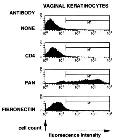
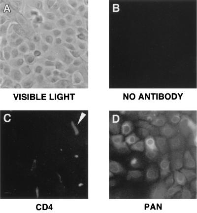
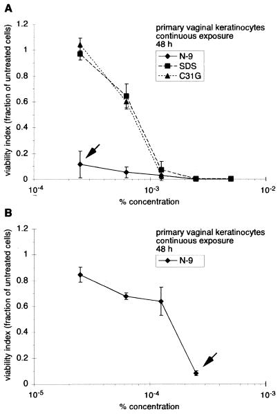
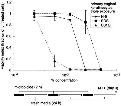
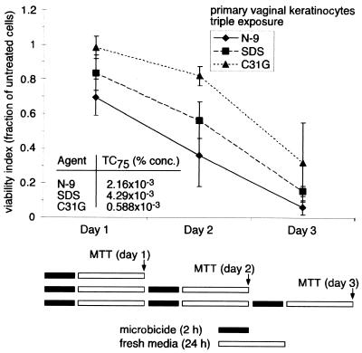
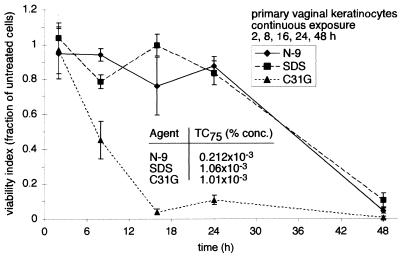
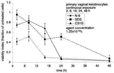
A broad-spectrum vaginal microbicide must be effective against a variety of sexually transmitted disease pathogens and be minimally toxic to the cell types found within the vaginal epithelium, including vaginal keratinocytes. We assessed the sensitivity of primary human vaginal keratinocytes to potential topical vaginal microbicides nonoxynol-9 (N-9), C31G, and sodium dodecyl sulfate (SDS). Direct immunofluorescence and fluorescence-activated cell sorting analyses demonstrated that primary vaginal keratinocytes expressed epithelial cell-specific keratin proteins. Experiments that compared vaginal keratinocyte sensitivity to each agent during a continuous, 48-h exposure demonstrated that primary vaginal keratinocytes were almost five times more sensitive to N-9 than to either C31G or SDS. To evaluate the effect of multiple microbicide exposures on cell viability, primary vaginal keratinocytes were exposed to N-9, C31G, or SDS three times during a 78-h period. In these experiments, cells were considerably more sensitive to C31G than to N-9 or SDS at lower concentrations within the range tested. When agent concentrations were chosen to result in an endpoint of 25% viability after three daily exposures, each exposure decreased cell viability at the same constant rate. When time-dependent sensitivity during a continuous 48-h exposure was examined, exposure to C31G for 18 h resulted in losses in cell viability not caused by either N-9 or SDS until at least 24 to 48 h. Cumulatively, these results reveal important variations in time- and concentration-dependent sensitivity to N-9, C31G, or SDS within populations of primary human vaginal keratinocytes cultured in vitro. These investigations represent initial steps toward both in vitro modeling of the vaginal microenvironment and studies of factors that impact the in vivo efficacy of vaginal topical microbicides.
Figures
Similar articles
-
Inactivation of human immunodeficiency virus type 1 by nonoxynol-9, C31G, or an alkyl sulfate, sodium dodecyl sulfate.Antiviral Res. 1999 Oct;43(3):157-73. doi: 10.1016/s0166-3542(99)00044-3. Antiviral Res. 1999. PMID: 10551374
-
Comparative in vitro sensitivities of human immune cell lines, vaginal and cervical epithelial cell lines, and primary cells to candidate microbicides nonoxynol 9, C31G, and sodium dodecyl sulfate.Antimicrob Agents Chemother. 2002 Jul;46(7):2292-8. doi: 10.1128/AAC.46.7.2292-2298.2002. Antimicrob Agents Chemother. 2002. PMID: 12069993 Free PMC article.
-
Prolonged exposure to the candidate microbicide C31G differentially reduces cellular sensitivity to agent re-exposure.Biomed Pharmacother. 2005 Sep;59(8):460-8. doi: 10.1016/j.biopha.2005.07.009. Biomed Pharmacother. 2005. PMID: 16154719
-
Clinical development of microbicides for the prevention of HIV infection.Curr Pharm Des. 2004;10(3):315-36. doi: 10.2174/1381612043386374. Curr Pharm Des. 2004. PMID: 14754390 Review.
-
Microbicides for prevention of transmission of sexually transmitted diseases.Curr Pharm Des. 2005;11(29):3731-46. doi: 10.2174/138161205774580633. Curr Pharm Des. 2005. PMID: 16305508 Review.
Cited by
-
Microbicides: a new hope for HIV prevention.Indian J Med Res. 2011 Dec;134(6):939-49. doi: 10.4103/0971-5916.92639. Indian J Med Res. 2011. PMID: 22310826 Free PMC article. Review.
-
Inactivation of HIV-1 in breast milk by treatment with the alkyl sulfate microbicide sodium dodecyl sulfate (SDS).Retrovirology. 2005 Apr 29;2:28. doi: 10.1186/1742-4690-2-28. Retrovirology. 2005. PMID: 15888210 Free PMC article.
-
Controlled Dye Aggregation in Sodium Dodecylsulfate-Stabilized Poly(methylmethacrylate) Nanoparticles as Fluorescent Imaging Probes.ACS Omega. 2018 Jul 31;3(7):7663-7672. doi: 10.1021/acsomega.8b00785. Epub 2018 Jul 11. ACS Omega. 2018. PMID: 30221237 Free PMC article.
-
In vitro surfactant structure-toxicity relationships: implications for surfactant use in sexually transmitted infection prophylaxis and contraception.PLoS One. 2011;6(5):e19850. doi: 10.1371/journal.pone.0019850. Epub 2011 May 16. PLoS One. 2011. PMID: 21603626 Free PMC article.
-
Innate and adaptive anti-HIV immune responses in the female reproductive tract.J Reprod Immunol. 2013 Mar;97(1):74-84. doi: 10.1016/j.jri.2012.10.010. J Reprod Immunol. 2013. PMID: 23432874 Free PMC article. Review.
References
-
- Calis S, Yulug N, Sumnu M, Ayhan A, Hincal A A. A non-antibiotic antimicrobial mixture (C31G): evaluation of the antimicrobial efficiency of C31G on vaginal cultures. Boll Chim Farm. 1992;131:335–338. - PubMed
-
- Cook R L, Rosenberg M J. Do spermicides containing nonoxynol-9 prevent sexually transmitted infections? A meta-analysis. Sex Transm Dis. 1998;25:144–150. - PubMed
-
- Harrison C, Chantler E. The effect of nonoxynol-9 and chlorhexidine on HIV and sperm in vitro. Int J STD AIDS. 1998;9:92–97. - PubMed
-
- Hermonat P L, Daniel R W, Shah K V. The spermicide nonoxynol-9 does not inactivate papillomavirus. Sex Transm Dis. 1992;19:203–205. - PubMed
-
- Howett M K, Neely E B, Christensen N D, Wigdahl B, Krebs F C, Malamud D, Patrick S D, Pickel M D, Welsh P A, Reed C A, Ward M G, Budgeon L R, Kreider J W. A broad-spectrum microbicide with virucidal activity against sexually transmitted viruses. Antimicrob Agents Chemother. 1999;43:314–321. - PMC - PubMed
Publication types
MeSH terms
Substances
Grants and funding
LinkOut - more resources
Full Text Sources
Other Literature Sources
