. 2000 Jun 15;14(12):1460-70.

Chaperone hsp27 inhibits translation during heat shock by binding eIF4G and facilitating dissociation of cap-initiation complexes

Affiliations

Chaperone hsp27 inhibits translation during heat shock by binding eIF4G and facilitating dissociation of cap-initiation complexes

R Cuesta et al. Genes Dev. .

Abstract

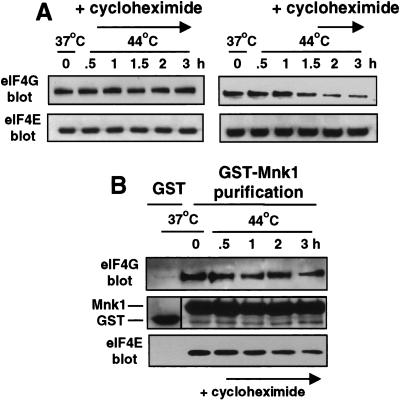
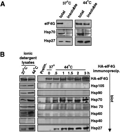
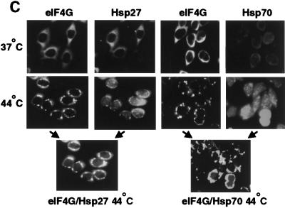
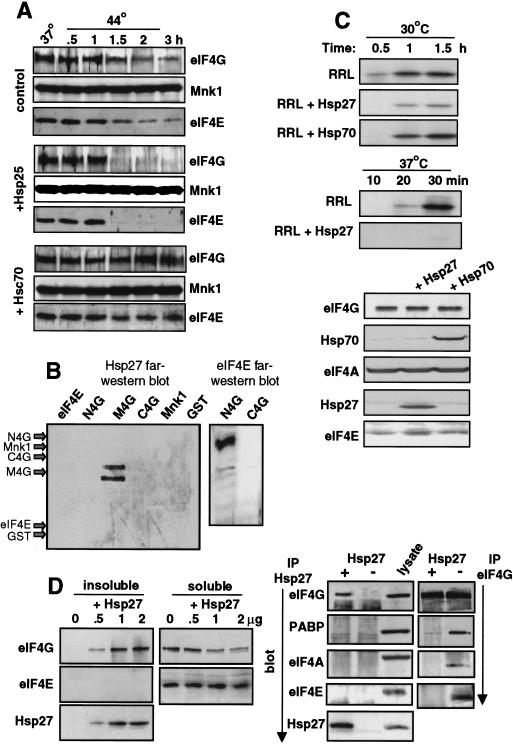
Inhibition of protein synthesis during heat shock limits accumulation of unfolded proteins that might damage eukaryotic cells. We demonstrate that chaperone Hsp27 is a heat shock-induced inhibitor of cellular protein synthesis. Translation of most mRNAs requires formation of a cap-binding initiation complex known as eIF4F, consisting of factors eIF4E, eIF4A, eIF4E kinase Mnk1, poly(A)-binding protein, and adaptor protein eIF4G. Hsp27 specifically bound eIF4G during heat shock, preventing assembly of the cap-initiation/eIF4F complex and trapping eIF4G in insoluble heat shock granules. eIF4G is a specific target of Hsp27, as eIF4E, eIF4A, Mnk1, poly(A)-binding protein, eIF4B, and eIF3 were not bound by Hsp27 and were not recruited into insoluble complexes. Dissociation of eIF4F was enhanced during heat shock by ectopic overexpression of Hsp25, the murine homolog of human Hsp27. Overexpression of Hsc70, a constitutive homolog of Hsp70, prevented loss of cap-initiation complexes and maintained eIF4G solubility. Purified Hsp27 specifically bound purified eIF4G in vitro, prevented in vitro translation, eliminated eIF4G interaction with protein binding factors, and promoted eIF4G insolubilization. These results therefore demonstrate that Hsp27 is a heat-induced inhibitor of eIF4F-dependent mRNA translation.


Figures

Figure 1
Figure 1
Heat shock does not significantly impair Mnk1 kinase activity. 293T cells were transfected with plasmids expressing GST−, GST–Mnk1, or GST–T2A2 (a kinase deficient mutant of Mnk1) (Waskiewicz et al. 1997). Cells were maintained at 37°C or heat-shocked at 44°C for 2 hr, or treated with 30 ng of human EGF/ml for 15 min (Feigenblum et al. 1998). Equal amounts of GST proteins were recovered from cell lysates by glutathione–Sepharose chromatography and incubated with purified, recombinant (nonphosphorylated) eIF4E and [γ-32P]ATP in an in vitro kinase reaction. (Top,middle) Proteins resolved by SDS-PAGE and stained with Coomassie blue. (Bottom) Same gel visualized by autoradiography. Bands were quantitated by digital densitometry and are typical of at least three independent experiments.
Figure 2
Figure 2
Heat shock blocks binding of Mnk1 to eIF4F. Cells were transiently transfected with plasmids expressing GST− or GST–Mnk1, then untreated, treated with 20 ng/ml of rapamycin for 2 hr, heat shocked at 44°C for 2 hr, or treated with rapamycin and heat-shocked simultaneously. Equal amounts of protein extracts were used. (A) GST–Mnk1 was recovered by glutathione–Sepharose chromatography. (B) Specific immunoprecipitation of eIF4E. Proteins were resolved by SDS-PAGE and immunoblotted for eIF4G, eIF4E, GST, or 4E-BP1. (C) Cells were labeled in vivo with 32PO4, eIF4E was recovered by immunoprecipitation and equal fractions were resolved by SDS-PAGE and autoradiography. Results are typical of at least three independent experiments and were quantitated by digital densitometry.
Figure 3
Figure 3
Heat shock induces insolubilization of eIF4G. 293T cells were maintained at 37°C or heat shocked at 44°C for the times shown. (A) Cell extracts were prepared using nonionic detergent (Triton X-100) or ionic detergent (SDS at 100°C) to solubilize proteins, equal amounts were resolved by SDS-PAGE and immunoblotted for eIF4G, eIF4E, eIF4A, eIF4B, eIF3, or PABP. Four subunits of eIF3 are shown. (B) Cells were labeled in vivo with 32PO4 during the time course of heat shock, eIF4E was recovered by immunoprecipitation and analyzed as described in the legend to Figure 2. (C) Cells were labeled in vivo with 50 μCi/ml of [35S]methionine for 30 min during heat shock as shown, equal amounts of protein extracts were resolved by SDS-PAGE and fluorographed. Results are typical of at least three independent experiments and were quantitated by digital densitometry.
Figure 4
Figure 4
Loss of eIF4F complexes parallel eIF4G insolubilization. 293T cells were transiently transfected with GST− or GST–Mnk1 expression vectors, maintained at 37°C, or heat-shocked for the times shown. Cells were extracted with nonionic detergent (Triton X-100) to purify soluble proteins. Equal amounts of extracts were used to recover GST–Mnk1 by glutathione–Sepharose chromatography (A), or immunoprecipitate eIF4E with specific antisera (B). Proteins were immunoblotted as shown. Results are typical of at least three independent experiments and were quantitated by digital densitometry.
Figure 5
Figure 5
Proteins synthesized during heat shock mediate insolubilization of eIF4G. (A) 293T cells were treated with cycloheximide at 50 μg/ml to prevent protein synthesis, then lysed in nonionic (Triton X-100) buffer to solubilize proteins at the indicated times. (B) 293T cells transfected with GST–Mnk1 were treated with 50 μg/ml cycloheximide as shown, lysed in Triton X-100 buffer, and GST–Mnk1 was purified by glutathione–Sepharose chromatography. Proteins were resolved by SDS-PAGE and immunoblotted with specific antisera as shown.
Figure 6
Figure 6
Chaperones associated with eIF4G. (A) 293T cells were heat shocked at 44°C for 2 hr. Cells were lysed in RIPA detergent buffer. Whole-cell lysates containing total cell protein (total) were normalized for equal protein levels, insoluble protein pellets were recovered by glycerol gradient centrifugation, pellets were resolubilized in SDS with heating to 100°C and proteins were resolved by SDS-PAGE and immunoblotted as indicated (insoluble). (B) 293T cells were transfected with an HA-epitope-tagged eIF4G expression vector and maintained at 37°C or heat-shocked at 44°C. (Left) Cells were lysed as above; equal amounts were resolved by SDS-PAGE and immunoblotted, as shown. (Right) eIF4G was immunoprecipitated with antisera to the HA-epitope, or preimmune serum (Preim.), and precipitates were resolved by SDS-PAGE and immunoblotted with antisera as shown. Results were quantified by digital densitometry. (C) HeLa cells were grown on cover slips, heat-shocked for 2 hr at 44°C or maintained at 37°C, fixed-permeabilized, and reacted with primary antibodies to eIF4G, Hsp27, or Hsp70, followed by staining with the following secondary antibodies: eIF4G, green fluorescence, Hsp27 or Hsp70, red fluorescence. Cells were visualized and photographed using a Zeiss Axiophot microscope. Coimaging analysis was performed by double-exposure using fluorescein and rhodamine-specific filters.
Figure 6
Figure 6
Chaperones associated with eIF4G. (A) 293T cells were heat shocked at 44°C for 2 hr. Cells were lysed in RIPA detergent buffer. Whole-cell lysates containing total cell protein (total) were normalized for equal protein levels, insoluble protein pellets were recovered by glycerol gradient centrifugation, pellets were resolubilized in SDS with heating to 100°C and proteins were resolved by SDS-PAGE and immunoblotted as indicated (insoluble). (B) 293T cells were transfected with an HA-epitope-tagged eIF4G expression vector and maintained at 37°C or heat-shocked at 44°C. (Left) Cells were lysed as above; equal amounts were resolved by SDS-PAGE and immunoblotted, as shown. (Right) eIF4G was immunoprecipitated with antisera to the HA-epitope, or preimmune serum (Preim.), and precipitates were resolved by SDS-PAGE and immunoblotted with antisera as shown. Results were quantified by digital densitometry. (C) HeLa cells were grown on cover slips, heat-shocked for 2 hr at 44°C or maintained at 37°C, fixed-permeabilized, and reacted with primary antibodies to eIF4G, Hsp27, or Hsp70, followed by staining with the following secondary antibodies: eIF4G, green fluorescence, Hsp27 or Hsp70, red fluorescence. Cells were visualized and photographed using a Zeiss Axiophot microscope. Coimaging analysis was performed by double-exposure using fluorescein and rhodamine-specific filters.
Figure 7
Figure 7
Effect of Hsp25/27 and Hsc/Hsp70 on eIF4F complex and translation activity. (A) 293T cells were transfected with vectors expressing GST–Mnk1 and either murine Hsp25 or human Hsc70. Cells were maintained at 37°C or heat-shocked at 44°C for the times shown, soluble proteins were prepared, and GST–Mnk1 was recovered. Proteins were resolved by SDS-PAGE and immunoblotted as shown. (B) Equal amounts (0.5 μg) of purified recombinant proteins as indicated were resolved by SDS-PAGE, transferred to membrane, and analyzed by far Western analysis by reacting with purified Hsp27 (left) or eIF4E (right), followed by antisera specific for Hsp27 or eIF4E, respectively, and ECL. Arrows indicate the electrophoretic positions of indicated proteins determined by staining the membrane with Coomassie blue (not shown). (C) Equal amounts of a luciferase reporter mRNA were translated in RRLs containing [35S]methionine, with and without addition of purified Hsp27 or Hsp70, at 30°C or 37°C. Translation products were resolved by SDS-PAGE. Western immunoblot of equal amounts of RRL with specific antisera as shown. (D) Purified recombinant Hsp27 was added to 50 μl of RRL at 37°C for 30 min, then insoluble and soluble fractions were prepared. (Left) All of the insoluble and one-third of the soluble fractions were resolved by SDS-PAGE and immunoblotted with antisera as shown. (Right) After removal of the insoluble protein fraction from control and Hsp27 containing samples, immunoprecipitation of Hsp27 or eIF4G was carried out using the soluble protein fraction, after normalization for eIF4G, followed by immunoblot analysis with antisera as shown. One-third volume of lysate was resolved as a control. Results are typical of at least three independent experiments and were quantitated by digital densitometry.

References

    1. Beretta L, Gingras A-C, Svitkin YV, Hall MN, Sonenberg N. Rapamycin blocks the phosphorylation of 4E-BP1 and inhibits cap-dependent initiation of translation. EMBO J. 1996;15:658–664. - PMC - PubMed
    1. Carper SW, Rocheleau T, Cimino D, Storm FK. Hsp 27 stimulates recovery of RNA and protein synthesis following a heat shock. J Cell Biochem. 1997;66:153–164. - PubMed
    1. Craig EA, Weissman JS, Horwich AL. Hsps and molecular chaperones: mediators of protein conformation and turnover in the cell. Cell. 1994;78:365–372. - PubMed
    1. Duncan RF. Translational control during heat shock. In: Hershey JWB, Mathews MB, Sonenberg N, editors. Translational control. Cold Spring Harbor, NY: Cold Spring Harbor Laboratory Press; 1996. pp. 271–294.
    1. Duncan RF, Hershey JWB. Protein synthesis and protein phosphorylation during heat stress, recovery, and adaptation. J Cell Biol. 1989;109:1467–1481. - PMC - PubMed

Publication types

MeSH terms

Substances