. 2000 Jun 15;14(12):1512-27.

daf-12 encodes a nuclear receptor that regulates the dauer diapause and developmental age in C. elegans

Affiliations

daf-12 encodes a nuclear receptor that regulates the dauer diapause and developmental age in C. elegans

A Antebi et al. Genes Dev. .

Abstract

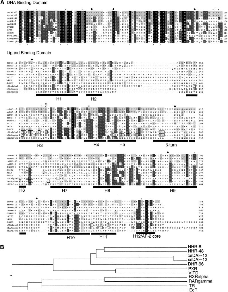
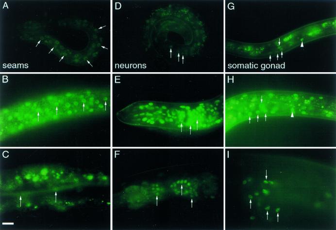
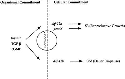
The daf-12 gene acts at the convergence of pathways regulating larval diapause, developmental age, and adult longevity in Caenorhabditis elegans. It encodes a nuclear receptor most closely related to two C. elegans receptors, NHR-8 and NHR-48, Drosophila DHR96, and vertebrate vitamin D and pregnane-X receptors. daf-12 has three predicted protein isoforms, two of which contain DNA- and ligand-binding domains, and one of which contains the ligand-binding domain only. Mutations cluster in DNA- and ligand-binding domains, but correspond to distinct phenotypic classes. DAF-12 is expressed widely in target tissues from embryo to adult, but is upregulated during midlarval stages. In the adult, expression persists in nervous system and somatic gonad, two tissues that regulate adult longevity. We propose that DAF-12 integrates hormonal signals in cellular targets to coordinate major life history traits.


Figures

Figure 1
Figure 1
(A) Tc1 transposon-induced mutations in daf-12 reveal a polymorphic 3.1-kb BglII fragment. Genomic DNA from backcrossed Tc1-induced alleles, m524 and m545, and their respective non-Daf revertants, m524m570 and m545m571, was digested with BglII, separated in 1% agarose, blotted onto nitrocellulose, and probed with radiolabeled Tc1. (B) Detection of a Tc1-associated mobility shift at the daf-12 locus. A Southern blot of BglII digested DNA was prepared as in A and hybridized with a radiolabeled 1.2-kb daf-12 probe. A 1.6-kb increase in size, due to Tc1 insertion, is seen in backcrossed and nonbackcrossed Tc1-tagged alleles (m524 and m545) but absent in parental mutator strains (RW7097, DR1164) and Tc1-excised nonDaf revertants (m524m570 and m524m571).
Figure 2
Figure 2
(A) Physical map of the daf-12 region. Rescuing cosmid, F11A1 (+). (B) cDNA structure (filled boxes) of 12A1, 12A3, and 12B isoforms, genomic organization, and corresponding positions of identified mutations at the daf-12 locus. Each isoform begins with the SL1 spliced leader (in white). Paired zinc fingers (Zn) of the DNA-binding domain (in white) and the ligand-binding domain (LBD) are indicated (in light gray). The 3′ untranslated region is shown in dark gray. Positions of the alternate splice donor and acceptor sites are shown for isoform 12A3 (numbering according to cosmid F11A1). Deletion (Δ), deletion/duplication (ΔD), splice acceptor mutation (^). Proposed initiator codons (AUG) and stop codons (●), and sites of polyA addition (AAA) are shown for each isoform. Allele names are followed by class in parentheses. The position of both lesions in alleles rh61rh411, rh61rh412, and rh62rh157 are shown. (C) cDNA structure (filled boxes) and genomic organization of daf-12 homologs nhr-8 and nhr-48. For nhr-8, full-length isoform 8A1 (clone cm14e12) lacks the SL1 spliced leader. Isoform 8A2 contains the SL1 spliced to an internal site in exon 1. Isoform 8B contains SL1 spliced to exon 2. For nhr-48, alternate exon 1 (14558/14562) and exon 7 (17691/17709) splice donors, and alternate exon 10 (18205/18238) splice acceptors for the various isoforms are indicated. A fifth 48B2 isoform (not shown), inferred from a partial cDNA clone, arises from the use of the exon 7 donor (17709) joined to the middle of exon 10 (18238). (Numbering is according to the ZK662 cosmid).
Figure 3
Figure 3
cDNA sequence, predicted protein products, and mutations in daf-12. Arrows indicate the starts of 12A1, 12A3, and 12B isoforms. The SL1 spliced leader is not shown. (▿) The position of introns; (v) bracket the amino acids missing from isoform 12A3. Base substitutions are indicated above the sequence, corresponding amino acid changes below, and premature stop codons indicated by a filled circle (●). The position of Tc1 insertion, m524::Tc1, is between T746 and A747 highlighted in bold. Underlined “M”, the predicted initiator methionines for the three isoforms. Deletion (Δ68) rh62rh157 deletes nucleotides 32347-32414. Deletion/duplication (ΔD) rh61rh411 deletes nucleotides 33058–33066, duplicates 33069–33090, and continues with 33067 (numbering according to cosmid F11A1). rh61rh412 deletes the underlined bases. rh257 splice acceptor mutation (^).
Figure 4
Figure 4
(A) Sequence alignments of the DBD and LBD of the ESCKA receptors with known nuclear receptors. Conserved cysteines that comprise Zn coordination sites of the DBD are starred. DAF-12 mutations are shown above the alignment (−1 = rh61rh412 frameshift). In the LBD, alignment with RAR-γ and RXR-α shows the positions of the 12 α helices (H1–H12) and β strands for comparison (Bourguet et al. 1995; Renaud et al. 1995; Wurtz et al. 1996). Helices 9–10 comprise a homodimerization interface in RXR (Bourguet et al. 1995). The predicted AF-2 domain with consensus hhXEhh (Danielian et al. 1992) lies in helix 12 (h = hydrophobic residue). In vitamin D and PXR receptors, forty amino acids that form an extended loop between helices 2 and 3 are omitted (indicated by ++). Boxed residues are ligand contact sites in TR and RAR receptors, as determined by X-ray crystallography (Renaud et al. 1995; Wagner et al. 1995; Wurtz et al. 1996). (Black shading) Residues conserved in nearly all nuclear receptors; (dark gray shading) residues conserved in a majority of the group shown; (light gray shading) residues matching DAF-12. The alignment includes Strongyloides stercoralis DAF-12 (Siddiqui et al., unpubl.; accession no. AAD37372), DHR96 (Fisk and Thummel 1995), human vitamin D (Baker et al. 1988), human PXR (Bertilsson et al. 1998), Drosophila ecdysone (Koelle et al. 1991), rat thyroid α1 (Izumo and Mahdavi 1988), human RAR-γ (Krust et al. 1989), and human RXR-α (Mangelsdorf et al. 1990). (B) Phylogenetic tree showing relationships between various nuclear receptors in the DBD. The tree was generated using the Megalign program of DNAStar, which derives a tree based on distance and then evaluates the tree based on parsimony criteria.
Figure 5
Figure 5
(A–C) Comparative heterochronic phenotypes at larval stage L3 in seam cells of wild type, class 3 allele rh61rh411, and class 1 allele rh61. Left lateral aspect. Scale bar, 10 μm. Seam cells and their immediate daughters express the GFP fusion to the Seam Cell Marker in their nuclei (Terns et al. 1997; J. Rothman, pers. comm.). (A) In wild type, epidermal seam cells along the lateral midline undergo a single stem cell division giving rise to two daughters (S3 program) at larval stage L3. Later the anterior daughter fuses with the hypodermal syncitium and the posterior daughter divides in subsequent stages. At larval stage L2 (not shown), seam cells undergo equational division followed by stem cell division, giving rise to 4 daughters (S2 program). (B) In rh61rh411 at larval stage L3, seam cells exhibit impenetrant repetition of S2 programs. (C) In rh61 seam cells exhibit penetrant repetition of S2 programs. (D–F) Mosaic dauer larvae form in some daf-12 mutants. (D) Wild-type dauer larva with full-length dauer alae, which are lateral cuticular ridges (arrows), and complete radial constriction. Cellular mosaicism is not seen. (E) daf-12(m25) and (F) daf-12(rh193) mosaic dauer larvae (grown at 15°), which occasionally show dauer alae and radial constriction in some cells (arrows) and partial dauer or non-dauer programs in others (arrowheads).
Figure 6
Figure 6
DAF-12::GFP expression pattern. Expression in epidermal seam cell nuclei (arrows) of early L1 (A), late L2 (B), and adult animals (C). Expression in various head neurons (arrows) of early L1 (D), late L2 (E), and adult (F) animals. Expression in somatic gonadal tissues of L3 dauer (G), late L2 non-dauer (H), and adult somatic gonadal tissues (I). In G and H, the arrows indicate the nuclei of somatic gonadoblasts, arrowheads the distal-tip cell. In I, arrows indicate various spermathecal nuclei. Lateral aspect, anterior is left. Scale bar, 10 μm. All photographs were exposed for equal times.
Figure 7
Figure 7
A model for daf-12 action. Insulin-like, TGF-β, and cGMP signaling act upstream of organismal commitments to diapause or reproductive growth. In favorable environments, the activity of these pathways stimulate reproductive growth, while unfavorable environments lead to diapause. Proposed hormonal signaling acts through daf-12 to select S3 or S3d alternatives. daf-12 together with other heterochronic genes acts in cells downstream of organismal commitments. daf-12a and a proposed redundant function, geneX, are required for S3 reproductive programs, whereas daf-12b activity is required for S3d dauer programs.

References

    1. Ambros V. A hierarchy of regulatory genes controls a larva-to-adult developmental switch in C. elegans. Cell. 1989;57:49–57. - PubMed
    1. Ambros V. Heterochronic genes. In: Riddle DL, Blumenthal T, Meyer B, Priess J, editors. C. elegans II. Cold Spring Harbor, N.Y: Cold Spring Harbor Laboratory Press; 1997. pp. 501–518. - PubMed
    1. Ambros V, Horvitz HR. Heterochronic mutants of the nematode Caenorhabditis elegans. Science. 1984;226:409–416. - PubMed
    1. Antebi A, Culotti JG, Hedgecock EM. daf-12 regulates developmental age and the dauer alternative in Caenorhabditis elegans. Development. 1998;125:1191–1205. - PubMed
    1. Antebi A, Norris C, Hedgecock E, Garriga G. Cell and growth cone migrations. In: Riddle DL, Blumenthal T, Meyer B, Priess J, editors. C. elegans II. Cold Spring Harbor, N.Y: Cold Spring Harbor Laboratory Press; 1997. pp. 583–609. - PubMed

Publication types

MeSH terms

LinkOut - more resources