. 2000 Jun 19;191(12):2065-74.
doi: 10.1084/jem.191.12.2065.

Alteration at a single amino acid residue in the T cell receptor alpha chain complementarity determining region 2 changes the differentiation of naive CD4 T cells in response to antigen from T helper cell type 1 (Th1) to Th2

Affiliations

Alteration at a single amino acid residue in the T cell receptor alpha chain complementarity determining region 2 changes the differentiation of naive CD4 T cells in response to antigen from T helper cell type 1 (Th1) to Th2

J M Blander et al. J Exp Med. .

Abstract

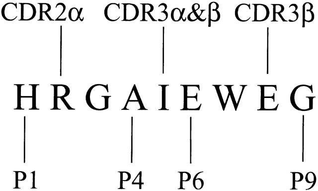
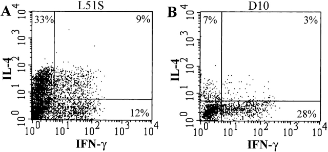
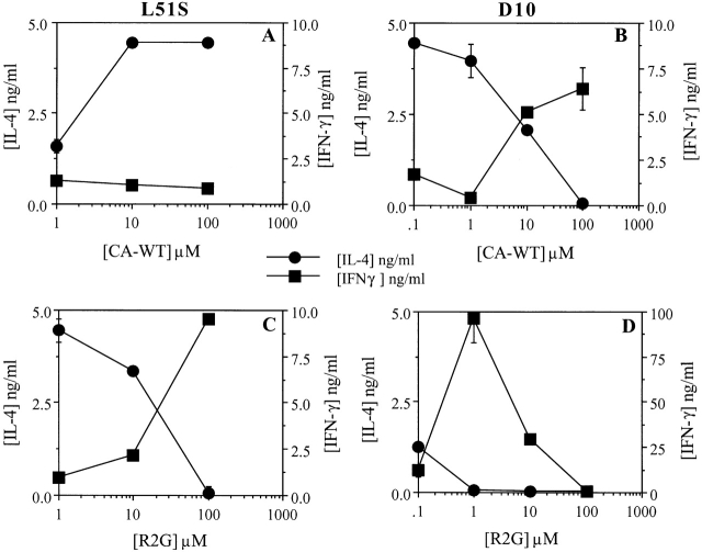
To study whether changes in the structure of a T cell receptor (TCR) at a single peptide-contacting residue could affect T cell priming with antigenic peptide, we made transgenic mice with a point mutation in the TCR alpha chain of the D10.G4.1 (D10) TCR and bred them to D10 beta chain transgenic mice. The mutation consisted of a leucine to serine substitution at position 51 (L51S), which we had already established contacted the second amino acid of the peptide such that the response to the reference peptide was reduced by approximately 100-fold. A mutation in the reference peptide CA134-146 (CA-WT) from the arginine at peptide position 2 to glycine (R2G) restored full response to this altered TCR. When we examined in vitro priming of naive CD4 T cells, we observed that the response to doses of CA-WT that induced T helper cell type 1 (Th1) responses in naive CD4 T cells from mice transgenic for the D10 TCR gave only Th2 responses in naive CD4 T cells derived from the L51S. However, when we primed the same T cells with the R2G peptide, we observed Th1 priming in both D10 and L51S naive CD4 T cells. We conclude from these data that a mutation in the TCR at a key position that contacts major histocompatibility complex-bound peptide is associated with a shift in T cell differentiation from Th1 to Th2.


Figures

Figure 1
Figure 1
A schematic representation of the CA-WT 9-mer peptide amino acid sequence showing both the D10 TCR contact and I-Ak MHC class II contact residues. The figure is adapted from reference 15. The peptide amino acid residues that contact I-Ak are at positions 1, 4, 6, and 9, and labeled P1, P4, P6, and P9. Those that contact the TCR are at positions 2, 5, and 8. The L51S mutation affects the CDR2 region of the D10 TCR α chain, which contacts position 2 in the CA-WT peptide.
Figure 2
Figure 2
CD4 T cells in L51S TCR transgenic mice are markedly reduced in numbers. CD4 enriched cells from the LNs of D10 (A and C) or L51S (B and D) TCR transgenic mice were stained with Quantum red–conjugated anti-CD4 and biotinylated 3D3 followed by PE-conjugated streptavidin (A and B). LN cells were also stained simultaneously with Quantum red–conjugated anti-CD4, FITC-conjugated anti-Vα2, and biotinylated anti-Vβ8.1,8.2 followed by PE-conjugated streptavidin. CD4+ T cells were gated and their expression of the transgenic TCR Vα2+Vβ8+ is shown (C and D). Log fluorescence intensity is shown on both axes.
Figure 3
Figure 3
Proliferation of CD4 T cells from both D10 and L51S TCR transgenic mice in response to varying doses of CA-WT or R2G peptide in the presence of irradiated syngeneic splenocytes. The x-axis shows the concentration of peptide (in μM). The y-axis shows the counts per minute (CPM) of [3H]thymidine incorporated as a measure of proliferation.
Figure 4
Figure 4
FACS® sorting of naive CD4 T cells from D10 or L51S TCR transgenic mice. Sterile sorting of naive transgenic CD4 T cells by FACS® based on the expression of the transgenic α chain (Vα2+) and the naive phenotype marker L-selectin (Mel-14+). CD4 T cell–enriched suspensions of spleen and LNs, D10 (A) and L51S (B), were stained with FITC-conjugated anti-Vα2 and biotinylated Mel-14 followed by PE-conjugated streptavidin. Naive transgenic Mel-14+Vα2+ cells were gated and collected. Log fluorescence intensity is shown on both axes.
Figure 5
Figure 5
Intracellular cytokine staining of naive L51S and D10 CD4 T cells shows the predominant population of IL-4 producers in L51S cultures. Naive CD4 T cell cultures were primed with 10 μM CA-WT peptide and restimulated on day 6 with PMA and ionomycin, followed by treatment with brefeldin A. Cultured L51S naive CD4 T cells (A), or cultured D10 naive CD4 T cells (B), were stained 6 h later with Quantum red–conjugated anti-CD4, fixed with 1% paraformaldehyde, permeabilized, and stained with FITC-conjugated anti–IFN-γ, and PE-conjugated anti-IL-4. CD4+ T cells were gated, and their intracellular staining for IL-4 and IFN-γ is shown. The log fluorescence intensity is shown on both axes.
Figure 6
Figure 6
L51S TCR transgenic naive CD4 T cells differentiate into Th2-like IL-4–producing cells when primed in vitro with CA-WT peptide, and produce IFN-γ when stimulated with R2G. Naive CD4 T cells were cultured in the presence of varying doses of CA-WT (A and B) or R2G peptide (C and D), irradiated syngeneic splenocytes, and 25 U/ml IL-2. The primary supernatants were collected on day 4 and the cells were restimulated with 10 μM R2G peptide, after which the secondary supernatants were collected on day 4 and ELISAs were performed in duplicate to measure the concentrations of IL-4 and IFN-γ. (A and C) L51S CD4 T cell cytokine profiles; (B and D) D10 CD4 T cell cytokine profiles. IL-4 and IFN-γ concentrations are shown in ng/ml quantities. The concentrations of peptide used for priming are shown on the x-axes.

References

    1. Germain R.N. Major histocompatibility complex-dependent antigen processing and peptide presentationproviding ligands for the clonal activation of T lymphocytes. Cell. 1994;76:287–299. - PubMed
    1. Janeway C.A., Jr., Bottomly K. Signals and signs for lymphocyte responses. Cell. 1994;76:275–285. - PubMed
    1. Mosmann T.R., Cherwinski H., Bond M.W., Giedlin M.A., Coffman R.L. Two types of murine helper T cell clones. I. Definition according to profiles of lymphokine activities and secreted proteins. J. Immunol. 1986;136:2348–2357. - PubMed
    1. Abbas A.K., Murphy K.M. Functional diversity of helper T lymphocytes. Nature. 1996;383:787–793. - PubMed
    1. Constant S.L., Bottomly K. Induction of Th1 and Th2 CD4+ T cell responsesthe alternative approaches. Annu. Rev. Immunol. 1997;15:297–322. - PubMed

Publication types

MeSH terms