. 2000 Jul 5;97(14):7709-14.
doi: 10.1073/pnas.130183897.

PapD-like chaperones provide the missing information for folding of pilin proteins

Affiliations

PapD-like chaperones provide the missing information for folding of pilin proteins

M M Barnhart et al. Proc Natl Acad Sci U S A. .

Abstract

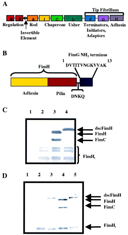
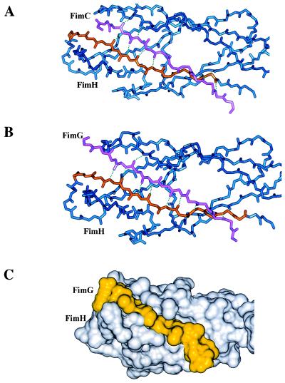
A fundamental question in molecular biology is how proteins fold into domains that can serve as assembly modules for building up large macromolecular structures. The biogenesis of pili on the surface of Gram-negative bacteria requires the orchestration of a complex process that includes protein synthesis, folding via small chaperones, secretion, and assembly. The results presented here support the hypothesis that pilus subunit folding and biogenesis proceed via mechanisms termed donor strand complementation and donor strand exchange. Here we show that the steric information necessary for pilus subunit folding is not contained in one polypeptide sequence. Rather, the missing information is transiently donated by a strand of a small chaperone to allow folding. Providing the missing information for folding, via a 13-amino acid peptide extension to the C-terminal end of a pilus subunit, resulted in the production of a protein that no longer required the chaperone to fold. This mechanism of small periplasmic chaperone function described here deviates from classical hsp60 chaperone-assisted folding.


Figures

Figure 1
Figure 1
Donor strand complementation of FimH in cis. Shown are schematic diagrams of the type 1 gene cluster (A) and dscFimH (B). Immunoblots developed with anti-FimCH antiserum of periplasmic extracts (C) after no expression of FimH (lane 1), FimH alone (lane 2), FimH + FimC (lane 3), or dscFimH (lane 4). A proportion of FimH truncation occurred under all conditions and was labeled FimHt. (D) Elution of FimH or dscFimH from mannose-Sepharose after incubation with periplasm containing FimC (lane 1), FimH alone (lane 2), dscFimH (lane 3), FimH + FimC (lane 4), or dscFimH + FimC (lane 5). The elutions were run on a SDS/PAGE gel followed by Western blotting using anti-FimCH antibodies.
Figure 2
Figure 2
Modeling of the N terminus of FimG into the FimH pilin domain. (A) Crystal structure of the FimH pilin domain (blue) of the FimCH complex highlighting the interaction between the F strand of FimH (orange) and the G1β strand of FimC (pink). (B) Pilin domain of FimH (blue) modeling the anti-parallel interaction between the F strand of FimH (orange) and the FimG donor strand (pink). (C) Connolly surface representation of the FimH pilin domain (white) with the FimG donor strand (yellow). Note the more extensive hydrogen bonding (dotted lines) with the FimG donor strand.
Figure 3
Figure 3
Urea denaturation curves of FimH and dscFimH. Shown is a Coomassie blue-stained SDS/PAGE gel of purified FimH and dscFimH (inset lanes 1 and 2, respectively). FimH (circles) and dscFimH (diamonds) were incubated with increasing concentrations of urea + 4 mM DTT, and the change in fluorescence at 350 nm was measured to monitor denaturation.
Figure 4
Figure 4
Refolding of dscFimH and FimH. The CD spectra were measured for native (solid line) and 9 M urea denatured (dashed line) FimH (A), dscFimH (B), and native FimCH (C). The CD spectra of denatured dscFimH after rapid dilution to 0.45 M urea (long and short dashed line) (B) and denatured FimH after rapid dilution to 0.45 M urea in the presence (long and short dashed line) (C) or absence of FimC (elicited no signal attributable to aggregation of FimH) were also determined. The ability of FimC to bind to denatured FimH that was subjected to rapid dilution was measured (D). FimC was either present in the diluent (left side) or added after dilution (right side). The ability of FimH separated from FimCH in 3 M urea to bind to FimC (left) or R8A FimC (right) when diluted to 0.45 M urea was measured (E).

Comment in

References

    1. Hung D L, Knight S D, Woods R M, Pinkner J S, Hultgren S J. EMBO J. 1996;15:3792–3805. - PMC - PubMed
    1. Soto G E, Hultgren S J. J Bacteriol. 1999;181:1059–1071. - PMC - PubMed
    1. Lindberg F, Tennent J M, Hultgren S J, Lund B, Normark S. J Bacteriol. 1989;171:6052–6058. - PMC - PubMed
    1. Roberts J A, Marklund B-I, Ilver D, Haslam D, Kaack M B, Baskin G, Louis M, Mollby R, Winberg J, Normark S. Proc Natl Acad Sci USA. 1994;91:11889–11893. - PMC - PubMed
    1. Jones C H, Pinkner J S, Nicholes A V, Slonim L N, Abraham S N, Hultgren S J. Proc Natl Acad Sci USA. 1993;90:8397–8401. - PMC - PubMed

Publication types

MeSH terms

LinkOut - more resources