. 2000 Jul 5;97(14):8045-50.
doi: 10.1073/pnas.130198797.

Identification and characterization of a newly recognized population of high-Na+, low-K+, low-density sickle and normal red cells

Affiliations

Identification and characterization of a newly recognized population of high-Na+, low-K+, low-density sickle and normal red cells

R M Bookchin et al. Proc Natl Acad Sci U S A. .

Abstract

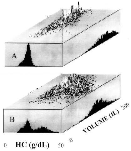
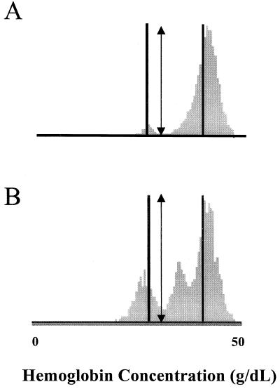
We describe a population of sickle cell anemia red cells (SS RBCs) ( approximately 4%) and a smaller fraction of normal RBCs (<0.03%) that fail to dehydrate when permeabilized to K(+) with either valinomycin or elevated internal Ca(2+). The nonshrinking, valinomycin-resistant (val-res) fractions, first detected by flow cytometry of density-fractionated SS RBCs, constituted up to 60% of the lightest, reticulocyte-rich (R1) cell fraction, and progressively smaller portions of the slightly denser R2 cells and discocytes. R1 val-res RBCs had a mean cell hemoglobin concentration of approximately 21 g of Hb per dl, and many had an elongated shape like "irreversibly sickled cells," suggesting a dense SS cell origin. Of three possible explanations for val-res cells, failure of valinomycin to K(+)-permeabilize the cells, low co-ion permeability, or reduced driving K(+) gradient, the latter proved responsible: Both SS and normal val-res RBCs were consistently high-Na(+) and low-K(+), even when processed entirely in Na-free media. Ca(2+) + A23187-induced K(+)-permeabilization of SS R1 fractions revealed a similar fraction of cal-res cells, whose (86)Rb uptake showed both high Na/K pump and leak fluxes. val-res/cal-res RBCs might represent either a distinct erythroid genealogy, or an "end-stage" of normal and SS RBCs. This paper focuses on the discovery, basic characterization, and exclusion of artifactual origin of this RBC fraction. Many future studies will be needed to clarify their mechanism of generation and full pathophysiological significance.


Figures

Figure 1
Figure 1
Progressive changes with time in the HC and V distributions of normal RBCs after K+-permeabilization with valinomycin. Histograms were obtained with the Bayer H*3 analyzer during a 1-h incubation at 37°C of RBCs suspended in buffer D (15 mM K) and treated with valinomycin. Note the progressive increase in HC and decrease in V, with no evident broadening of cell distribution. CV, coefficient of variation. Samples taken at the times indicated were diluted in the same buffer containing sphering agent and were read immediately without RNA staining.
Figure 2
Figure 2
Effects of K+-permeabilization with valinomycin on the HC distributions of the light fractions of SS RBCs. After density fractionation, the three lightest fractions of SS RBCs, R1, R2, and LD (light discocyte) (see Methods for density boundaries) were treated as in Fig. 1. Upper and Lower histograms show the HC distributions before and after a 30-min incubation with valinomycin, respectively.
Figure 3
Figure 3
Effects of K+-permeabilization with valinomycin on the HC distributions of the reticulocyte and nonreticulocyte components of the lightest, R1, fraction of SS RBCs. The treatment was the same as in Fig. 2, except that the samples of RBC suspension were diluted in buffer D containing RNA stain for the reticulocytes and incubated 15 min before adding the sphering agent. In the histograms, the reticulocytes are shown in gray and nonreticulocyte RBCs in black.
Figure 4
Figure 4
86Rb influx measurements in cal-res and non-cal-res portions of the lightest, R1, fraction of SS RBCs, in the presence and absence of ouabain and high (1 mM) bumetanide. Before measurements, the isolated, dehydrated non-cal-res cells were replenished with KCl and rehydrated. A and B show two patterns of influx seen in four experiments. ○, cal-res RBCs; ●, cal-res RBCs + ouabain; □, non-cal-res RBCs; ■, non-cal-res RBCs + ouabain. The experiment in C is typical of several showing the effects of added bumetanide on ouabain-treated cal-res and non-cal-res RBCs. □, cal-res RBCs + ouabain; ■, cal-res RBCs + ouabain + bumetanide; ▿, non-cal-res RBCs + ouabain; ▾, non-cal-res RBCs + ouabain + bumetanide.
Figure 5
Figure 5
Cell volume and HC histograms and three-dimensional cytograms of the SS R1 RBCs before (A) and after (B) K+-permeabilization with valinomycin. The different RBC volume distributions corresponding to the valinomycin-sensitive and the val-res fractions can be distinguished on the cytogram, despite their overlap on the volume histogram: note the broad volume distribution of the lighter (val-res) cells, compared with the cells that have dehydrated.
Figure 6
Figure 6
Photomicrograph of the val-res component of the R1 fraction of SS RBCs. Note the presence of many RBCs with the typical elongated shapes of ISC.
Figure 7
Figure 7
Effects of K+-permeabilization with valinomycin on the HC distributions of the lightest fraction of normal RBCs from two individuals. Solid vertical lines show HC gates at 28 g of Hb per dl and 41 g of Hb per dl. The arrows indicate a gate of 32 g of Hb per dl, chosen to distinguish the most valinomycin-resistant fraction from the valinomycin-sensitive majority of RBCs. Histograms A and B exemplify the heterogeneity of distribution of the normal val-res populations and show that in some samples (B) a distinct population of intermediately val-res cells can be resolved.

References

    1. Raftos J E, Bookchin R M, Lew V L. J Physiol (London) 1998;499:17–25. - PMC - PubMed
    1. Raftos J E, Bookchin R M, Lew V L. J Physiol (London) 1996;491:773–777. - PMC - PubMed
    1. Ortiz O E, Lew V L, Bookchin R M. Blood. 1986;67:710–715. - PubMed
    1. Sorette M P, Shiffer K, Clark M R. Blood. 1992;80:249–254. - PubMed
    1. Etzion Z, Lew V L, Bookchin R M. Am J Physiol. 1996;271:C1111–C1121. - PubMed

Publication types