. 2000 Jun 20;97(13):7124-9.
doi: 10.1073/pnas.97.13.7124.

Structure-based discovery of an organic compound that binds Bcl-2 protein and induces apoptosis of tumor cells

Affiliations

Structure-based discovery of an organic compound that binds Bcl-2 protein and induces apoptosis of tumor cells

J L Wang et al. Proc Natl Acad Sci U S A. .

Abstract

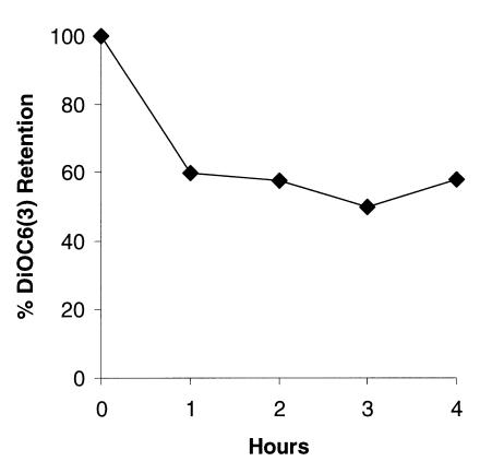
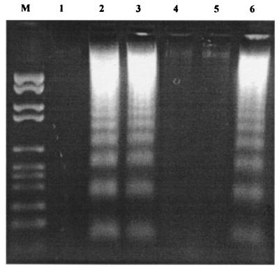
Bcl-2 and related proteins are key regulators of apoptosis or programmed cell death implicated in human disease including cancer. We recently showed that cell-permeable Bcl-2 binding peptides could induce apoptosis of human myeloid leukemia in vitro and suppress its growth in severe combined immunodeficient mice. Here we report the discovery of HA14-1, a small molecule (molecular weight = 409) and nonpeptidic ligand of a Bcl-2 surface pocket, by using a computer screening strategy based on the predicted structure of Bcl-2 protein. In vitro binding studies demonstrated the interaction of HA14-1 with this Bcl-2 surface pocket that is essential for Bcl-2 biological function. HA14-1 effectively induced apoptosis of human acute myeloid leukemia (HL-60) cells overexpressing Bcl-2 protein that was associated with the decrease in mitochondrial membrane potential and activation of caspase-9 followed by caspase-3. Cytokine response modifier A, a potent inhibitor of Fas-mediated apoptosis, did not block apoptosis induced by HA14-1. Whereas HA14-1 strongly induced the death of NIH 3T3 (Apaf-1(+/+)) cells, it had little apoptotic effect on Apaf-1-deficient (Apaf-1(-/-)) mouse embryonic fibroblast cells. These data are consistent with a mechanism by which HA14-1 induces the activation of Apaf-1 and caspases, possibly by binding to Bcl-2 protein and inhibiting its function. The discovery of this cell-permeable molecule provides a chemical probe to study Bcl-2-regulated apoptotic pathways in vivo and could lead to the development of new therapeutic agents.


Figures

Figure 1
Figure 1
(A) Structure of HA14-1 (ethyl 2-amino-6-bromo-4-(1-cyano-2-ethoxy-2-oxoethyl)-4H-chromene-3-carboxylate), a nonpeptidic organic compound that binds Bcl-2 protein. Note that HA14-1 has two chiral centers (marked by asterisks) located at the C4 position and the carbon atom attached with the cyano group, respectively. (B) Structural model for the complex of HA14-1 with the Bcl-2 surface pocket as predicted by computer docking calculation.
Figure 2
Figure 2
Binding of HA14-1 to Bcl-2 protein in vitro as measured by a competitive fluorescence polarization assay. The data points represent the mean of three independent experiments.
Figure 3
Figure 3
Effect of HA14-1 on the viability of HL-60 cells. The cells were incubated with HA14-1 at different concentrations for 4 h. The cell viability was determined by using MTS method in 96-well plate.
Figure 4
Figure 4
Apoptosis induced by HA14-1 in HL-60 cells as indicated by DNA ladders (A) and cleavage of caspase-9, caspase-3 and PARP (B). DNA ladders were visualized on 2% of agarose gel after the cells were treated with 50 μM HA14-1 compound at the designated time. M, DNA marker. Lanes: 1, control; 2, 1 h of HA14-1 treatment; 3, 2 h of HA14-1 treatment; 4, 3 h of HA14-1 treatment; 5, 4 h of HA14-1 treatment; 6, preincubation with zVAD-fmk for 2 h before the treatment with HA14-1 for 4 h. The cleavage of caspase-3, -9, and PARP was shown by Western blot analysis using polyclonal rabbit antibodies.
Figure 5
Figure 5
Decrease in mitochondrial membrane potential caused by HA14-1. HL-60 cells were cultured in the presence of 50 μM HA14-1 for different time points. Changes in mitochondrial membrane potential after HA14-1 treatment were examined by using DiOC6(3) fluorescence dye.
Figure 6
Figure 6
Apoptosis of lung cancer H1299 cells induced by HA14-1 cannot be blocked by CrmA. The effects of HA14-1 and Fas antibody as a positive control were tested in human lung cancer H1299 cells transfected with either plasmid containing neomycin resistance gene (in lanes 1–3) or the same vector containing CrmA gene (in lanes 4–6). Cells were treated with Fas antibody or HA14-1 for 24 h and were analyzed for DNA ladders on 2% agarose gel. Lanes: 1 and 4, untreated control; 2 and 5, treatment with Fas antibody; 3 and 6, treatment with 50 μM HA14-1.
Figure 7
Figure 7
Effect of HA14-1 on Apaf-1−/− and Apaf-1+/+ cells. Apaf-1-deficient mouse embryonic fibroblast (Apaf-1−/−) and NIH 3T3 (Apaf-1+/+) cells were treated with 50 μM HA14-1 for 24 h. Percent cell death was determined by using Trypan blue staining. To examine the role of caspases in the death of Apaf-1+/+ cells induced by HA14-1, cells were preincubated with zVAD-fmk for 2 h before the treatment of the compound.

References

    1. Tsujimoto Y, Gorham J, Cossman J, Jaffe E, Croce C M. Science. 1985;229:1390–1393. - PubMed
    1. Adams J, Cory S. Science. 1998;281:1322–1326. - PubMed
    1. Chao D, Korsmeyer S. Annu Rev Immunol. 1998;16:395–419. - PubMed
    1. Thompson C B. Science. 1995;267:1456–1462. - PubMed
    1. Reed J C. Curr Opin Oncol. 1999;1:68–75. - PubMed

Publication types