. 2000 Jun 20;97(13):7301-6.
doi: 10.1073/pnas.97.13.7301.

Identification and characterization of a p53 homologue in Drosophila melanogaster

Affiliations

Identification and characterization of a p53 homologue in Drosophila melanogaster

S Jin et al. Proc Natl Acad Sci U S A. .

Abstract

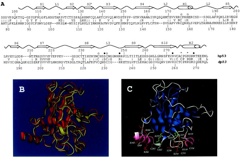
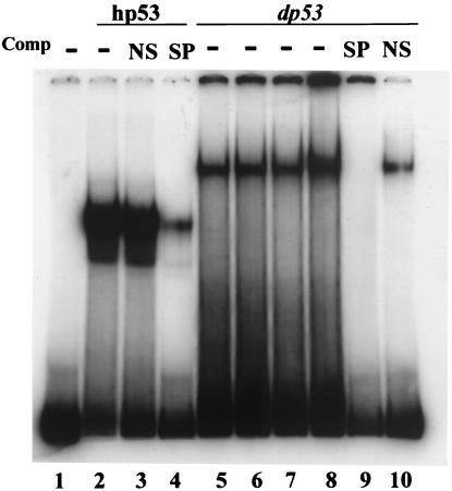
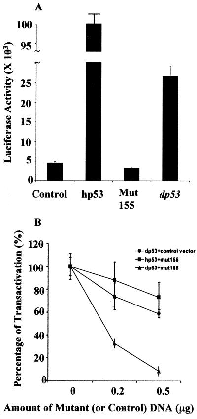
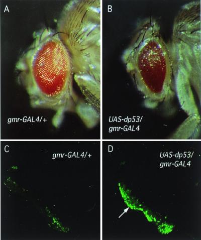
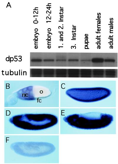
The tumor suppressor gene p53 in mammalian cells plays a critical role in safeguarding the integrity of genome. It functions as a sequence-specific transcription factor. Upon activation by a variety of cellular stresses, p53 transactivates downstream target genes, through which it regulates cell cycle and apoptosis. However, little is known about p53 in invertebrates. Here we report the identification and characterization of a Drosophila p53 homologue gene, dp53. dp53 encodes a 385-amino acid protein with significant homology to human p53 (hp53) in the region of the DNA-binding domain, and to a lesser extent the tetramerization domain. Purified dp53 DNA-binding domain protein was shown to bind to the consensus hp53-binding site by gel mobility analysis. In transient transfection assays, expression of dp53 in Schneider cells transcriptionally activated promoters that contained consensus hp53-responsive elements. Moreover, a mutant dp53 (Arg-155 to His-155), like its hp53 counterpart mutant, exerted a dominant-negative effect on transactivation. Ectopic expression of dp53 in Drosophila eye disk caused cell death and led to a rough eye phenotype. dp53 is expressed throughout the development of Drosophila with highest expression levels in early embryogenesis, which has a maternal component. Consistent with this, dp53 RNA levels were high in the nurse cells of the ovary. It appears that p53 is structurally and functionally conserved from flies to mammals. Drosophila will provide a useful genetic system to the further study of the p53 network.


Figures

Figure 1
Figure 1
(A) Sequence and structure comparison of the Drosophila and human p53 DNA-binding domains. Sequence alignment of the dp53 and hp53 DNA-binding domains as produced by psi-blast. The secondary structure elements of hp53 are shown above (S, β-strand; L, loop; H, α-helix). Residues involved in DNA binding (*, contacting bases; ●, contacting phosphate backbone) and zinc binding (○) also are indicated (6). (B) Superimposition of the crystal structure of hp53 (yellow cartoon) DNA-binding domain and the model of the dp53 domain (red cartoon) predicted by program modeller. (C) Protein structure model of the dp53 DNA-binding domain. Color scheme: red, residues preserved between the human and Drosophila sequences; green, conservative substitutions; orange, preserved Zn-coordinating residues; and yellow, nonconservative substitutions. B and C were rendered by program dino (http://www.biozentrum.unibas.ch/∼x-ray/dino).
Figure 2
Figure 2
dp53 binds to consensus hp53-binding sequence. Gel mobility-shift assay was performed by using 32P-end-labeled consensus p53-binding oligonucleotide. hp53 DNA-binding domain (amino acids 94–292) or dp53 N terminus plus DNA-binding domain (amino acids 1–297) were purified from bacteria. hp53 protein (100 ng; lanes 2, 3, and 4) or different amount of dp53 protein (lane 5, 50 ng; lane 6, 100 ng; lane 7, 150 ng; lane 8, 200 ng; and lanes 9 and 10, 100 ng) were added into the reactions without competitor, with nonspecific competitor (NS, 500 ng) or with specific competitor (SP, 500 ng) as indicated. Lane 1 contained the probe only.
Figure 3
Figure 3
dp53 transcriptionally activates promoter with hp53 responsive elements in Schneider cells. (A) Expression of dp53 activates pG13. (B) a mutant dp53 (Arg-155 to His-155) has a dominant-negative effect. In A, 0.2 μg of pG13 was cotransfected into Schneider cells with 0.2 μg of control vector (control) or with expression vectors of hp53, mutant dp53 (Mut155), dp53 as indicated. In B, 0.2 μg of pG13 and 0.2 μg of expression vectors of wild-type p53 (dp53 or hp53, as indicated) were cotransfected into Schneider cells with the indicated amount (0, 0.2 μg, 0.5 μg) of mutant dp53 (mut155) expression vector or control vector. In each case, transcription activation by wild-type p53 without the cotransfection of mutant dp53 (or control vector) is arbitrarily set as 100%. Percentage of activation is the ratio of the fold activation with the cotransfection of mutant dp53 (or control vector) divided by the fold activation without the cotransfection of mutant dp53.
Figure 4
Figure 4
Increased cell death in the fly retina induced by dp53. dp53 was overexpressed in the fly retina under the control of a photoreceptor-specific promoter (gmr) using the UAS/GAL4 binary expression system (B and D). gmr-GAL4/+ served as wild-type control (A and C). Note the reduced size and the roughness of the eye caused by the overexpression of dp53 (B). (C and D) Third instar eye imaginal discs were subjected to acridine orange staining. Note the increased amount of cell death in the discs from animals overexpressing dp53 (D, arrow).
Figure 5
Figure 5
(A) dp53 RNA is expressed throughout development. Total RNA from the indicated developmental stages was subjected to an RNase-protection assay using a dp53-specific probe. Note the elevated levels of dp53 RNA in females and early embryos. Tubulin RNA was used as loading control. (BF) In situ hybridization was performed on wild-type embryos using a dp53-specific RNA probe. (B) Stage 10 egg chamber. Note presence of dp53 RNA in the nurse cells (nc) of the egg chamber. No signal was observed in the somatic follicle cells (fc). o, oocyte. (C) Blastoderm embryo probed with antisense RNA; the signal inside the egg indicates a maternal component of dp53 RNA. (D and E) Embryos before and after germ band retraction, showing a ubiquitous expression of dp53. (E) Embryo probed with sense RNA as a control for the specificity of the probe.

References

    1. Levine A J. Cell. 1997;88:323–331. - PubMed
    1. Hollstein M, Sidransky D, Vogelstein B, Harris C C. Science. 1991;253:49–53. - PubMed
    1. Oliner J D, Kinzler K W, Meltzer P S, George D L, Vogelstein B. Nature (London) 1992;358:80–83. - PubMed
    1. Kussie P H, Gorina S, Marechal V, Elenbaas B, Moreau J, Levine A J, Pavletich N P. Science. 1996;274:948–953. - PubMed
    1. Roth J, Dobbelstein M, Freedman D A, Shenk T, Levine A J. EMBO J. 1998;17:554–564. - PMC - PubMed

Publication types

Substances

Associated data