

Inhibition of sonic hedgehog autoprocessing in cultured mammalian cells by sterol deprivation
- PMID: 10860995
- PMCID: PMC16541
- DOI: 10.1073/pnas.97.13.7307
Inhibition of sonic hedgehog autoprocessing in cultured mammalian cells by sterol deprivation
Abstract
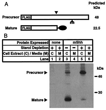
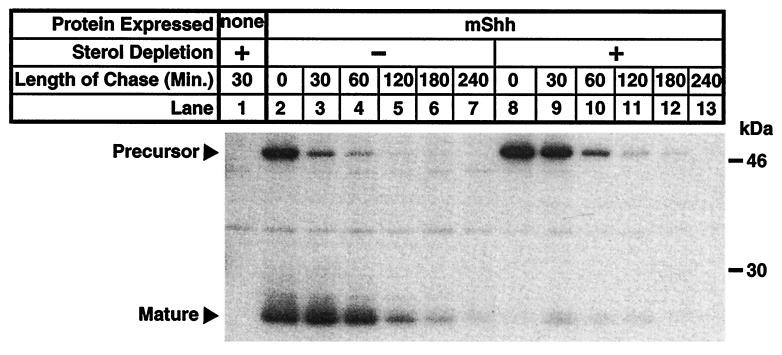
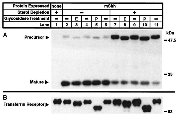
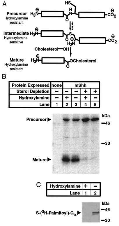
Sonic hedgehog (Shh) is a signaling molecule that is important for defining patterning in the developing vertebrate central nervous system. After translation, Shh autoproteolyzes and covalently attaches cholesterol to the newly formed carboxyl terminus, a modification crucial for normal Shh signaling. Presented here is evidence that acute severe sterol deprivation in cultured Chinese hamster ovary cells expressing mouse Shh (mShh) inhibits autoprocessing of the protein. These conditions allowed the first detailed kinetic analysis of mShh autoprocessing and turnover rates revealing that cells rapidly degrade both precursor and mature mShh regardless of sterol content and sterol deprivation increases the rate of precursor degradation. Inhibition of mShh autoprocessing also allowed the determination of the subcellular localization of mShh precursor which accumulates in a pre-medial Golgi intracellular compartment. Finally, the precursor form of mShh that results from autoprocessing inhibition appears to accumulate as an amide rather than a stable thioester.
Figures
Similar articles
-
A cell-based bioluminescence reporter assay of human Sonic Hedgehog protein autoprocessing to identify inhibitors and activators.J Biol Chem. 2022 Dec;298(12):102705. doi: 10.1016/j.jbc.2022.102705. Epub 2022 Nov 16. J Biol Chem. 2022. PMID: 36400200 Free PMC article.
-
Proteolytic processing yields two secreted forms of sonic hedgehog.Mol Cell Biol. 1995 Apr;15(4):2294-303. doi: 10.1128/MCB.15.4.2294. Mol Cell Biol. 1995. PMID: 7891723 Free PMC article.
-
Cholesterol modification of hedgehog signaling proteins in animal development.Science. 1996 Oct 11;274(5285):255-9. doi: 10.1126/science.274.5285.255. Science. 1996. PMID: 8824192
-
Cholesterol modification of proteins.Biochim Biophys Acta. 2000 Dec 15;1529(1-3):188-202. doi: 10.1016/s1388-1981(00)00148-7. Biochim Biophys Acta. 2000. PMID: 11111088 Review.
-
Life, death and Sonic hedgehog.Bioessays. 2000 Jun;22(6):499-502. doi: 10.1002/(SICI)1521-1878(200006)22:6<499::AID-BIES1>3.0.CO;2-F. Bioessays. 2000. PMID: 10842302 Review.
Cited by
-
Cholesterol modification of Hedgehog family proteins.J Clin Invest. 2002 Sep;110(5):591-6. doi: 10.1172/JCI16506. J Clin Invest. 2002. PMID: 12208857 Free PMC article. Review. No abstract available.
-
Desmosterolosis-phenotypic and molecular characterization of a third case and review of the literature.Am J Med Genet A. 2011 Jul;155A(7):1597-604. doi: 10.1002/ajmg.a.34040. Epub 2011 Jun 10. Am J Med Genet A. 2011. PMID: 21671375 Free PMC article. Review.
-
Cholesterol synthesis-related enzyme oxidosqualene cyclase is required to maintain self-renewal in primary erythroid progenitors.Cell Prolif. 2011 Oct;44(5):441-52. doi: 10.1111/j.1365-2184.2011.00771.x. Cell Prolif. 2011. PMID: 21951287 Free PMC article.
-
Immunohistochemical analysis of Sonic hedgehog signalling in normal human urinary tract development.J Anat. 2007 Nov;211(5):620-9. doi: 10.1111/j.1469-7580.2007.00808.x. Epub 2007 Sep 11. J Anat. 2007. PMID: 17850284 Free PMC article.
-
Molecular mechanisms of Sonic hedgehog mutant effects in holoprosencephaly.Proc Natl Acad Sci U S A. 2005 Nov 22;102(47):17026-31. doi: 10.1073/pnas.0507848102. Epub 2005 Nov 10. Proc Natl Acad Sci U S A. 2005. PMID: 16282375 Free PMC article.
References
-
- Hammerschmidt M, Brook A, McMahon A P. Trends Genet. 1997;13:14–21. - PubMed
-
- Ekker S C, Ungar A R, Greenstein P, von Kessler D P, Porter J A, Moon R T, Beachy P A. Curr Biol. 1995;5:944–955. - PubMed
-
- Kos L, Chiang C, Mahon K A. Mech Dev. 1998;70:25–34. - PubMed
-
- Pearse R V, II, Tabin C J. J Exp Zool. 1998;282:677–690. - PubMed
-
- Marti E, Takada R, Bumcrot D A, Sasaki H, McMahon A P. Development (Cambridge, UK) 1995;121:2537–2547. - PubMed
Publication types
MeSH terms
Substances
Grants and funding
LinkOut - more resources
Full Text Sources
