

Chromosome instability contributes to loss of heterozygosity in mice lacking p53
- PMID: 10861008
- PMCID: PMC16558
- DOI: 10.1073/pnas.97.13.7405
Chromosome instability contributes to loss of heterozygosity in mice lacking p53
Abstract
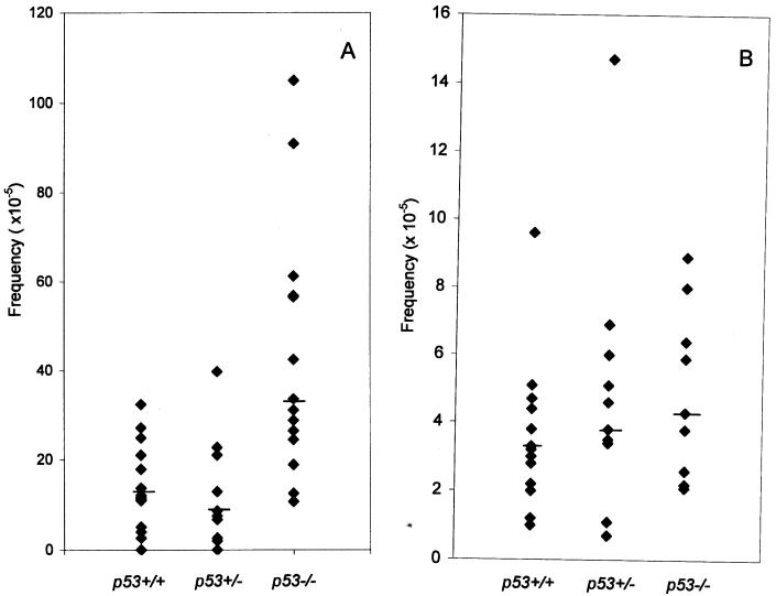
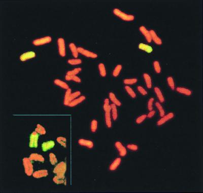
The p53 tumor suppressor protein participates in multiple cellular processes including cell cycle checkpoints and programmed cell death. In cell lines, loss of p53 function is associated with increased genetic instability including aneuploidy, gene amplification, and point mutation. Although similar genetic instability often accompanies the progression of malignancy in tumors, its role in tumor initiation in normal cells is not clear. To study whether or not loss of p53 leads to genetic instability in normal cells in vivo, we have examined mechanisms of loss of heterozygosity (LOH) at the Aprt (adenine phosphoribsyltransferase) and flanking loci in normal fibroblasts and T lymphocytes of p53-deficient mice. Somatic cell variants that arose in vivo as a consequence of genetic or epigenetic alterations abolishing Aprt function were selected and expanded in vitro by virtue of their resistance to 2,6-diaminopurine (DAP). We observed that p53 null mice produced about three times as many DAP-resistant fibroblast colonies than wild-type mice, but the frequency of DAP-resistant T lymphocyte colonies was not significantly changed. Mitotic recombination, but not point mutation, partly accounted for the increase in the frequency of DAP-resistant fibroblasts. Most significantly, chromosome loss/duplication and interstitial deletion, which were extremely rare events in the wild-type mice, represented a significant proportion of LOH events in both fibroblasts and T lymphocytes of p53 null mice. Also, increased interstitial deletion was observed in fibroblasts of p53 heterozygous mice. These data suggest that increased genetic variation, including chromosome instability, starts at the initiation stage of tumorigenesis when functional p53 is absent or reduced.
Figures
References
-
- Levine A J. Cell. 1997;88:323–331. - PubMed
-
- Agarwal M L, Taylor W R, Chernov M V, Chernova O B, Stark G R. J Biol Chem. 1998;273:1–4. - PubMed
-
- Wang X W, Yeh H, Schaeffer L, Roy R, Moncollin V, Egly J M, Wang Z, Friedberg E C, Evans M K, Taffe B G, et al. Nat Genet. 1995;10:188–195. - PubMed
-
- Offer H, Wolkowicz R, Matas D, Blumenstein S, Livneh Z, Rotter V. FEBS Lett. 1999;450:197–204. - PubMed
Publication types
MeSH terms
Substances
Grants and funding
LinkOut - more resources
Full Text Sources
Molecular Biology Databases
Research Materials
Miscellaneous
