. 2000 Jun 15;43(4):379-88.
doi: 10.1002/1097-4695(20000615)43:4<379::aid-neu6>3.0.co;2-0.

Rhythmic coupling among cells in the suprachiasmatic nucleus

Affiliations

Rhythmic coupling among cells in the suprachiasmatic nucleus

C S Colwell. J Neurobiol. .

Abstract

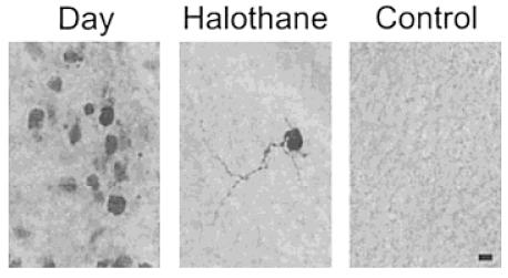
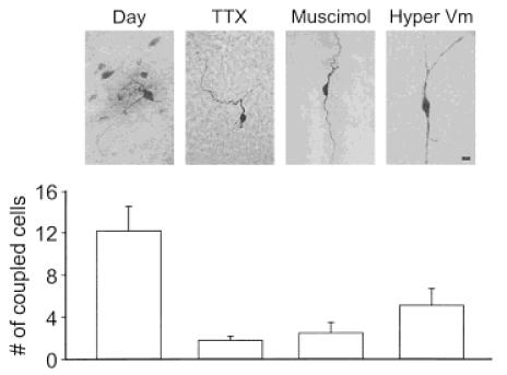
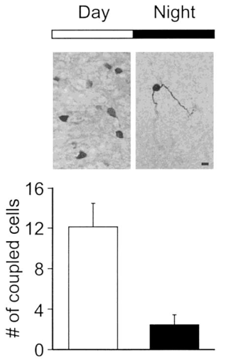
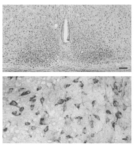
In mammals, the part of the nervous system responsible for most circadian behavior can be localized to a pair of structures in the hypothalamus known as the suprachiasmatic nucleus (SCN). Previous studies suggest that the basic mechanism responsible for the generation of these rhythms is intrinsic to individual cells. There is also evidence that the cells within the SCN are coupled to one another and that this coupling is important for the normal functioning of the circadian system. One mechanism that mediates coordinated electrical activity is direct electrical connections between cells formed by gap junctions. In the present study, we used a brain slice preparation to show that developing SCN cells are dye coupled. Dye coupling was observed in both the ventrolateral and dorsomedial subdivisions of the SCN and was blocked by application of a gap junction inhibitor, halothane. Dye coupling in the SCN appears to be regulated by activity-dependent mechanisms as both tetrodotoxin and the GABA(A) agonist muscimol inhibited the extent of coupling. Furthermore, acute hyperpolarization of the membrane potential of the original biocytin-filled neuron decreased the extent of coupling. SCN cells were extensively dye coupled during the day when the cells exhibit synchronous neural activity but were minimally dye coupled during the night when the cells are electrically silent. Immunocytochemical analysis provides evidence that a gap-junction-forming protein, connexin32, is expressed in the SCN of postnatal animals. Together the results are consistent with a model in which gap junctions provide a means to couple SCN neurons on a circadian basis.


Figures

Figure 1
Figure 1
Multiple SCN cells are stained after intracellular injection of the tracer biocytin. In these experiments, single SCN neurons are voltage clamped with a whole-cell patch electrode that contains biocytin (2 mg/mL). After recording, the presence of biocytin within cells was visualized by HRP-streptavidin and reaction with DAB. Left: In most cases, multiple cells were found to stain for biocytin following this procedure. Middle: This dye coupling was prevented by the application of the gap junction blocker halothane (0.1%). Right: No staining was observed when biocytin-loaded electrodes were placed into the slice without patching a cell. Scale bar = 10 μm.
Figure 2
Figure 2
Dye coupling among SCN cells was activity-dependent. To determine if the transfer of dye between SCN cells may be actively regulated, TTX or muscimol was placed in the bath while the original neuron was being filled with biocytin. Both treatments inhibit the spontaneous electrical activity exhibited by these cells as well as chemical synaptic transmission. Both treatments inhibited dye coupling as indicated by images and corresponding cell counts. Similar results (labeled “Hyper Vm”) were obtained when the electrical activity of the SCN neuron originally filled with biocytin was inhibited by holding the cell’s resting membrane potential at −80 mV. Under these conditions, the neurons do not generate spontaneous action potentials but still receive synaptic input from surrounding cells. All three treatments produced a significant ( p < .05) decrease in the number of coupled cells. Scale bar = 10 μm.
Figure 3
Figure 3
Dye coupling among SCN cells exhibited a circadian rhythm. SCN neurons are known to undergo a daily rhythm of electrical activity with spontaneous activity high during the day and low during the night. Although most SCN were dye coupled during the day (ZT 1–6), only a few SCN neurons during the night appeared to be dye coupled. The day/night difference in the number of coupled cells was significant ( p < .01).
Figure 4
Figure 4
Connexin 32 protein is expressed in the SCN. Cx32 appears to be one of the main Cx isoforms responsible for the formation of gap junctions between neurons. Accordingly, a polyclonal antibody against Cx32 (Zymed, 1:500 dilution) was used to stain SCN tissue from 14-dayold animals perfused during the day (ZT 2). Top: Low magnification (100×) view of Cx32 expression in the SCN. Scale bar = 100 μm. Bottom: Higher magnification (400×) view of labeled cells within the SCN. Scale bar = 10 μm.

References

    1. Balsalobre A, Damiola F, Schibler U. A serum shock induces circadian gene expression in mammalian tissue culture cells. Cell. 1998;93:929–937. - PubMed
    1. Bloomfield SA, Xin D, Osborne T. Light-induced modulation of coupling between AII amacrine cells in the rabbit retina. Vis Neurosci. 1997;14:565–576. - PubMed
    1. Bouskila Y, Dudek FE. Synchronous neuronal activity in the suprachiasmatic nucleus independent of chemical synaptic transmission. Proc Natl Acad Sci USA. 1993;90:3207–3210. - PMC - PubMed
    1. Card JP, Brecha N, Karten HJ, Moore RY. Immunocytochemical localization of vasoactive intestinal peptide-containing cells and processes in the suprachiasmatic nucleus of the rat. J Neurosci. 1981;1:1289–1303. - PMC - PubMed
    1. Cepeda C, Colwell CS, Itri JN, Chandler SH, Levine MS. Dopaminergic modulation of NMDA-induced whole-cell currents in neostriatal neurons in slices: contribution of calcium conductances. J Neurophysiol. 1998;79:82–94. - PubMed

Publication types

LinkOut - more resources