. 2000 Jun;105(12):1731-40.
doi: 10.1172/JCI8472.

Natural antibodies with the T15 idiotype may act in atherosclerosis, apoptotic clearance, and protective immunity

Affiliations

Natural antibodies with the T15 idiotype may act in atherosclerosis, apoptotic clearance, and protective immunity

P X Shaw et al. J Clin Invest. 2000 Jun.

Abstract

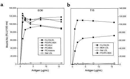
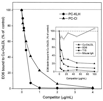
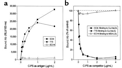
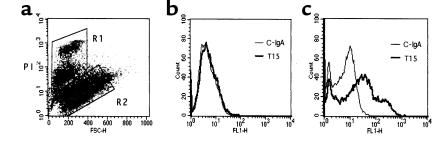
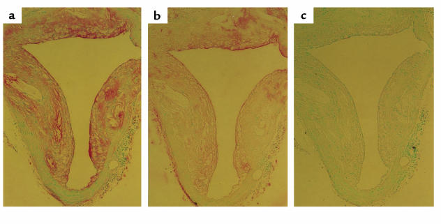
The immune response to oxidized LDL (OxLDL) may play an important role in atherogenesis. Working with apoE-deficient mice, we isolated a panel of OxLDL-specific B-cell lines that secrete IgM Abs that specifically bind to oxidized phospholipids such as 1-palmitoyl-2-(5-oxovaleroyl)-sn-glycero-3-phosphorylcholine (POVPC). These Abs block uptake of OxLDL by macrophages, recognize similar oxidation-specific epitopes on apoptotic cells, and are deposited in atherosclerotic lesions. The Abs were found to be structurally and functionally identical to classic "natural" T15 anti-PC Abs that are of B-1 cell origin and are reported to provide optimal protection from virulent pneumococcal infection. These findings suggest that there has been natural selection for B-1 cells secreting oxidation-specific/T15 antibodies, both for their role in natural immune defense and for housekeeping roles against oxidation-dependent neodeterminants in health and disease.


Figures

Figure 1
Figure 1
The DNA sequences and VDJ splice sites of the somatic VH rearrangements expressed in the EO6 B-cell hybridoma and the T(EPC)15 plasmacytoma, and their relationship to the most homologous germ-line (GL) VH, DH, and JH gene segments. Sequence analysis for the 372 encoding nucleotides of the EO6 VH rearrangement did not reveal a single nucleotide variation suggestive of somatic hypermutation. The canonical T15/EO6 VDJ rearrangement has been described as an archetype for primary homology-directed recombination, which more frequently occurs during B-lymphogenesis in the fetal liver. CDR: complementarity-determining region.
Figure 2
Figure 2
Reduced amino acid sequence alignment of EO autoantibodies with the classic anti-PC Ab T15. (a) Variable region of Ig light-chain sequences of EO autoantibodies are aligned against Vκ22/Jκ5 gene rearrangement. (b) Variable regions of heavy-chain sequences of EO autoantibodies are aligned against S107.1/DFL16.1/JH1 rearrangement. Both VH and VL show 100% homology to the germline genes of T15.
Figure 3
Figure 3
Binding of EO6 and T15 to oxidation-specific epitopes of LDL and PC. The indicated antigens were plated on microtiter wells at the indicated concentrations overnight at 4°C. EO6 (a) or T15 (b) was added at 5 μg/mL followed by the AP-conjugated goat anti-mouse IgM (for EO6) or IgA (for T15) secondary Abs. The amount of bound Abs was expressed as RLU/100 ms. Nat LDL, native LDL.
Figure 4
Figure 4
Inhibition of EO6 binding to Cu-OxLDL by PC, as sodium salt (PC-Cl) or PC-KLH conjugate. Cu-OxLDL (10 μg/mL) was plated on microtiter wells overnight at 4°C. EO6 (10 μg/mL) was added to wells in the absence or presence of the indicated concentrations of competitors, and the amount of bound EO6 was detected by AP-conjugated goat anti-mouse IgM. The amount of bound EO6 was expressed as the percent of EO6 binding to Cu-OxLDL in the absence of competitor. Inset: Inhibition of EO6 binding to Cu-OxLDL by both EO6 and T15. Cu-OxLDL (10 μg/mL) was coated on microtiter wells overnight at 4°C. Biotinylated EO6 (10 μg/mL) was added to microtiter wells in the absence or presence of the indicated concentrations of competitors. The amount of biotinylated EO6 bound to Cu-OxLDL was then detected by AP-conjugated NeutrAvidin®. The amount of biotinylated EO6 bound to the antigen in the absence of competitor was expressed as a percentage of the control. Nonspecific mouse IgA was used as isotype control for T15.
Figure 5
Figure 5
Binding of EO6 and T15 to pneumococcal C-PS. (a) C-PS was plated on microtiter wells at the indicated concentrations overnight at 4°C. Each Ab (5 μg/mL) was added to the plate and detected by AP-conjugated secondary Abs (goat anti-mouse IgM for EO6 and EO14, goat anti-mouse IgA for T15). The amount of bound Ab was expressed as RLU/100 ms. (b) Cu-OxLDL or MDA-LDL (10 μg/mL) was plated as antigen, and the indicated Ab was added to microtiter wells in the absence or presence of the indicated concentrations of C-PS. Bound Ab was detected by AP-conjugated secondary Abs (goat anti-mouse IgM for EO6 and EO14, goat anti-mouse IgA for T15), and was expressed as percent control of Ab binding to its antigen without competitor.
Figure 6
Figure 6
T15 IgA recognizes apoptotic cells. FACS® analysis of T15 Ab and control IgA (C-IgA; nonspecific monoclonal) binding to apoptotic PAECs. (a) Apoptosis-induced PAECs were gated into two populations according to PI intensity and FSC as described (7). Region 2 (R2): normal cells and cells in very early apoptosis. Region 1 (R1): cells with dim and bright PI staining (apoptotic). (b) Binding of control IgA and T15 to R2 cells. (c) Binding of control IgA and T15 to R1 cells.
Figure 7
Figure 7
Presence of T15 idiotypic Abs in atherosclerotic lesions. Atherosclerotic segments of aorta of LDLR–/– mice were prepared and stained as described in Methods. Epitopes recognized are indicated by a red color; the nuclei are counterstained with methyl green. (a) Section immunostained with a combined anti-mouse IgG/IgM antisera, indicating the presence of Ig in lesion. (b) An adjacent section immunostained with biotinylated AB1-2 (anti-T15 idiotype). A similar pattern demonstrates that some Ig deposited in the lesion represents Abs of the T15 idiotype. (c) An adjacent section stained with biotinylated nonimmune IgG was free of specific staining.

Comment in

References

    1. Steinberg, D., and Witztum, J.L. 1999. Lipoproteins, lipoprotein oxidation, and atherogenesis. In Molecular basis of cardiovascular disease. K.R. Chien, editor. W.B. Saunders Co. Philadelphia, Pennsylvania, USA. 458–475.
    1. Palinski W, et al. ApoE-deficient mice are a model of lipoprotein oxidation in atherogenesis. Demonstration of oxidation-specific epitopes in lesions and high titers of autoantibodies to malondialdehyde-lysine in serum. Arterioscler Thromb. 1994;14:605–616. - PubMed
    1. Palinski W, et al. Cloning of monoclonal autoantibodies to epitopes of oxidized lipoproteins from apolipoprotein E-deficient mice. Demonstration of epitopes of oxidized low density lipoprotein in human plasma. J Clin Invest. 1996;98:800–814. - PMC - PubMed
    1. Palinski W, Miller E, Witztum JL. Immunization of low density lipoprotein (LDL) receptor-deficient rabbits with homologous malondialdehyde-modified LDL reduces atherogenesis. Proc Natl Acad Sci USA. 1995;92:821–825. - PMC - PubMed
    1. Freigang S, Hörkkö S, Miller E, Witztum JL, Palinski W. Immunization of LDL receptor-deficient mice with homologous malondialdehyde-modified and native LDL reduces progression of atherosclerosis by mechanisms other than induction of high titers of antibodies to oxidative neoepitopes. Arterioscler Thromb Vasc Biol. 1998;18:1972–1982. - PubMed

Publication types

MeSH terms