. 2000 Jun;105(12):1769-77.
doi: 10.1172/JCI8892.

Enteroaggregative Escherichia coli expresses a novel flagellin that causes IL-8 release from intestinal epithelial cells

Affiliations

Enteroaggregative Escherichia coli expresses a novel flagellin that causes IL-8 release from intestinal epithelial cells

T S Steiner et al. J Clin Invest. 2000 Jun.

Abstract

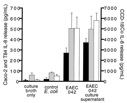
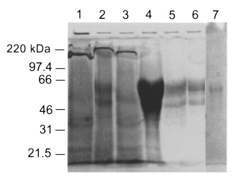
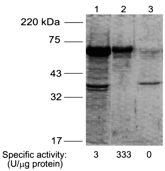
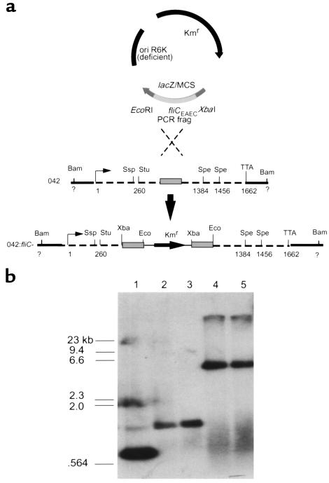
Enteroaggregative Escherichia coli (EAEC) is an emerging cause of acute and persistent diarrhea worldwide. EAEC infections are associated with intestinal inflammation and growth impairment in infected children, even in the absence of diarrhea. We previously reported that prototype EAEC strains rapidly induce IL-8 production by Caco-2 intestinal epithelial cells, and that this effect is mediated by a soluble, heat-stable factor released by these bacteria in culture. We herein report the cloning, sequencing, and expression of this biologically active IL-8-releasing factor from EAEC, and its identification as a flagellin that is unique among known expressed proteins. Flagella purified from EAEC 042 and several other EAEC isolates potently release IL-8 from Caco-2 cells; an engineered aflagellar mutant of 042 does not release IL-8. Finally, cloned EAEC flagellin expressed in nonpathogenic E. coli as a polyhistidine-tagged fusion protein maintains its proinflammatory activity. These findings demonstrate a major new means by which EAEC may cause intestinal inflammation, persistent diarrhea, and growth impairment that characterize human infection with these organisms. Furthermore, they open new approaches for diagnosis and vaccine development. This novel pathogenic mechanism of EAEC extends an emerging paradigm of bacterial flagella as inflammatory stimuli.


Figures

Figure 1
Figure 1
IL-8 release from intestinal cell lines in response to EAEC. Five-day-old monolayers of Caco-2 (black bars), T84 (gray bars), or CCD-18Co (white bars) cells were exposed for 3 hours to live EAEC 042, filtrates of saturated overnight cultures of EAEC 042, nonpathogenic (control) E. coli (HB101, K12, or GDI 20), or culture broth alone. The separate Y-axes reflect the tenfold greater amount of IL-8 release by CCD-18Co fibroblasts compared with Caco-2 or T84 colonic carcinoma cells. Error bars represent SEM.
Figure 2
Figure 2
Gel (9% SDS-PAGE) of sequentially purified, IL-8–releasing protein fractions from EAEC 042 culture supernatants. Samples were concentrated by membrane ultrafiltration to a volume of 25 μL and run simultaneously, with the exception of lane 7, which was run in a separate gel. Lane 1: Culture supernatant from overnight growth of 042. Lane 2: DEAE-cellulose batch adsorption and elution with NaCl. Lane 3: Sixty percent ammonium sulfate precipitation. Lane 4: Gel permeation chromatography in the presence of 6 M guanidine-HCl. Lane 5: Pooled, active gel permeation fractions, dialyzed to remove guanidine. Lane 6: Strong anion exchange and subsequent hydrophobic interaction chromatography. Lane 7: Hydroxyapatite chromatography.
Figure 3
Figure 3
Gel (9% SDS-PAGE) of EAEC flagellar isolation. Samples were concentrated by acetone precipitation before loading. Lane 1: Flagella sheared from EAEC 042 and pelleted by ultracentrifugation. Lane 2: The approximately 65-kDa band, cut and eluted from a previous gel, dialyzed to remove SDS, and reVrun in SDS-containing loading buffer. Lane 3: The approximately 35-kDa band similarly eluted, dialyzed, and rerun. Specific activity was defined as the amount of sample producing half-maximal IL-8 release from Caco-2 cells at 3 hours of exposure.
Figure 4
Figure 4
Construction of 042:fliC. (a) Restriction maps of pJP5603 (3.1 kb) with the cloned 588-bp PCR fragment of fliCEAEC (gray bar), wild-type fliCEAEC (dashed line), and the map of the desired transconjugant. Restriction sites and locations are shown (Bam, BamHI; Ssp, SspI; Stu, StuI; Spe, SpeI; Xba, XbaI; Eco, EcoRI). (b) Southern hybridization of 042 and 042:fliC using the 588-bp PCR fragment from the 042 genome as a probe. Lane 1: Probe, 1 ng. Lane 2: 042, SpeI/StuI cut. Lane 3: 042, SpeI/SspI cut. Lane 4: 042:fliC, SpeI/StuI cut. Lane 5: 042:fliC, SpeI/SspI cut. Kmr, kanamycin resistance cassette; TTA, stop codon; ori R6K, origin of replication.
Figure 5
Figure 5
Motility and IL-8–releasing activity of 042 and 042:fliC. (a) Growth at 72 hours on swarm plates without kanamycin. Bacteria picked from the periphery (swarming area) of the lower left plate had lost kanamycin resistance. (b) Electron microscopy with negative staining for flagella. Bar = 1 μm. (c) IL-8 release from Caco-2 cells incubated for 3 hours with culture supernatants of EAEC 042 grown in 1% tryptone (n = 5), 1% tryptone with 3% NaCl (n = 2), culture supernatants (sup) (n = 5), or live cultures (n = 3) of 042:fliC grown in 1% tryptone. P < 0.05 by Kruskal-Wallis test.
Figure 6
Figure 6
Expression and purification of FliC-EAEC. (a) BL21(DE3)pLysS transformed with pfliC or pCRT7/NT-E3 (expression control) were harvested from mid–log phase cultures at the indicated times after addition of IPTG. Ten microliters of bacterial lysate was loaded per well for 9% SDS-PAGE, and transferred to PVDF membrane after gel was run. The membrane was probed with Ni2+-HRP to detect polyhistidine-containing proteins, stripped with 0.2 N NaOH, and reprobed with polyclonal anti-flagellar antiserum. A lane containing 2.5 μg of EAEC 042 flagella (A) was included in the gel as a control. For IL-8 release, purified flagella (72 ng) or bacterial lysate (1 μg) was added to 500-μL wells of Caco-2 cells in the same experiment, and supernatant was assayed after 3 hours of incubation. (b) BL21(DE3)pLysS:pfliC was harvested 2 hours after addition of IPTG to mid–log phase culture. Lysates were purified by nickel affinity chromatography, visualized on 9% SDS-PAGE gels, and transferred to a PVDF membrane that was probed with Ni2+-HRP. Lane 1: EAEC 042 flagella, 1.4 μg. Lane 2: Crude lysate, 15 μg. Lane 3: Flow-through from nickel column, 12.5 μg. Lane 4: Eluate from column, 10.8 μg. For IL-8 release, 350 ng of flagella, 1 μg of bacterial lysate, and 1 μg of eluted flagellin were tested.
Figure 7
Figure 7
Comparison of deduced amino acid sequences of flagellins from EAEC 042 (EAEC), S. dysenteriae (SD), S. flexneri (SF), and E. coli K12 (K12). Trypsin peptides identified by mass spectrometry from the initial purification product of EAEC 042 supernatants are underlined. Amino acids in the EAEC flagellin differing from those in S. dysenteriae are shown above the line. The arrows represent the locations of the forward and reverse primers used to generate the 588-bp fliC probe. FliC-EAEC accession AF 194946.

Comment in

References

    1. Nataro J, Steiner T, Guerrant R. Enteroaggregative Escherichia coli. Emerg Infect Dis. 1998;4:251–261. - PMC - PubMed
    1. Nataro J, et al. Patterns of adherence of diarrheagenic Escherichia coli to HEp-2 cells. Pediatr Infect Dis J. 1987;6:829–831. - PubMed
    1. Cohen MB, et al. Colonization by enteroaggregative Escherichia coli in travelers with and without diarrhea. J Clin Microbiol. 1993;31:351–353. - PMC - PubMed
    1. Gascon J, et al. Enteroaggregative Escherichia coli strains as a cause of traveler’s diarrhea: a case-control study. J Infect Dis. 1998;177:1409–1412. - PubMed
    1. Mayer HB, Wanke CA. Enteroaggregative Escherichia coli as a possible cause of diarrhea in an HIV-infected patient. N Engl J Med. 1995;332:273–274. - PubMed

Publication types