. 2000 Jul;74(14):6242-50.
doi: 10.1128/jvi.74.14.6242-6250.2000.

An enzymatic footprinting analysis of the interaction of 40S ribosomal subunits with the internal ribosomal entry site of hepatitis C virus

Affiliations

An enzymatic footprinting analysis of the interaction of 40S ribosomal subunits with the internal ribosomal entry site of hepatitis C virus

V G Kolupaeva et al. J Virol. 2000 Jul.

Abstract

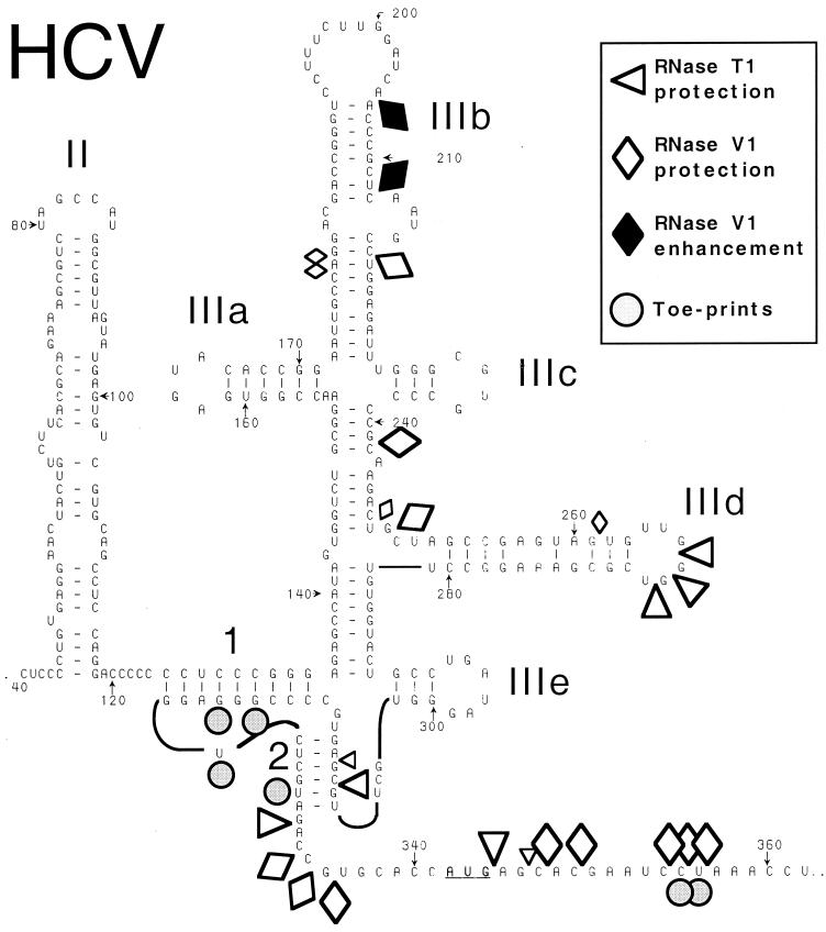
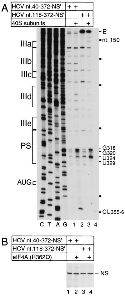
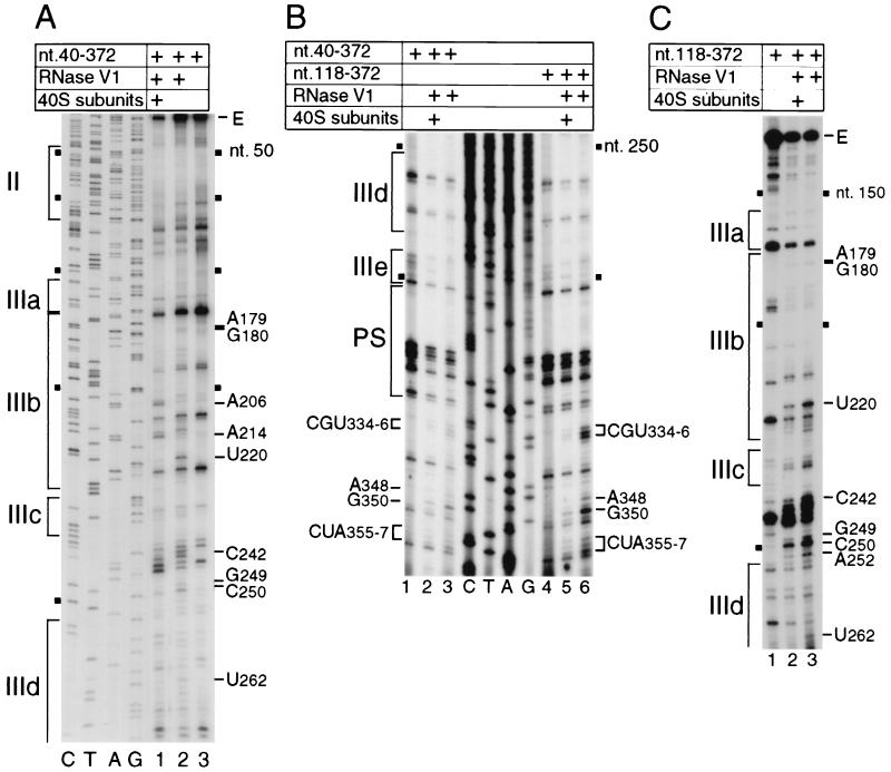
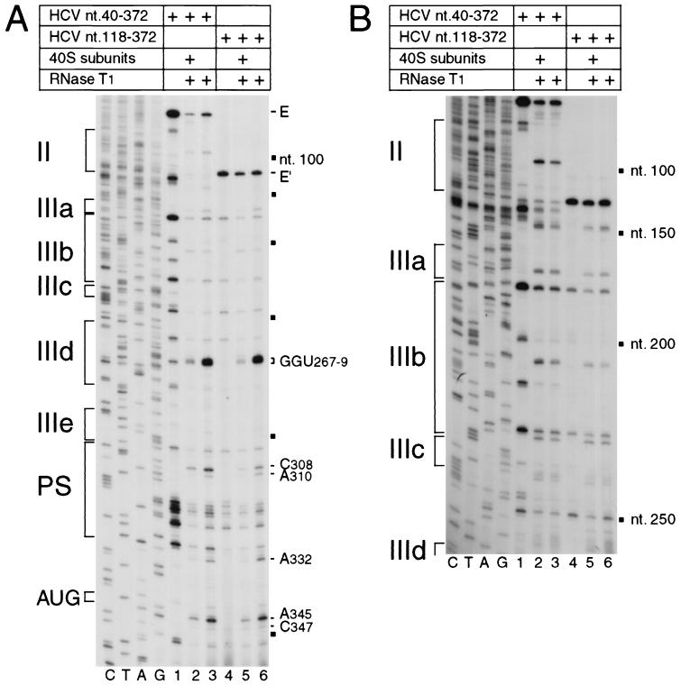
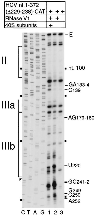
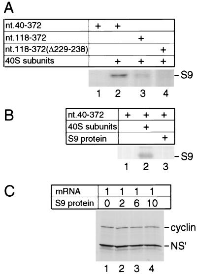
Hepatitis C virus translation is initiated on a approximately 330-nucleotide (nt)-long internal ribosomal entry site (IRES) at the 5' end of the genome. In this process, a 43S preinitiation complex (comprising a 40S ribosomal subunit, eukaryotic initiation factor 3 (eIF3), and a ternary [eIF2-GTP-initiator tRNA] complex) binds the IRES in a precise manner so that the initiation codon is placed at the ribosomal P site. This binding step involves specific interactions between the IRES and different components of the 43S complex. The 40S subunit and eIF3 can bind to the IRES independently; previous analyses revealed that eIF3 binds specifically to an apical half of IRES domain III. Nucleotides in the IRES that are involved in the interaction with the 40S subunit were identified by RNase footprinting and mapped to the basal half of domain III and in domain IV. Interaction sites were identified in locations that have been found to be essential for IRES function, including (i) the apical loop residues GGG(266-268) in subdomain IIId and (ii) the pseudoknot. Extensive protection from RNase cleavage also occurred downstream of the pseudoknot in domain IV, flanking both sides of the initiation codon and corresponding in length to that of the mRNA-binding cleft of the 40S subunit. These results indicate that the 40S subunit makes multiple interactions with the IRES and suggest that only nucleotides in domain IV are inserted into the mRNA-binding cleft of the 40S subunit.


Figures

FIG. 1
FIG. 1
Schematic representation of the secondary structure of the HCV IRES (based on references and 41), showing sites in nt 40-372.NS′ mRNA that are protected from cleavage by RNases T1 and V1 or at which cleavage is enhanced following binding of a 40S ribosomal subunit. These sites and the position of toeprints caused by bound 40S subunits are indicated by symbols shown at the upper right. Smaller symbols indicate weaker protection from cleavage. Domain IV (nt 331 to 354) is shown as an unstructured linear sequence for greater clarity. The initiation codon (AUG342-344) is underlined. The nomenclature used to describe subdomains of the IRES is from reference ; the helices that constitute the pseudoknot are labeled 1 and 2.
FIG. 2
FIG. 2
Influence of domain II on IRES function. (A) Toeprint analysis of binary ribosomal complex formation on the HCV IRES. Ribosomal 40S subunits were incubated with HCV nt 40-372.NS′ or nt 118-372.NS′ mRNA under standard reaction conditions and then analyzed by primer extension. Full-length nt 118-372.NS′ cDNA is marked E′; other cDNA products terminated at the sites indicated on the right. Reference lanes C, T, A, and G depict HCV sequences; IRES subdomains IIIa, IIIb, IIIc, IIId, IIIe, and PS (pseudoknot) are indicated on the left; HCV nucleotides are indicated by black squares at 50-nt intervals from nt 150 to 350 on the right. (B) Translation in RRL of HCV nt 40-372.NS′ mRNA (lanes 1 and 2) or nt 118-372.NS′ mRNA (lane 3 and 4) in the presence (lanes 2 and 4) or absence (lanes 1 and 3) of a 10-fold molar excess of mutant eIF4A(R362Q). Translation products were analyzed by autoradiography after electrophoresis on a 12% polyacrylamide gel. The position of the NS′ translation product is indicated.
FIG. 3
FIG. 3
RNase V1 footprinting of the binary 40S subunit-HCV IRES complex. The gels show polyacrylamide-urea gel fractionation of cDNA products obtained after primer extension. (A) Sensitivity of HCV nt 40-372 RNA upstream of nt 272 to cleavage (lanes 1 and 2) either alone (lane 2) or bound by a 40S subunit (lane 1); (B) sensitivity of HCV nt 40-372 RNA (lanes 1 to 3) or nt 118-372 RNA (lanes 4 to 6) upstream of nt 360 to cleavage (lanes 2, 3, 5, and 6) either alone (lanes 3 and 6) or bound by a 40S subunit (lanes 2 and 5); (C) sensitivity of HCV nt 118-372 RNA upstream of nt 265 to cleavage (lanes 2 and 3) either alone (lane 3) or bound by a 40S subunit (lane 2). cDNA products obtained after primer extension of untreated HCV RNA are shown in lane 3 of panel A, lanes 1 and 4 of panel B, and lane 1 of panel C. A dideoxynucleotide sequence generated with the same primer (shown in lanes C, T, A, and G in panels A and B) was run in parallel on each gel. IRES subdomains II, IIIa, IIIb, IIIc, IIId, IIIe, and PS (pseudoknot), as appropriate, are indicated on the left of each panel; HCV nucleotides are indicated by black squares at 50-nt intervals, and the positions of protected residues are indicated on the sides of each panel.
FIG. 4
FIG. 4
RNase T1 footprinting of the binary 40S subunit-HCV IRES complex. Polyacrylamide-urea gel fractionation of cDNA products obtained after primer extension shows the sensitivity of HCV nt 40-372 RNA (lanes 1 to 3) or nt 118-372 RNA (lanes 4 to 6) upstream of nt 360 (A) or of nt 250 (B) to cleavage (lanes 1, 2, 5, and 6) either alone (lanes 3 and 6) or bound by a 40S subunit (lanes 2 and 5). cDNA products obtained after primer extension of untreated HCV RNA are shown in lanes 1 and 4 of both panels. A dideoxynucleotide sequence generated with the same primer and run in parallel is shown to the left of both panels. IRES subdomains II, IIIa, IIIb, IIIc, IIId, IIIe, and PS (pseudoknot) are indicated on the left of each panel; HCV nucleotides are indicated by black squares at 50-nt intervals, and the positions of protected residues are marked on the right of each panel. The position of the initiation codon AUG is indicated to the left of panel A.
FIG. 5
FIG. 5
RNase V1 footprinting of the binary ribosomal complex formed on HCV nt 40-372 RNA lacking subdomain IIIc (nt 229 to 238). Polyacrylamide-urea gel fractionation of cDNA products obtained after primer extension shows the sensitivity of the HCV RNA upstream of nt 255 to cleavage (lanes 1 and 2) either alone (lane 2) or bound by a 40S subunit (lane 1). cDNA products obtained after primer extension of untreated HCV RNA are shown in lane 3. A dideoxynucleotide sequence generated with the same primer and run in parallel is shown to the left; HCV nucleotides are indicated by black squares at 50-nt intervals from nt 100 to 250. IRES subdomains II, IIIa, and IIIb are indicated on the left, and the positions of protected residues are indicated to the right. The position of the nt 229-238 deletion is indicated by an asterisk.
FIG. 6
FIG. 6
Ribosomal protein S9 requires the context of the ribosomal 40S subunit to bind to the HCV IRES. (A and B) UV cross-linking of native S9 as a constituent of 40S subunits (A, lanes 2 and 3; B, lane 2) and (B) UV cross-linking of recombinant S9 (B, lane 3) to [32P]UTP-labeled HCV nt 40-372 RNA (A, lanes 1 and 2; B, lanes 1 to 3), nt 118 to 372 RNA (A, lane 3) or nt 118-372(Δ229-238) RNA (panel A, lane 4). Samples were treated with RNases after irradiation, and proteins were resolved by gel electrophoresis. The position of S9 is indicated to the right of both panels. (C) Effect of recombinant S9 added with dicistronic cyclin-CSFV IRES-NS′ mRNA at the indicated molar ratios to translation reaction mixtures on CSFV IRES-mediated NS′ translation.

References

    1. Becher P, Orlich M, Shannon A D, Horner G, König M, Thiel J-J. Phylogenetic analysis of pestiviruses from domestic and wild ruminants. J Gen Virol. 1997;78:1357–1366. - PubMed
    1. Brown E A, Zhang H, Ping L-H, Lemon S M. Secondary structure of the 5′ nontranslated regions of hepatitis C virus and pestivirus genomic RNAs. Nucleic Acids Res. 1992;20:5041–5045. - PMC - PubMed
    1. Browning K S, Leung D W, Clark J M. Protection of satellite tobacco necrosis virus ribonucleic acid by wheat germ 40S and 80S ribosomes. Biochemistry. 1980;19:2276–2283. - PubMed
    1. Buratti E, Tisminetzky S, Zotti M, Baralle F E. Functional analysis of the interaction between HCV 5′ UTR and putative subunits of eukaryotic translation initiation factor eIF3. Nucleic Acids Res. 1998;26:3179–3187. - PMC - PubMed
    1. Clarke B. Molecular virology of hepatitis C virus. J Gen Virol. 1997;78:2397–2410. - PubMed

Publication types

Associated data