. 2000 Jul;74(14):6316-23.
doi: 10.1128/jvi.74.14.6316-6323.2000.

Interdependence of hemagglutinin glycosylation and neuraminidase as regulators of influenza virus growth: a study by reverse genetics

Affiliations

Interdependence of hemagglutinin glycosylation and neuraminidase as regulators of influenza virus growth: a study by reverse genetics

R Wagner et al. J Virol. 2000 Jul.

Abstract

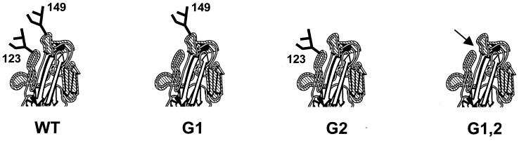
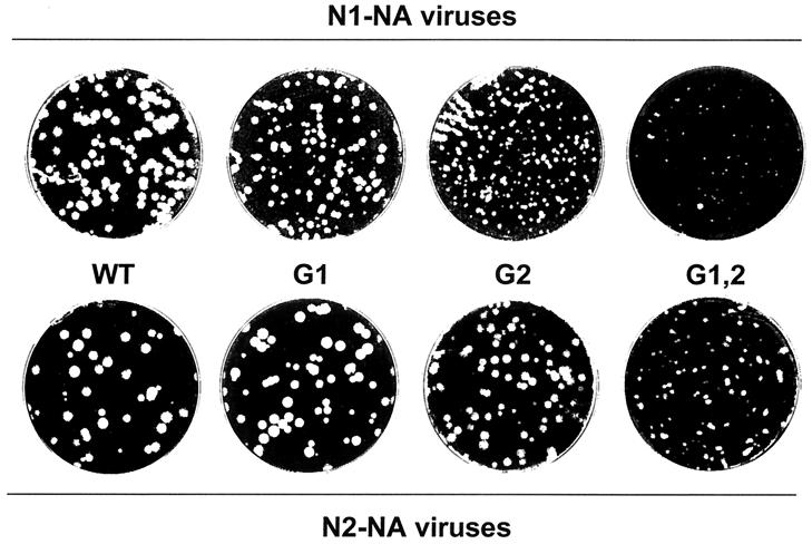
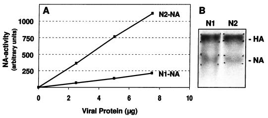
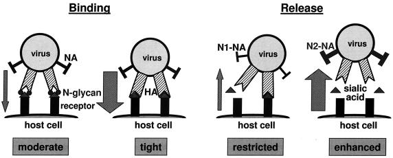
The hemagglutinin (HA) of fowl plague virus A/FPV/Rostock/34 (H7N1) carries two N-linked oligosaccharides attached to Asn123 and Asn149 in close vicinity to the receptor-binding pocket. In previous studies in which HA mutants lacking either one (mutants G1 and G2) or both (mutant G1,2) glycosylation sites had been expressed from a simian virus 40 vector, we showed that these glycans regulate receptor binding affinity (M. Ohuchi, R. Ohuchi, A. Feldmann, and H. D. Klenk, J. Virol. 71:8377-8384, 1997). We have now investigated the effect of these mutations on virus growth using recombinant viruses generated by an RNA polymerase I-based reverse genetics system. Two reassortants of influenza virus strain A/WSN/33 were used as helper viruses to obtain two series of HA mutant viruses differing only in the neuraminidase (NA). Studies using N1 NA viruses revealed that loss of the oligosaccharide from Asn149 (mutant G2) or loss of both oligosaccharides (mutant G1,2) has a pronounced effect on virus growth in MDCK cells. Growth of virus lacking both oligosaccharides from infected cells was retarded, and virus yields in the medium were decreased about 20-fold. Likewise, there was a reduction in plaque size that was distinct with G1,2 and less pronounced with G2. These effects could be attributed to a highly impaired release of mutant progeny viruses from host cells. In contrast, with recombinant viruses containing N2 NA, these restrictions were much less apparent. N1 recombinants showed lower neuraminidase activity than N2 recombinants, indicating that N2 NA is able to partly overrule the high-affinity binding of mutant HA to the receptor. These results demonstrate that N-glycans flanking the receptor-binding site of the HA molecule are potent regulators of influenza virus growth, with the glycan at Asn149 being dominant and that at Asn123 being less effective. In addition, we show here that HA and NA activities need to be highly balanced in order to allow productive influenza virus infection.


Figures

FIG. 1
FIG. 1
Head region of FPV HA. N-linked oligosaccharides adjacent to the receptor-binding pocket are indicated. Mutants G1 and G2 lack the glycosylation sites at Asn123 and Asn149, respectively. Both sites are absent in mutant G1,2. The arrow marks the entrance to the receptor-binding pocket.
FIG. 2
FIG. 2
Restriction analysis of the HA cDNA derived from recombinant viruses. (A) Scheme of the HA cDNA used for the generation of recombinants. Positions of endonuclease recognition motifs introduced as genetic tag sites for individual mutants are indicated. The binding sites of specific oligonucleotide primers used in RT-PCR are indicated by arrows. nt, nucleotide. (B) RT-PCR analysis of RNA isolated from wild-type (WT), G1, G2, and G1,2 recombinant viruses of the N1 series. RT-PCR products were incubated with endonucleases as indicated and separated on an agarose gel. RNA isolated from FPV was used as a control.
FIG. 3
FIG. 3
Analysis of the glycosylation pattern of HA from recombinant viruses. HA was immunoprecipitated from 35S-labeled viruses of the N2 group. One half of the material was treated with PNGase F (+), while the other half remained untreated (−). The protein profile was analyzed by SDS-PAGE, and bands were visualized by fluorography.
FIG. 4
FIG. 4
Growth curves of recombinants in MDCK cells. Cell monolayers were infected at an MOI of 0.001 with recombinant viruses, and supernatants were monitored for HA titers at the time points indicated. (A) Viruses of the N2 series. (B) Viruses of the N1 series. ■, wild type; ▴, G1; ⧫, G2; ●, G1,2.
FIG. 5
FIG. 5
Analysis of cell-to-cell spread of recombinant viruses by plaque assay. MDCK cell monolayers were infected with wild-type viruses (WT) or recombinant viruses of the N1 NA and N2 NA groups as indicated. Monolayers were covered with an agarose-containing overlay for 3 days and stained with crystal violet. Dilutions were chosen individually for each virus stock in order to obtain optimal plaque pictures.
FIG. 6
FIG. 6
Comparison of specific NA activities of WT/N1 and WT/N2 viruses. (A) Different amounts of purified virus were incubated with MU-NANA for 20 min at 37°C. The reaction was stopped, and NA activity was calculated by measuring the fluorescence of the liberated methylumbelliferone. The data are means of three experiments. They indicate that WT/N2 has a higher NA activity than WT/N1. (B) Analysis of equal amounts of purified WT/N1 (N1) and WT/N2 (N2) virions labeled with [3H]glucosamine by SDS-PAGE under nonreducing conditions. HA and NA bands were excised from the gel, and incorporated radioactivity was determined by liquid scintillation counting. The data show that both virus preparations contain equal amounts of HA and NA.
FIG. 7
FIG. 7
Virus elution from chicken erythrocytes. Twofold dilutions of recombinant viruses of the N2 NA group (A) and the N1 NA group (B) were incubated with equal volumes of chicken erythrocytes at 4°C for 1 h. Samples were then transferred to 37°C, and the reduction in HA titers was recorded periodically. Results are presented as percentages of the initial HA titer at 4°C. ■, wild type; ▴, G1; ⧫, G2; ●, G1,2.
FIG. 8
FIG. 8
Release of recombinant viruses from MDCK cells. MDCK cells were infected at an MOI of 5 with recombinant viruses and incubated at 37°C overnight. One hour before virus harvest, VCNA was added to the culture medium of one half of the samples. Titers of progeny viruses released into the medium were determined by plaque assay. Levels of virus release in the absence of VCNA are presented as a percentage of the virus titers released after VCNA treatment.
FIG. 9
FIG. 9
Regulation of virus binding and release by HA glycosylation and neuraminidase activity. Receptor affinity is controlled by the oligosaccharides adjacent to the receptor-binding site on HA. The efficiency of release depends on the activity of NA.

References

    1. Aytay S, Schulze I T. Single amino acid substitutions in the hemagglutinin can alter the host range and receptor binding properties of H1 strains of influenza A virus. J Virol. 1991;65:3022–3028. - PMC - PubMed
    1. Castrucci M R, Kawaoka Y. Biologic importance of neuraminidase stalk length in influenza A virus. J Virol. 1993;67:759–764. - PMC - PubMed
    1. Colman P. Structure and function of the neuraminidase. In: Nicholson K G, Webster R G, Hay A J, editors. Textbook of influenza. London, England: Blackwell Science; 1998. pp. 65–73.
    1. Compans R W, Dimmock N J, Meier-Ewert H. Effect of antibody to neuraminidase on the maturation and hemagglutinating activity of an influenza A2 virus. J Virol. 1969;4:528–534. - PMC - PubMed
    1. Connor R J, Kawaoka Y, Webster R G, Paulson J C. Receptor specificity in human, avian, and equine H2 and H3 influenza virus isolates. Virology. 1994;205:17–23. - PubMed

Publication types

MeSH terms

Substances

LinkOut - more resources