. 2000 Jul;74(14):6408-17.
doi: 10.1128/jvi.74.14.6408-6417.2000.

Human papillomavirus type 16 E6 induces self-ubiquitination of the E6AP ubiquitin-protein ligase

Affiliations

Human papillomavirus type 16 E6 induces self-ubiquitination of the E6AP ubiquitin-protein ligase

W H Kao et al. J Virol. 2000 Jul.

Abstract

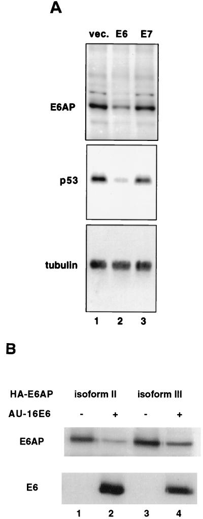
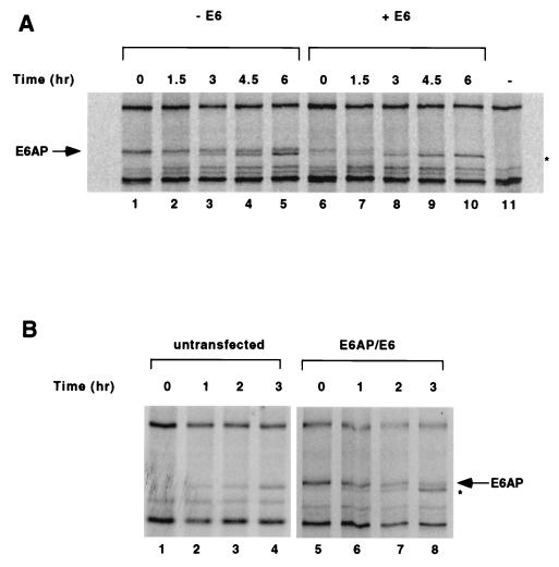
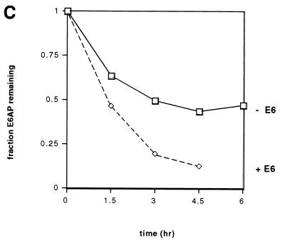
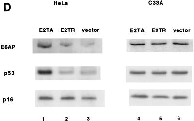
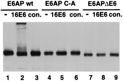
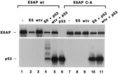
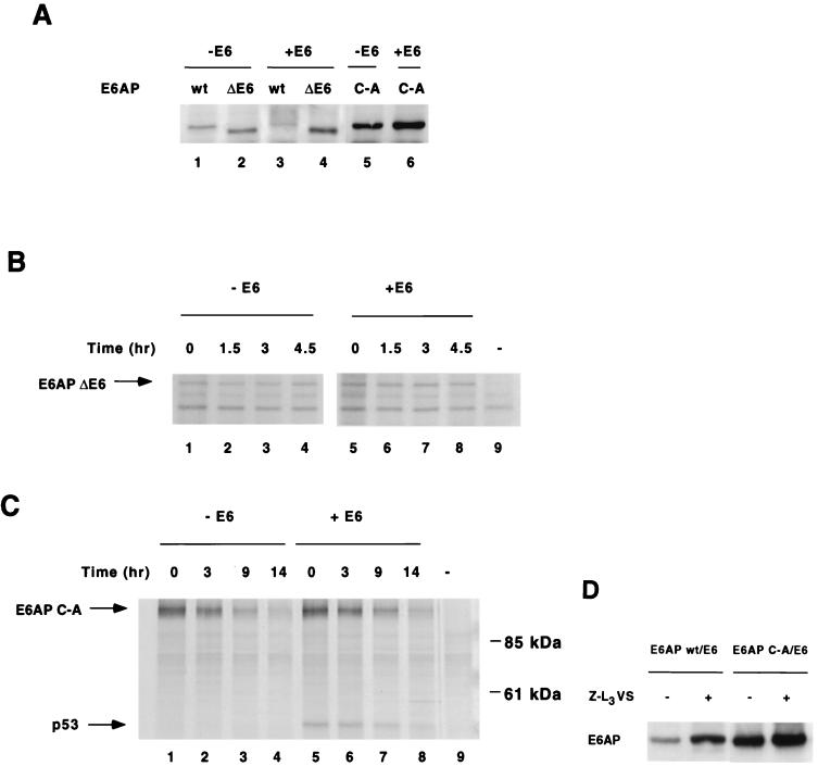
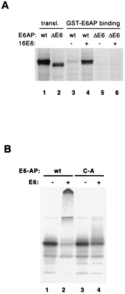
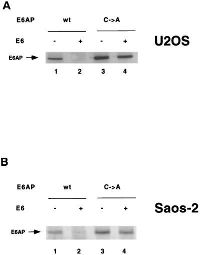
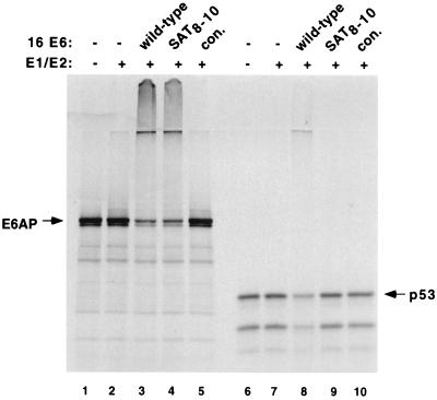
The E6 protein of the high-risk human papillomaviruses (HPVs) and the cellular ubiquitin-protein ligase E6AP form a complex which causes the ubiquitination and degradation of p53. We show here that HPV16 E6 promotes the ubiquitination and degradation of E6AP itself. The half-life of E6AP is shorter in HPV-positive cervical cancer cells than in HPV-negative cervical cancer cells, and E6AP is stabilized in HPV-positive cancer cells when expression of the viral oncoproteins is repressed. Expression of HPV16 E6 in cells results in a threefold decrease in the half-life of transfected E6AP. E6-mediated degradation of E6AP requires (i) the binding of E6 to E6AP, (ii) the catalytic activity of E6AP, and (iii) activity of the 26S proteasome, suggesting that E6-E6AP interaction results in E6AP self-ubiquitination and degradation. In addition, both in vitro and in vivo experiments indicate that E6AP self-ubiquitination results primarily from an intramolecular transfer of ubiquitin from the active-site cysteine to one or more lysine residues; however, intermolecular transfer can also occur in the context of an E6-mediated E6AP multimer. Finally, we demonstrate that an E6 mutant that is able to immortalize human mammary epithelial cells but is unable to degrade p53 retains its ability to bind and degrade E6AP, raising the possibility that E6-mediated degradation of E6AP contributes to its ability to transform mammalian cells.


Figures

FIG. 1
FIG. 1
HPV16 E6 lowers E6AP levels in vivo. (A) E6 but not E7 lowers levels of endogenous E6AP in C33A cells. C33A cells were cotransfected with puromycin resistance plasmid along with empty vector (lane 1), vector expressing E6 (lane 2), or vector expressing E7 (lane 3). After selection of puromycin-resistant cells, protein extracts were resolved by SDS-PAGE and analyzed by immunoblotting with rabbit polyclonal E6AP antibody (upper panel), anti-p53 antibody (middle panel), or antitubulin antibody (lower panel). (B) HPV16 E6 decreases the steady-state levels of transfected E6AP full-length isoforms. C33A cells were transfected with plasmids encoding AU1-tagged HPV16 E6 (lanes 2 and 4) and HA-tagged E6AP isoforms II (lanes 1 and 2) or III (lanes 3 and 4). Cells not receiving the E6 plasmid were transfected with an equivalent amount of empty vector. After cell lysis, protein extracts were resolved by SDS-PAGE and immunoblotted with the 12CA5 antibody to detect HA-tagged E6AP or with the AU1 antibody to detect AU1-tagged E6.
FIG. 2
FIG. 2
HPV16 E6 accelerates the degradation of E6AP. (A) Pulse-chase analysis of E6AP isoform I in the absence (lanes 1 to 5) or presence (lanes 6 to 10) of E6. C33A cells were transfected with HA-E6AP isoform I either with or without AU1-16E6. As a negative control, one plate of cells was transfected with an equivalent amount of empty vector (lane 11). At 48 h posttransfection, cells were labeled with [35S]methionine and chased with nonradioactive media for the indicated times. Cell extracts were harvested, and equal amounts of each sample were immunoprecipitated overnight with the 12CA5 antibody and a mixture of protein A- and protein G-agarose beads. Immunoprecipitated proteins were resolved by SDS-PAGE and visualized by autoradiography. (B) Pulse-chase analysis of untransfected C33A cells (lanes 1 to 4) or cells transfected with HA-E6AP isoform I and AU1-E6 (lanes 5 to 8). The band immediately below E6AP in panels A and B (marked by asterisk) increases with time even in the absence of transfected HA-E6AP (Fig. 2B, lanes 1 to 4) and is likely an endogenous protein not related to E6AP that cross-reacts with the 12CA5 antibody. (C) Quantitation of data shown in panel A. Intensities of the gel bands were quantified using the NIH Image 1.60/68k program.
FIG. 2
FIG. 2
HPV16 E6 accelerates the degradation of E6AP. (A) Pulse-chase analysis of E6AP isoform I in the absence (lanes 1 to 5) or presence (lanes 6 to 10) of E6. C33A cells were transfected with HA-E6AP isoform I either with or without AU1-16E6. As a negative control, one plate of cells was transfected with an equivalent amount of empty vector (lane 11). At 48 h posttransfection, cells were labeled with [35S]methionine and chased with nonradioactive media for the indicated times. Cell extracts were harvested, and equal amounts of each sample were immunoprecipitated overnight with the 12CA5 antibody and a mixture of protein A- and protein G-agarose beads. Immunoprecipitated proteins were resolved by SDS-PAGE and visualized by autoradiography. (B) Pulse-chase analysis of untransfected C33A cells (lanes 1 to 4) or cells transfected with HA-E6AP isoform I and AU1-E6 (lanes 5 to 8). The band immediately below E6AP in panels A and B (marked by asterisk) increases with time even in the absence of transfected HA-E6AP (Fig. 2B, lanes 1 to 4) and is likely an endogenous protein not related to E6AP that cross-reacts with the 12CA5 antibody. (C) Quantitation of data shown in panel A. Intensities of the gel bands were quantified using the NIH Image 1.60/68k program.
FIG. 3
FIG. 3
Endogenous E6AP is more rapidly degraded in HPV-positive HeLa cells than in HPV-negative C33A cells. (A) Comparison of endogenous E6AP levels in HeLa and C33A cells. A total of 50 μg of protein extract from HeLa and C33A cells were protein extracts were resolved by SDS-PAGE and analyzed by immunoblotting with anti-E6AP antibody (upper panel) or anti-actin (lower panel). (B) Pulse-chase analysis of E6AP in HeLa and C33A cells. Cells were labeled with [35S]methionine and chased with nonradioactive media for the indicated times. Cell extracts were harvested, and equal amounts of each sample were immunoprecipitated with polyclonal anti-E6AP, separated by SDS-PAGE, and visualized by autoradiography. (C) Quantitation of data in panel B. (D) Stabilization of E6AP protein levels in HeLa cells, but not in C33A cells, transfected with BPV1 E2. Cells were transfected with 1 μg of pBabe-puro plus 5 μg of either BPV1 E2TA or E2TR expression plasmids or else vector alone. Cells were maintained under puromycin-containing medium for 5 days. After harvesting of the cell extracts, 50 μg of protein per sample was resolved by SDS-PAGE and immunoblotted with anti-E6AP (upper panel), anti-p53 (middle panel), or anti-p16 (lower panel).
FIG. 3
FIG. 3
Endogenous E6AP is more rapidly degraded in HPV-positive HeLa cells than in HPV-negative C33A cells. (A) Comparison of endogenous E6AP levels in HeLa and C33A cells. A total of 50 μg of protein extract from HeLa and C33A cells were protein extracts were resolved by SDS-PAGE and analyzed by immunoblotting with anti-E6AP antibody (upper panel) or anti-actin (lower panel). (B) Pulse-chase analysis of E6AP in HeLa and C33A cells. Cells were labeled with [35S]methionine and chased with nonradioactive media for the indicated times. Cell extracts were harvested, and equal amounts of each sample were immunoprecipitated with polyclonal anti-E6AP, separated by SDS-PAGE, and visualized by autoradiography. (C) Quantitation of data in panel B. (D) Stabilization of E6AP protein levels in HeLa cells, but not in C33A cells, transfected with BPV1 E2. Cells were transfected with 1 μg of pBabe-puro plus 5 μg of either BPV1 E2TA or E2TR expression plasmids or else vector alone. Cells were maintained under puromycin-containing medium for 5 days. After harvesting of the cell extracts, 50 μg of protein per sample was resolved by SDS-PAGE and immunoblotted with anti-E6AP (upper panel), anti-p53 (middle panel), or anti-p16 (lower panel).
FIG. 4
FIG. 4
E6-dependent E6AP ubiquitination requires the active-site cysteine and the E6-binding domain of E6AP. Partially purified baculovirus-expressed epitope tagged wild-type E6AP (wt; lanes 1 to 3), E6AP carrying the active-site cysteine to alanine mutation (C-A; lanes 4 to 6), and E6AP with an internal deletion of 18 amino acids corresponding to the E6 binding domain (ΔE6; lanes 7 to 9) were incubated with E1 protein, E2 protein (A. thaliana Ubc8), ubiquitin, and ATP either without (−) or with baculovirus-expressed HPV16 E6 protein (16E6) or an equivalent control fraction from cells infected with wild-type baculovirus alone (con.). E6AP was detected by SDS-PAGE and immunoblotting with antibody against the HA epitope.
FIG. 5
FIG. 5
HPV16 E6 stimulates the degradation of both E6AP and p53 in an insect cell system. High Five insect cells (derived from Tricoplusia ni; Invitrogen) were infected with combinations of baculoviruses expressing wild-type E6AP (wt; lanes 1 to 5) or the active-site cysteine-to-alanine mutation of E6AP (C-A; lanes 7 to 11), HPV16 E6, p53, and nonrecombinant virus (wtv) as indicated. Lane 6 represents cells infected only with p53-expressing virus. Cell extracts were made 36 h postinfection and analyzed by SDS-PAGE and immunoblotting with anti-p53 antibody and rabbit polyclonal E6AP antibody.
FIG. 6
FIG. 6
E6-mediated E6AP degradation in human cells requires E6-E6AP interaction, E6AP catalytic activity, and proteasome function. (A) Effect of E6 on steady-state levels of E6AP wild-type, ΔE6, and C-A. HA-E6AP isoform I (wild-type, ΔE6, or C-A mutant) was transfected with or without AU1-E6 into C33A cells. Cells were harvested 48 h posttransfection and immunoblotted for HA-E6AP with the 12CA5 antibody. (B and C) E6 expression does not affect the degradation rate of transfected E6AP ΔE6 (B) or C-A mutants (C) in C33A cells. Pulse-chase analysis of E6AP ΔE6 and C-A in the absence or presence of E6 was performed as in Fig. 2. Endogenous p53 immunoprecipitates with E6AP C-A in the presence of E6 as shown. (D) Steady-state levels of E6AP wild-type and C-A mutant in the presence of the proteasome inhibitor Z-L3VS. C33A cells were cotransfected with AU1-E6 and either HA-E6AP wild type or C-A. At 36 h posttransfection, transfected cells were treated for 14 h with either 50 μM Z-L3VS in DMSO or DMSO alone as a negative control. Cells were harvested, separated by SDS-PAGE and immunoblotted with the 12CA5 antibody.
FIG. 7
FIG. 7
HPV16 E6 induces degradation of a mixture of wild-type and mutant E6AP and mediates E6AP dimerization in vitro. (A) E6AP dimerization in the presence of E6. In vitro-translated, 35S-labeled wild-type (lanes 3 and 4) and ΔE6 (lanes 5 and 6) forms of E6AP were incubated with GST-E6AP protein in the presence or absence of E6 and then immobilized on glutathione-Sepharose beads. The beads were washed and analyzed by SDS-PAGE, followed by autoradiography. (B) Degradation of wild-type and mutant E6AP in vitro. E6AP wild type (wt) and the active-site Cys-to-Ala mutant (C-A) were translated in rabbit reticulocyte lysate in the presence of [35S]methionine. The translation reactions were incubated for 15 min at room temperature with E1 protein, E2 protein (A. thaliana Ubc8), ubiquitin, ATP, and baculovirus-expressed HPV16 E6 protein (+) or an equivalent control fraction (−). Reaction products were analyzed by SDS-PAGE and autoradiography.
FIG. 8
FIG. 8
Expression of HPV16 E6 induces degradation of wild-type and mutant E6AP in Saos-2 (A) and U2OS (B) cells. HA-E6AP (wild-type or C-A mutant) was transfected with or without AU1-tagged E6 into tissue culture cells. Cells were harvested at 48 h posttransfection and immunoblotted for HA-E6AP with the 12CA5 antibody. U2OS cells were transfected with E6AP isoform I; Saos-2 cells were transfected with E6AP isoform II.
FIG. 9
FIG. 9
HPV16E6 SAT8–10 mutant ubiquitinates E6AP and not p53 in vitro. Wild-type E6AP (95-kDa protein) and p53 were translated in rabbit reticulocyte lysate in the presence of [35S]methionine. The translation reactions were incubated for 30 min at room temperature without (−) or with E1 protein, E2 protein (A. thaliana Ubc8), ubiquitin, or ATP in the absence (−) or presence of partially purified baculovirus-expressed HPV16 E6, HPV16E6 SAT8–10 mutant, or an equivalent control fraction from cells infected with wild-type virus alone (con.). Reaction products were analyzed by SDS-PAGE and autoradiography.

Similar articles

Cited by

References

    1. Andersson S, Davis D L, Dahlback H, Jornvall H, Russell D W. Cloning, structure, and expression of the mitochondrial cytochrome P-450 sterol 26-hydroxylase, a bile acid biosynthetic enzyme. J Biol Chem. 1989;264:8222–8229. - PubMed
    1. Band V, De Caprio J A, Delmolino L, Kulesa V, Sager R. Loss of p53 protein in human papillomavirus type 16 E6-immortalized human mammary epithelial cells. J Virol. 1991;65:6671–6676. - PMC - PubMed
    1. Beer-Romero P, Glass S, Rolfe M. Antisense targeting of E6AP elevates p53 in HPV-infected cells but not in normal cells. Oncogene. 1997;14:595–602. - PubMed
    1. Bogyo M, McMaster J S, Gaczynska M, Tortorella D, Goldberg A L, Ploegh H. Covalent modification of the active site threonine of proteasomal beta subunits and the Escherichia coli homolog HslV by a new class of inhibitors. Proc Natl Acad Sci USA. 1997;94:6629–6634. - PMC - PubMed
    1. Boyer S N, Wazer D E, Band V. E7 protein of human papilloma virus-16 induces degradation of retinoblastoma protein through the ubiquitin-proteasome pathway. Cancer Res. 1996;56:4620–4624. - PubMed

Publication types