. 2000 Jul;74(14):6459-68.
doi: 10.1128/jvi.74.14.6459-6468.2000.

Functional significance of the interaction of hepatitis A virus RNA with glyceraldehyde 3-phosphate dehydrogenase (GAPDH): opposing effects of GAPDH and polypyrimidine tract binding protein on internal ribosome entry site function

Affiliations

Functional significance of the interaction of hepatitis A virus RNA with glyceraldehyde 3-phosphate dehydrogenase (GAPDH): opposing effects of GAPDH and polypyrimidine tract binding protein on internal ribosome entry site function

M Yi et al. J Virol. 2000 Jul.

Abstract

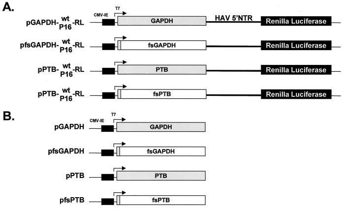
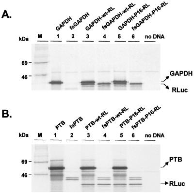
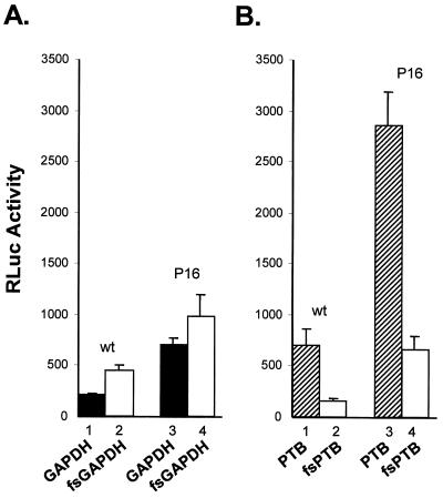
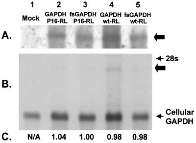
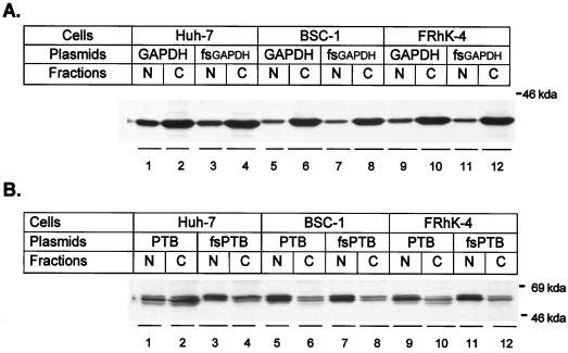
Glyceraldehyde 3-phosphate dehydrogenase (GAPDH), a cellular enzyme involved in glycolysis, binds specifically to several viral RNAs, but the functional significance of this interaction is uncertain. Both GAPDH and polypyrimidine tract binding protein (PTB) bind to overlapping sites in stem-loop IIIa of the internal ribosome entry site (IRES) of Hepatitis A virus (HAV), a picornavirus. Since the binding of GAPDH destabilizes the RNA secondary structure, we reasoned that GAPDH may suppress the ability of the IRES to direct cap-independent translation, making its effects antagonistic to the translation-enhancing activity of PTB (D. E. Schultz, C. C. Hardin, and S. M. Lemon, J. Biol. Chem. 271:14134-14142, 1996). To test this hypothesis, we constructed plasmids containing a dicistronic transcriptional unit in which the HAV IRES was placed between an upstream GAPDH-coding sequence and a downstream Renilla luciferase (RLuc) sequence. Transfection with this plasmid results in overexpression of GAPDH and in RLuc production as a measure of IRES activity. RLuc activity was compared with that from a control, null-expression plasmid that was identical except for a frameshift mutation within the 5' GAPDH coding sequence. In transfection experiments, GAPDH overexpression significantly suppressed HAV IRES activity in BSC-1 and FRhK-4 cells but not in Huh-7 cells, which have a significantly greater cytoplasmic abundance of PTB. GAPDH suppression of HAV translation was greater with the wild-type HAV IRES than with the IRES from a cell culture-adapted virus (HM175/P16) that has reproducibly higher basal translational activity in BSC-1 cells. Stem-loop IIIa RNA from the latter IRES had significantly lower affinity for GAPDH in filter binding experiments. Thus, the binding of GAPDH to the IRES of HAV suppresses cap-independent viral translation in vivo in African green monkey kidney cells. The enhanced replication capacity of cell culture-adapted HAV in such cells may be due in part to reduced affinity of the viral IRES for GAPDH.


Figures

FIG. 1
FIG. 1
Schematic representation of GAPDH and PTB expression constructs. Transcription is under control of a composite T7/CMV promoter in all plasmids. (A) Organization of dicistronic plasmids that express GAPDH or PTB from the 5′ reading frame by a cap-dependent translation mechanism and the reporter RLuc from the 3′ reading frame under the translational control of the HAV IRES (wt or HM175/P16), which is placed within the intercistronic space. Frameshift (fs) deletion mutants (“null-expression” vectors) express only a small, N-terminal fragment of GAPDH or PTB but differ by only a few nucleotides from the other constructs (shaded boxes indicate open reading frame segments that undergo translation). (B) Organization of monocistronic plasmids expressing GAPDH or PTB and of related frameshift mutants.
FIG. 2
FIG. 2
SDS-PAGE of protein products of in vitro-coupled transcription and translation reactions carried out in rabbit reticulocyte lysates programmed with the monocistronic and dicistronic plasmids depicted in Fig. 1. (A) Products of translation of the GAPDH expression vectors. The positions of the GAPDH and RLuc proteins are shown on the right. No GAPDH product is evident from translation of fsGAPDH constructs. (B) Translation products from the PTB expression vectors. PTB appeared as a doublet band of about 57 kDa. No PTB product is evident from translation of fsPTB constructs. The intensity of the image in panel B is reduced relative to that in panel A to demonstrate the doublet nature of the PTB bands. Equivalent amounts of RLuc were produced from the two sets of constructs.
FIG. 3
FIG. 3
GAPDH suppresses HAV IRES-dependent translation in BSC-1 cells. BSC-1 cells were transfected with dicistronic plasmids expressing GAPDH (solid bars) or the frameshift GAPDH mutant (open bars) (A) and PTB (shaded bars) or the frameshift PTB mutant (open bars) (B). RLuc was expressed from each construct under control of the wt or HM175/P16 (P16) IRES located within the intercistronic space. At 48 h following transfection, cells were lysed and assayed for RLuc activity. The results shown are means of two independent transfection experiments, each carried out in triplicate (a total of six transfections). The error bars indicate SD.
FIG. 4
FIG. 4
Northern analysis of the poly(A) RNA fraction recovered from BSC-1 cells transfected previously with the GAPDH expression vectors (lanes 2 and 4), their related null-expression mutants (lanes 3 and 5), or mock-transfected cells (lane 1). (A and B) The 24-h (A) and 3-h (B) exposures of the blot, respectively. The thick arrows indicate the location of the dicistronic GAPDH-IRES-RLuc transcript. (C) Normalized ratio of the hybridization signal specific for the endogenous GAPDH transcript (loading control) relative to the dicistronic GAPDH mRNA. N/A, not applicable, since there is no dicistronic transcript present.
FIG. 5
FIG. 5
Effects of GAPDH overexpression (A) or PTB expression (B) on either the wt (bars 1, 3, and 5) or the HM175/P16 (bars 2, 4, and 6) IRES elements in three different cell lines: BSC-1 (bars 1 and 2), FRhK-4 (bars 3 and 4), and Huh-7 (bars 5 and 6). RLuc activities in cells transfected with dicistronic expression constructs containing either the wt or P16 IRES were normalized to those present in cells transfected in parallel with the corresponding fsGAPDH or fsPTB null-expression plasmids. The results shown are means of the values obtained in two independent transfection experiments, each done in triplicate (a total of six transfections). Error bars indicate SD.
FIG. 6
FIG. 6
Enhanced chemiluminescence visualization of immunoblots of nuclear (N) and cytoplasmic (C) extracts prepared from cells that had been transfected 48 h previously with pGAPDH (A) or pPTB (B) or the related null-expression plasmids, pfsGAPDH and pfsPTB. Immunoblots were reacted with either anti-GAPDH antibody (A) or anti-PTB antibody (B).
FIG. 7
FIG. 7
Results of filter binding experiments assessing the affinities of GAPDH (A) or PTB (B) proteins to synthetic RNA probes representing corresponding stem-loop IIIa segments of wt HM175 virus (solid line) or the cell culture-adapted HM175/P16 virus (dashed lines). The 32P-labeled RNA probes were mixed with purified GAPDH or His6-PTB recombinant proteins in a binding buffer and then passed through sequential filters that either specifically retain protein and protein-RNA complexes or RNA. The results shown represent the fraction of probe bound to protein at different protein concentrations.

Similar articles

Cited by

References

    1. Binn L N, Lemon S M, Marchwicki R H, Redfield R R, Gates N L, Bancroft W H. Primary isolation and serial passage of hepatitis A virus strains in primate cell cultures. J Clin Microbiol. 1984;20:28–33. - PMC - PubMed
    1. Blyn L B, Towner J S, Semler B L, Ehrenfeld E. Requirement of poly(rC) binding protein 2 for translation of poliovirus RNA. J Virol. 1997;71:6243–6246. - PMC - PubMed
    1. Borman A, Howell M T, Patton J G, Jackson R J. The involvement of a spliceosome component in internal initiation of human rhinovirus RNA translation. J Gen Virol. 1993;74:1775–1788. - PubMed
    1. Borovjagin A, Pestova T, Shatsky I. Pyrimidine tract binding protein strongly stimulates in vitro encephalomyocarditis virus RNA translation at the level of preinitiation complex formation. FEBS Lett. 1994;351:299–302. - PubMed
    1. Brown E A, Day S P, Jansen R W, Lemon S M. The 5′ nontranslated region of hepatitis A virus RNA: secondary structure and elements required for translation in vitro. J Virol. 1991;65:5828–5838. - PMC - PubMed

Publication types

MeSH terms

Substances