. 2000 Jul;74(14):6476-84.
doi: 10.1128/jvi.74.14.6476-6484.2000.

Infection of polarized cultures of human intestinal epithelial cells with hepatitis A virus: vectorial release of progeny virions through apical cellular membranes

Affiliations

Infection of polarized cultures of human intestinal epithelial cells with hepatitis A virus: vectorial release of progeny virions through apical cellular membranes

C A Blank et al. J Virol. 2000 Jul.

Abstract

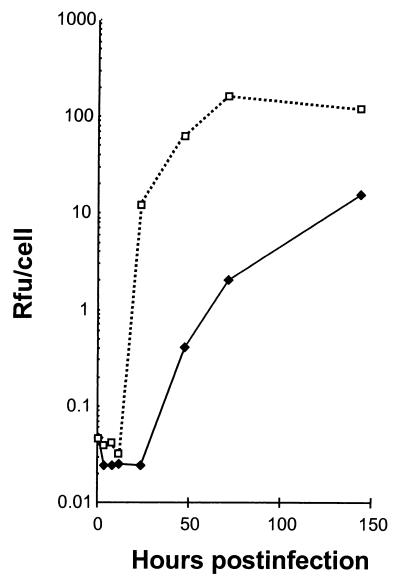
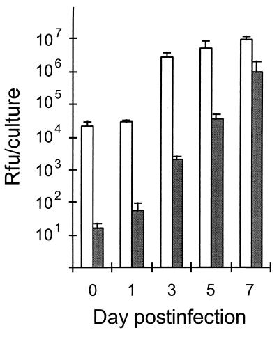
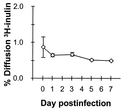
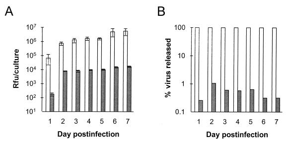
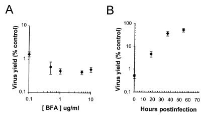
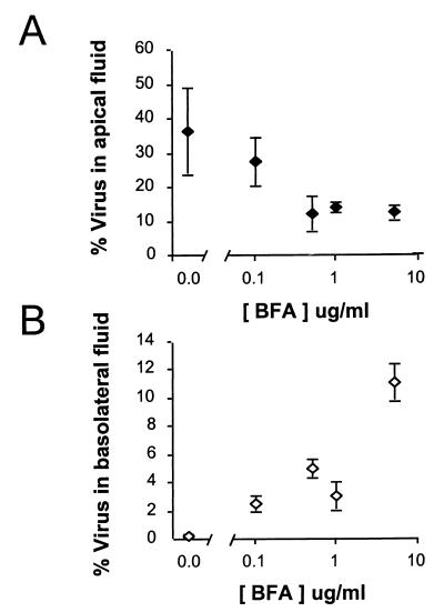
Although hepatitis A virus (HAV) is typically transmitted by the fecal-oral route, little is known of its interactions with cells of the gastrointestinal tract. We studied the replication of HAV in polarized cultures of Caco-2 cells, a human cell line which retains many differentiated functions of small intestinal epithelial cells. Virus uptake was 30- to 40-fold more efficient when the inoculum was placed on the apical rather than the basolateral surface of these cells, suggesting a greater abundance of the cellular receptor for HAV on the apical surface. Infection proceeded without cytopathic effect and did not influence transepithelial resistance or the diffusion of inulin across cell monolayers. Nonetheless, there was extensive release of progeny virus, which occurred almost exclusively into apical supernatant fluids (36.4% +/- 12.5% of the total virus yield compared with 0.23% +/- 0.13% release into basolateral fluids). Brefeldin A caused a profound inhibition of HAV replication, but also selectively reduced apical release of virus. These results indicate that polarized human epithelial cell cultures undergo vectorial infection with HAV and that virus release is largely restricted to the apical membrane. Virus release occurs in the absence of cytopathic effect and may involve cellular vesicular transport mechanisms.


Figures

FIG. 1
FIG. 1
Replication of cell culture-adapted HAV under one-step conditions in cultures of Caco-2 cells grown on an impermeable plastic surface (♦; MOI = 4.9 RFU/cell) and in nonpolarized BS-C-1 cells (□; MOI = 4.5 RFU/cell).
FIG. 2
FIG. 2
Indirect immunofluorescence detection of HAV antigen in infected Caco-2 cells. (A) Normal uninfected Caco-2 monolayer with DAPI nuclear counterstain. (B) Caco-2 cells 8 days following infection with HAV at an MOI of 6.0. The inset shows a high-power view of the cytoplasmic distribution of punctate HAV-specific fluorescence in two cells (no counterstain). (C) Caco-2 cells 12 days following infection with HAV. Viral antigen was visualized with a monoclonal antibody reactive with the virus capsid.
FIG. 3
FIG. 3
Cell-associated HAV following inoculation of polarized Caco-2 cells on either apical (open bars) or basolateral (shaded bars) cell surfaces at an MOI of approximately 6 RFU/cell. The results shown represent the means of the cell-associated virus content of three replicate infected cultures (± standard deviation).
FIG. 4
FIG. 4
Permeability of Caco-2 cells to [3H]inulin following infection with HAV. Duplicate monolayer cultures of polarized Caco-2 cells were infected by apical inoculation of virus under the conditions employed for the experiment shown in Fig. 2. At the intervals noted following infection, the apical-to-basolateral diffusion of [3H]inulin was measured over a 90-min period. The results shown represent the mean hourly rate (± range) of [3H]inulin diffusion. A rate of inulin diffusion of less than 1%/h across the monolayer indicates the presence of intact junctional complexes. Diffusion across the porous support in the absence of cells approximated 61%/h.
FIG. 5
FIG. 5
Vectorial release of HAV from apically infected polarized Caco-2 cell monolayers. Cells were inoculated as in Fig. 2 and washed prior to refeeding following the 2-h adsorption period. Apical and basolateral supernatant fluids were collected at 24-h intervals for virus titration and replaced with fresh media. (A) Virus content of apical (open bars) and basolateral (shaded bars) supernatant fluids. The values shown represent the mean virus titer of fluids from three replicate infected cultures (± standard deviation). (B) Proportion of all released virus (apical plus basolateral supernatant fluid virus) released into apical (open bars) or basolateral (shaded bars) fluids. The results shown represent the means of three replicate cultures at each time point, as in panel A.
FIG. 6
FIG. 6
Brefeldin A (BFA) inhibits HAV replication in Caco-2 cells. (A) Replicate cultures of apically inoculated Caco-2 cells were fed with medium containing various concentrations of brefeldin A beginning immediately after the 2-h period of viral adsorption. Brefeldin A was replenished every 6 h, and the medium was replaced daily. The results shown represent the mean total virus yield (cell-associated plus apical and basolateral fluid virus [± range]) at 72 h postinfection as a percentage of the yield from control cells infected in the absence of brefeldin A. (B) Effect of late addition of brefeldin A to apically inoculated Caco-2 cells. Cells were infected as in panel A with brefeldin A (10 μg/ml) added at various times postinfection. As in panel A, the results shown represent the mean total virus yield (± range) at 72 h postinfection as a percentage of virus yield from cells infected in the absence of brefeldin A.
FIG. 7
FIG. 7
Brefeldin A (BFA) inhibits apical release of progeny HAV. Cells were treated with various concentrations of brefeldin A beginning immediately after removal of an apical HAV inoculum, as in Fig. 6A. The proportion of the total virus yield (cell-associated plus apical and basolateral fluid virus) that was present in the apical (A) or basolateral (B) supernatant fluids 72 h postinfection was calculated for each culture. The results shown represent the mean (± range) results for replicate cultures treated with the drug and the mean (± standard deviation) of five cultures maintained in the absence of brefeldin A. Samples of apical and basolateral fluids from cells treated with 10 μg of brefeldin A per ml could not be assayed for HAV due to residual antiviral activity.
FIG. 8
FIG. 8
Monensin inhibits apical release of HAV from polarized Caco-2 cells. Cells were treated with the indicated concentration of monensin beginning 72 h after apical infection with HAV. The quantity of virus released into the apical supernatant fluids was then assessed after a 6-h incubation period and is shown as the percentage of virus present in the cell lysate (± range). The titer of virus present in the lysates of cells treated with the highest concentration of monensin (10−7 M) was 93% (range, 87 to 100%) of that present in untreated cells, indicating there was no antiviral effect over this short incubation period.

References

    1. Amerongen H M, Wilson G A, Fields B N, Neutra M R. Proteolytic processing of reovirus is required for adherence to intestinal M cells. J Virol. 1994;68:8428–8432. - PMC - PubMed
    1. Asher L V S, Binn L N, Mensing T L, Marchwicki R H, Vassell R A, Young G D. Pathogenesis of hepatitis A in orally inoculated owl monkeys (Aotus trivergatus) J Med Virol. 1995;47:260–268. - PubMed
    1. Blau D M, Compans R W. Polarization of viral entry and release in epithelial cells. Semin Virol. 1996;7:245–253.
    1. Clayson E T, Compans R W. Entry of simian virus 40 is restricted to apical surfaces of polarized epithelial cells. Mol Cell Biol. 1988;8:3391–3396. - PMC - PubMed
    1. Clayson E T, Jones-Brando L V, Compans R W. Release of simian virus 40 virions from epithelial cells is polarized and occurs without cell lysis. J Virol. 1989;63:2278–2288. - PMC - PubMed

Publication types

LinkOut - more resources