

Enhanced infectivity of an R5-tropic simian/human immunodeficiency virus carrying human immunodeficiency virus type 1 subtype C envelope after serial passages in pig-tailed macaques (Macaca nemestrina)
- PMID: 10864663
- PMCID: PMC112159
- DOI: 10.1128/jvi.74.14.6501-6510.2000
Enhanced infectivity of an R5-tropic simian/human immunodeficiency virus carrying human immunodeficiency virus type 1 subtype C envelope after serial passages in pig-tailed macaques (Macaca nemestrina)
Abstract
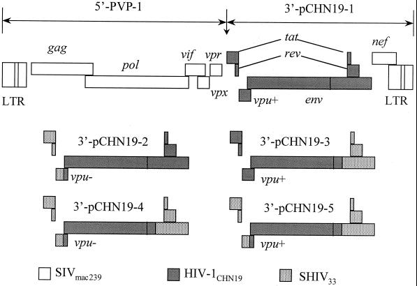
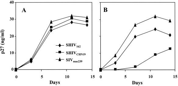
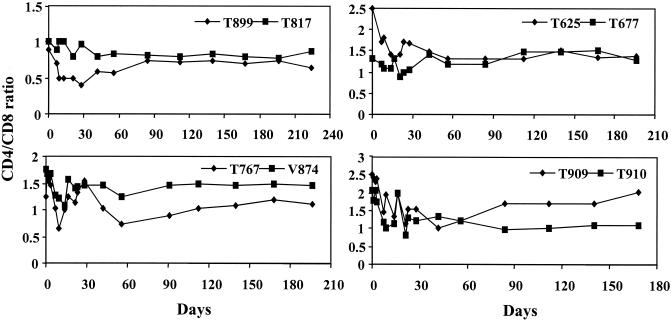
The increasing prevalence of human immunodeficiency virus type 1 (HIV-1) subtype C infection worldwide calls for efforts to develop a relevant animal model for evaluating strategies against the transmission of the virus. A chimeric simian/human immunodeficiency virus (SHIV), SHIV(CHN19), was generated with a primary, non-syncytium-inducing HIV-1 subtype C envelope from a Chinese strain in the background of SHIV(33). Unlike R5-tropic SHIV(162), SHIV(CHN19) was not found to replicate in rhesus CD4(+) T lymphocytes. SHIV(CHN19) does, however, replicate in CD4(+) T lymphocytes of pig-tailed macaques (Macaca nemestrina). The observed replication competence of SHIV(CHN19) requires the full tat/rev genes and partial gp41 region derived from SHIV(33). To evaluate in vivo infectivity, SHIV(CHN19) was intravenously inoculated, at first, into two pig-tailed and two rhesus macaques. Although all four animals became infected, the virus replicated preferentially in pig-tailed macaques with an earlier plasma viral peak and a faster seroconversion. To determine whether in vivo adaptation would enhance the infectivity of SHIV(CHN19), passages were carried out serially in three groups of two pig-tailed macaques each, via intravenous blood-bone marrow transfusion. The passages greatly enhanced the infectivity of the virus as shown by the increasingly elevated viral loads during acute infection in animals with each passage. Moreover, the doubling time of plasma virus during acute infection became much shorter in passage 4 (P4) animals (0.2 day) in comparison to P1 animals (1 to 2 days). P2 to P4 animals all became seropositive around 2 to 3 weeks postinoculation and had a decline in CD4/CD8 T-cell ratio during the early phase of infection. In P4 animals, a profound depletion of CD4 T cells in the lamina propria of the jejunum was observed. Persistent plasma viremia has been found in most of the infected animals with sustained viral loads ranging from 10(3) to 10(5) per ml up to 6 months postinfection. Serial passages did not change the viral phenotype as confirmed by the persistence of the R5 tropism of SHIV(CHN19) isolated from P4 animals. In addition, the infectivity of SHIV(CHN19) in rhesus peripheral blood mononuclear cells was also increased after in vivo passages. Our data indicate that SHIV(CHN19) has adapted well to grow in macaque cells. This established R5-tropic SHIV(CHN19)/macaque model would be very useful for HIV-1 subtype C vaccine and pathogenesis studies.
Figures
References
-
- Abebe A, Demissie D, Goudsmit J, Brouwer M, Kuiken C L, Pollakis G, Schuitemaker H, Fontanet A L, Rinke de Wit T F. HIV-1 subtype C syncytium- and non-syncytium-inducing phenotypes and coreceptor usage among Ethiopian patients with AIDS. AIDS. 1999;13:1305–1311. - PubMed
-
- Agy M B, Frumkin L R, Corey L, Coombs R W, Wolinsky S M, Koehler J, Morton W R, Katze M G. Infection of Macaca nemestrina by human immunodeficiency virus type-1. Science. 1992;257:103–106. - PubMed
-
- Agy M B, Schmidt A, Florey M J, Kennedy B J, Schaefer G, Katze M G, Corey L, Morton W R, Bosch M L. Serial in vivo passage of HIV-1 infection in Macaca nemestrina. Virology. 1997;238:336–343. - PubMed
-
- Almond N M, Heeney J L. AIDS vaccine development in primate models. AIDS. 1998;12(Suppl. A):S133–S140. - PubMed
-
- Bjorndal A, Sonnerborg A, Tscherning C, Albert J, Fenyo E M. Phenotypic characteristics of human immunodeficiency virus type 1 subtype C isolates of Ethiopian AIDS patients. AIDS Res Hum Retrovir. 1999;15:647–653. - PubMed
Publication types
MeSH terms
Substances
Grants and funding
LinkOut - more resources
Full Text Sources
Molecular Biology Databases
Research Materials
