. 2000 Jul;74(14):6581-91.
doi: 10.1128/jvi.74.14.6581-6591.2000.

Norwalk virus open reading frame 3 encodes a minor structural protein

Affiliations

Norwalk virus open reading frame 3 encodes a minor structural protein

P J Glass et al. J Virol. 2000 Jul.

Abstract

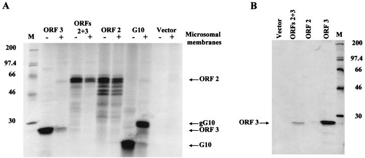
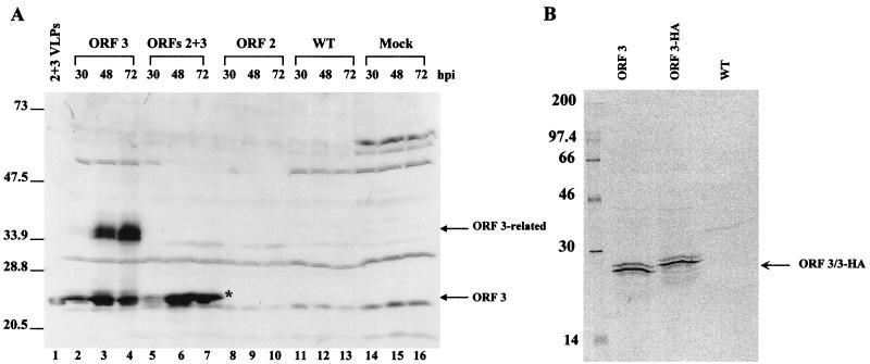
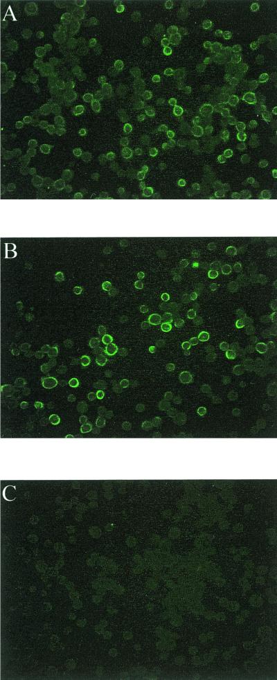
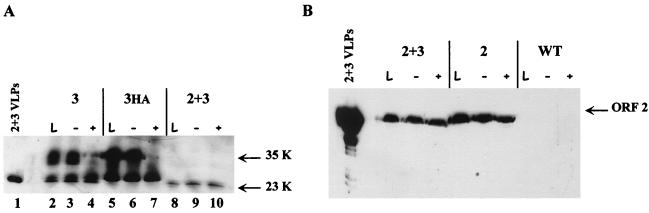
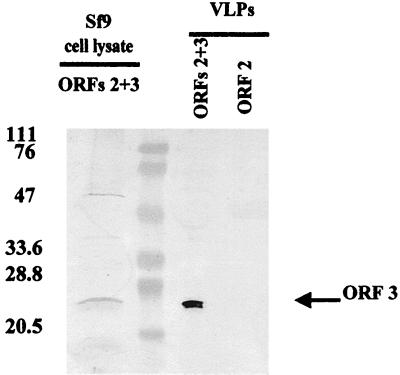
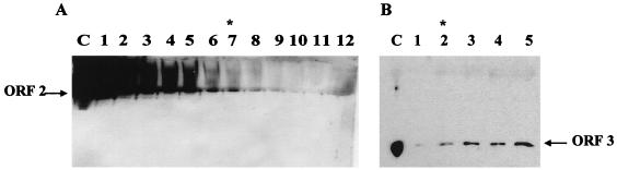
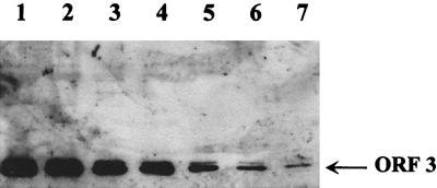
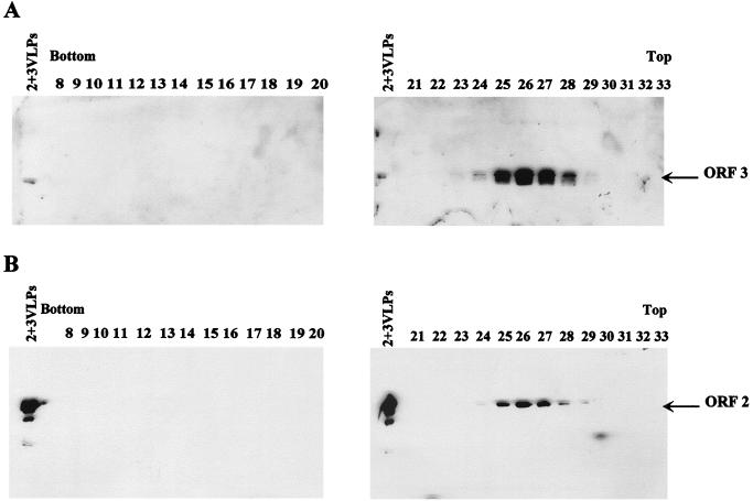
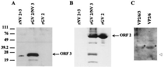
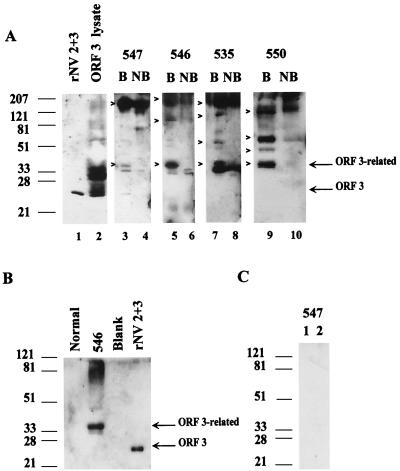
Norwalk virus (NV) is a causative agent of acute epidemic nonbacterial gastroenteritis in humans. The inability to cultivate NV has required the use of molecular techniques to examine the genome organization and functions of the viral proteins. The function of the NV protein encoded by open reading frame 3 (ORF 3) has been unknown. In this paper, we report the characterization of the NV ORF 3 protein expressed in a cell-free translation system and in insect cells and show its association with recombinant virus-like particles (VLPs) and NV virions. Expression of the ORF 3 coding region in rabbit reticulocyte lysates resulted in the production of a single protein with an apparent molecular weight of 23,000 (23K protein), which is not modified by N-linked glycosylation. The ORF 3 protein was expressed in insect cells by using two different baculovirus recombinants; one recombinant contained the entire 3' end of the genome beginning with the ORF 2 coding sequences (ORFs 2+3), and the second recombinant contained ORF 3 alone. Expression from the construct containing both ORF 2 and ORF 3 resulted in the expression of a single protein (23K protein) detected by Western blot analysis with ORF 3-specific peptide antisera. However, expression from a construct containing only the ORF 3 coding sequences resulted in the production of multiple forms of the ORF 3 protein ranging in size from 23,000 to 35,000. Indirect-immunofluorescence studies using an ORF 3 peptide antiserum showed that the ORF 3 protein is localized to the cytoplasm of infected insect cells. The 23K ORF 3 protein was consistently associated with recombinant VLPs purified from the media of insect cells infected with a baculovirus recombinant containing the entire 3' end of the NV genome. Western blot analysis of NV purified from the stools of NV-infected volunteers revealed the presence of a 35K protein as well as multiple higher-molecular-weight bands specifically recognized by an ORF 3 peptide antiserum. These results indicate that the ORF 3 protein is a minor structural protein of the virion.


Figures

FIG. 1
FIG. 1
The ORF 3 protein is produced by cell-free translation of ORF 3 and ORFs 2+3 transcripts. (A) Analysis of proteins produced by translation of synthetic mRNAs using RRLs in the absence (−) or presence (+) of canine pancreatic microsomal membranes. The translated proteins were labeled using Tran35S-label. An aliquot (10 μl/25 μl) of the translation reaction mixture was analyzed on an SDS–15% polyacrylamide gel. (B) Immunoprecipitation of the ORF 3 protein from cell-free translation reaction mixtures using the ORF 3 C-terminal peptide antiserum. The remaining aliquot of the translation reaction mixtures in panel A was immunoprecipitated in RIPA buffer, and the entire sample was analyzed by autoradiography of an SDS–15% polyacrylamide gel. Lane M contains 14C-methylated protein molecular weight markers (Amersham); lane ORFs 2+3 contains the entire 3′ end of the genome beginning with the ORF 2 coding sequences; lane G10 contains rotavirus gene 10; lane Vector contains pGem7Zf(+) vector with no insert. The ORF 2, ORF 3, G10, and glycosylated G10 (gG10) protein products are designated.
FIG. 2
FIG. 2
Expression of the ORF 3 protein in infected insect cells. Sf9 cells were either mock infected (Mock) or infected with wild-type baculovirus (WT), the ORF 2 baculovirus recombinant, the ORFs 2+3 baculovirus recombinant, or the ORF 3 baculovirus recombinant. (A) Kinetics of ORF 3 expression in infected insect cells. Cells were harvested at 30, 48, and 72 h p.i. by lysis in SDS-PAGE sample buffer. The samples were separated by SDS-PAGE in a 15% polyacrylamide gel, and Western blot analysis was performed using the rabbit ORF 3 C-terminal peptide antiserum at a dilution of 1:500. The asterisk highlights the 23K ORF 3 protein band that separates into two bands on some gels. (B) Immunoprecipitation of the ORF 3 protein from infected insect cells. Cells infected with wild-type baculovirus (WT), the ORF 3 or the HA-tagged ORF 3 (ORF 3-HA) baculovirus recombinants were harvested in RIPA buffer at 65 h p.i. and immunoprecipitated using the ORF 3 C-terminal peptide antiserum.
FIG. 3
FIG. 3
Immunofluorescence analysis of ORF 3 protein expression in infected insect cells. Sf9 cells were infected with the ORF 3 baculovirus recombinant (A), the ORFs 2+3 baculovirus recombinant (B), or wild-type baculovirus (C) and seeded onto 96-well plates. At 32 h p.i., the cells were fixed in methanol and rehydrated in PBS. The ORF 3 protein was detected using the ORF 3 C-terminal peptide antiserum (dilution of 1:100) followed by a fluorescein isothiocyanate-conjugated goat anti-rabbit serum (dilution of 1:1,000). Fluorescence was detected using an Olympus IX-70 inverted-system microscope. Images were captured using the DC3-30 color camera and Image Pro Plus software.
FIG. 4
FIG. 4
The 35K ORF 3 protein is phosphorylated. Sf9 cells were infected with wild-type baculovirus (WT) or the ORF 2, ORFs 2+3, ORF 3, or ORF 3HA baculovirus recombinants. Cells were harvested at 65 h p.i. in M-Per buffer plus protease inhibitors. An aliquot of the clarified lysate was removed for analysis (lanes L). The remaining sample was adjusted to 1× CIAP buffer and incubated in the absence (−) or presence (+) of CIAP (20 U). Western blot analysis was performed using the ORF 3 C-terminal peptide antiserum (1:500) (A) or the anti-rNV serum (1:1,000) (B). rNV 2+3 VLPs were included as a positive control for Western blot analysis.
FIG. 5
FIG. 5
The ORF 3 protein is associated with rNV VLPs. Recombinant rNV VLPs were purified from supernatants of Sf9 cells infected with the ORF 2 (rNV 2 VLPs) or the ORFs 2+3 (rNV 2+3 VLPs) baculovirus recombinants. VLPs sequentially purified over both CsCl and sucrose gradients were analyzed by SDS-PAGE and Western blotting using the rabbit ORF 3 C-terminal peptide antiserum (1:500).
FIG. 6
FIG. 6
Western blot analysis of the ORF 2 protein and the ORF 3 protein in an ORFs 2+3 VLP preparation. (A) Detection of the ORF 2 protein. Purified VLPs were diluted to a loading concentration of 2 μg and serially diluted twofold for separation on an SDS–15% polyacrylamide gel. Western blot analysis was performed using MAb 8301 at a dilution of 1:1000. (B) Detection of the ORF 3 protein. A higher concentration of purified VLPs was loaded, and ORF 3 was detected using the rabbit ORF 3 peptide antiserum (1:500). Asterisks denote the lowest level at which each protein was clearly detectable. The lanes designated C are ORFs 2+3 VLPs run as a control for ORF 2 and ORF 3 detection. (A) Lanes: 1, 2 μg; 2, 1 μg; 3, 0.5 μg; 4, 0.25 μg; 5, 0.125 μg; 6, 0.0625 μg; 7, 0.03125 μg; 8, 0.015625 μg; 9, 0.0078 μg; 10, 0.0039 μg; 11, 0.00195 μg; 12, 0.00097 μg. (B) Lanes: 1, 15 μg; 2, 17 μg; 3, 20 μg; 4, 22 μg; 5, 25 μg.
FIG. 7
FIG. 7
Western blot analysis of DI2/3 VLPs using the ORF 3 C-terminal peptide antiserum. VLPs were diluted to a loading concentration of 1 μg and serially diluted twofold for separation on an SDS-polyacrylamide gel. Western blot analysis was performed using the rabbit ORF 3 C-terminal peptide antiserum (1:500). Lanes: 1, 1 μg; 2, 0.5 μg; 3, 0.25 μg; 4, 0.125 μg; 5, 0.0625 μg; 6, 0.03125 μg; 7, 0.015625 μg. The DI2/3 particles were as pure as previously described VLP preparations (3).
FIG. 8
FIG. 8
The ORF 3 protein comigrates with the ORF 2 protein in VLPs in sucrose gradients. Western blot analysis of the sucrose gradient fractions from the DI2/3 VLP purification is shown. Proteins were detected using the rabbit ORF 3 C-terminal peptide antiserum (A) or the ORF 2 MAb 3912 (B). Fractions numbers and gradient orientation are indicated. Each gel contained a VLP control, 2+3VLPs (left lanes).
FIG. 9
FIG. 9
The ORF 3 protein associates with GV ORF 2 VLPs but not rotavirus VP2/6 VLPs. (A) GV VLPs (25 μg) were analyzed in an SDS–15% polyacrylamide gel. Western blot analysis was first performed using the rabbit ORF 3 C-terminal peptide antiserum. (B) The blot was then stripped in ECL stripping buffer as specified by the manufacturer (Amersham) and reprobed with the rabbit anti-rGV serum (1:500); the rGV ORF 2 and degradation products are detected by this serum. (C) Rotavirus VLPs (25 μg) were loaded for separation in an SDS–15% polyacrylamide gel and analyzed by Western blotting using the ORF 3 C-terminal peptide antiserum. Lane designations are as follows: rGV 2, GV ORF 2 VLPs; rGV 2/NV 3, dually infected GV ORF 2 and NV ORF 3 VLPs; rNV 2+3, NV ORFs 2+3 VLPs; VP2/6/3, dually infected rotavirus 2/6- and NV ORF 3 VLPs; VP2/6, rotavirus 2/6-VLPs. The open arrowhead designates where the ORF 3 protein would migrate if present. The asterisks show the location of rotavirus VP6 that is detected nonspecifically by the secondary antibody because of the large amount of protein on the gel.
FIG. 10
FIG. 10
Western blot analysis of partially purified virions from NV-infected volunteers using the ORF 3 peptide antiserum. (A) Analysis of samples from volunteers 547, 546, 535, and 550. Boiled (B) and not-boiled (NB) samples were analyzed to examine both continuous and discontinuous epitopes. Controls included ORFs 2+3 VLPs and ORF 3 baculovirus recombinant-infected cell lysate. The ORF 3-related proteins are designated on the right and by arrowheads. (B) Partially purified sample from an uninfected volunteer (Normal) prepared identically to the infected volunteer samples in panel A, partially purified NV from volunteer 546 (independent purification), and rNV 2+3 VLPs analyzed by Western blot analysis using the ORF 3 C-terminal peptide antiserum. (C) Two independent partial purifications of NV from stools of volunteer 547, analyzed by Western blotting using a rabbit preimmune serum (1:500).

Similar articles

Cited by

References

    1. Atmar R L, Metcalf T G, Neill F H, Estes M K. Detection of enteric viruses in oysters by using the polymerase chain reaction. Appl Environ Microbiol. 1993;59:631–635. - PMC - PubMed
    1. Au K-S, Mattion N M, Estes M K. A subviral particle binding domain on the rotavirus nonstructural glycoprotein NS28. Virology. 1993;194:665–673. - PubMed
    1. Ball J M, Hardy M E, Atmar R L, Conner M E, Estes M K. Oral immunization with recombinant Norwalk virus-like particles induces a systemic and mucosal immune response in mice. J Virol. 1998;72:1345–1353. - PMC - PubMed
    1. Ball J M, Tian P, Zeng C Q-Y, Morris A, Estes M K. Age-dependent diarrhea is induced by a viral nonstructural glycoprotein. Science. 1996;272:101–104. - PubMed
    1. Brierley I. Ribosomal frameshifting viral RNAs. J Gen Virol. 1995;76:1885–1892. - PubMed

Publication types

MeSH terms

LinkOut - more resources