. 2000 Jun;130(4):867-74.
doi: 10.1038/sj.bjp.0703396.

Activation of protein kinase B by the A(1)-adenosine receptor in DDT(1)MF-2 cells

Affiliations

Activation of protein kinase B by the A(1)-adenosine receptor in DDT(1)MF-2 cells

R Germack et al. Br J Pharmacol. 2000 Jun.

Abstract

In this study the effect of insulin and A(1)-adenosine receptor stimulation on protein kinase B (PKB) activation has been investigated in the hamster vas deferens smooth muscle cell line DDT(1)MF-2. Increases in PKB phosphorylation were determined by Western blotting using an antibody that detects PKB phosphorylation at Ser(473). Insulin, a recognized activator of PKB, stimulated a concentration-dependent increase in PKB phosphorylation in DDT(1)MF-2 cells (EC(50) 5+/-1 pM). The selective A(1)-adenosine receptor agonist N(6)-cyclopentyladenosine (CPA) stimulated time and concentration-dependent increases in PKB phosphorylation in DDT(1)MF-2 cells (EC(50) 1.3+/-0.5 nM). CPA-mediated increases in PKB phosphorylation were antagonized by the A(1)-adenosine receptor selective antagonist 1,3-dipropylcyclopentylxanthine (DPCPX) yielding an apparent K(D) value of 2.3 nM. Pre-treatment of DDT(1)MF-2 cells with pertussis toxin (PTX, 100 ng ml(-1) for 16 h), to block G(i)/G(o)-dependent pathways, abolished CPA (1 microM) induced phosphorylation of PKB. In contrast, responses to insulin (100 nM) were resistant to PTX pre-treatment. The phosphatidylinositol 3-kinase (PI-3K) inhibitors wortmannin (IC(50) 10.3+/-0.6 nM) and LY 294002 (IC(50) 10.3+/-1.2 microM) attenuated the phosphorylation of PKB elicited by CPA (1 microM) in a concentration-dependent manner. Wortmannin (30 nM) and LY 294002 (30 microM) also blocked responses to insulin (100 nM). Removal of extracellular Ca(2+) and chelation of intracellular Ca(2+) with BAPTA had no significant effect on CPA-induced PKB phosphorylation. Similarly, pretreatment (30 min) with inhibitors of protein kinase C (Ro 31-8220; 10 microM), tyrosine kinase (genistein; 100 microM), mitogen-activated protein (MAP) kinase kinase (PD 98059; 50 microM) and p38 MAPK (SB 203580; 20 microM) had no significant effect on CPA-induced PKB phosphorylation. In conclusion, these data demonstrate that A(1)-adenosine receptor stimulation in DDT(1)MF-2 cells increases PKB phosphorylation through a PTX and PI-3K-sensitive pathway.


Figures

Figure 1
Figure 1
Insulin and N6-cyclopentyladenosine-mediated phosphorylation of PKB in DDT1MF-2 cells. Levels of PKB phosphorylation were determined in cells maintained in 10% (vv−1) foetal bovine serum or in cells that were serum-starved for 16 h in DMEM containing 0.1% bovine serum albumin. DDT1MF-2 cells were treated with vehicle (Ctr), insulin (100 nM) or CPA (1 μM) for 10 min. Cell lysates (20 μg) were resolved by SDS–PAGE and transferred to nitrocellulose before being probed with antisera specific for phosphorylated PKB (Ser473). Similar results were obtained in two further experiments.
Figure 2
Figure 2
Insulin-mediated increase in the phosphorylation of PKB in DDT1MF-2 cells. (a) DDT1MF-2 cells were treated with vehicle (Ctr) or the indicated concentrations of insulin for 10 min. Cell lysates (20 μg) were resolved by SDS–PAGE and transferred to nitrocellulose before being probed with antisera specific for phosphorylated PKB (Ser473). A representative immunoblot showing insulin-mediated increases in PKB phosphorylation is shown in the upper panel. Combined results obtained from five independent experiments (mean±s.e.mean) are shown in the lower panel. Data are presented as the percentage increase above Ctr PKB phosphorylation (100%). *Significantly different (P<0.05) from the Ctr level of phosphorylation of PKB. (b) Effect of the PI-3K inhibitors wortmannin and LY 294002 on insulin-induced PKB phosphorylation. DDT1MF-2 cells were pre-incubated (30 min) with wortmannin (30 nM) and LY 294002 (30 μM) before stimulating with 100 nM insulin for 10 min. Similar results were obtained in two further experiments.
Figure 3
Figure 3
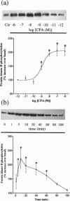
Concentration and time-dependent effects of CPA on the phosphorylation of PKB in DDT1MF-2 cells. (a) DDT1MF-2 cells were treated with vehicle (Ctr) or the indicated concentrations CPA for 10 min and (b) with vehicle (Ctr) or CPA (1 μM) for the indicated periods of time. Cell lysates (20 μg) were resolved by SDS–PAGE and transferred to nitrocellulose before being probed with antisera specific for phosphorylated PKB (Ser473). Representative immunoblots are shown in the upper panels. Combined results obtained from five (a) and four (b) independent experiments (mean±s.e.mean) are shown in the lower panels. Data are presented as the percentage increase above Ctr PKB phosphorylation (100%). *Significantly different (P<0.05) from the Ctr level of phosphorylation of PKB.
Figure 4
Figure 4
Inhibition of CPA-mediated increases in PKB phosphorylation by the selective A1-adenosine receptor antagonist DPCPX. DDT1MF-2 cells were preincubated for 30 min with the indicated concentrations of the selective adenosine A1 receptor antagonist DPCPX before stimulating with 10 nM CPA. Cell lysates (20 μg) were resolved by SDS–PAGE and transferred to nitrocellulose before being probed with antisera specific for phosphorylated PKB (Ser473). A representative immunoblot showing inhibition by DPCPX of the increase in PKB phosphorylation obtained with 10 nM CPA is depicted in the upper panel. Combined results obtained from three independent experiments (mean±s.e.mean) are shown in the lower panel. Data are expressed as the percentage of the control response to 10 nM CPA.
Figure 5
Figure 5
Effect of PTX on insulin and CPA-mediated PKB phosphorylation in DDT1MF-2 cells. Representative immunoblot showing the effects of pre-treating DDT1MF-2 cells for 16 h with PTX (100 ng ml−1) to block Gi/Go-dependent pathways before stimulating with 1 μM CPA or 100 nM insulin. Cell lysates (20 μg) were resolved by SDS–PAGE and transferred to nitrocellulose before being probed with antisera specific for phosphorylated PKB (Ser473). Similar results were obtained in three other independent experiments.
Figure 6
Figure 6
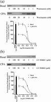
Effect of the PI-3K inhibitors wortmannin and LY 294002 on CPA-induced PKB phosphorylation in DDT1MF-2 cells. DDT1MF-2 cells were preincubated for 30 min with the indicated concentrations of the PI-3K inhibitors (a) wortmannin and (b) LY 294002 before stimulating with 1 μM CPA for 10 min. Cell lysates (20 μg) were resolved by SDS–PAGE and transferred to nitrocellulose before being probed with antisera specific for phosphorylated PKB (Ser473). Representative immunoblots showing the inhibition by wortmannin (a) and LY 294002 (b) of the basal and CPA-induced levels of PKB phosphorylation are shown in the upper panels. Combined results obtained from four independent experiments (mean±s.e.mean) are shown in the lower panels. Data are presented as the percentage increase above basal PKB phosphorylation (100%) in the absence of PI-3K inhibitor. *Significantly different (P<0.05) from the level of PKB phosphorylation in response to 1 μM CPA alone.
Figure 7
Figure 7
The effect of Ca2+ removal and inhibitors of various protein kinases on CPA-induced PKB phosphorylation in DDT1MF-2 cells. Measurements of 1 μM CPA-induced PKB phosphorylation (10 min stimulation) were made (a) in the presence (1.3 mM CaCl2) and absence of extracellular Ca2+ (nominally Ca2+-free Hanks/HEPES buffer containing 0.1 mM EGTA) to prevent Ca2+ influx and using BAPTA (cells pre-incubated for 30 min with 50 μM BAPTA/AM) in the absence of extracellular Ca2+ (nominally Ca2+-free Hanks/HEPES buffer containing 0.1 mM EGTA) to chelate Ca2+-released from intracellular stores and (b,c) in cells pre-incubated for 30 min with genistein (100 μM), Ro 31-8220 (10 μM), PD 98059 (50 μM) and SB 203580 (20 μM). Similar results were obtained in four independent experiments.

References

    1. BERRIDGE M.J. Inositol triphosphate and calcium signalling. Nature. 1993;361:315–325. - PubMed
    1. BUTLER A.P., MARTINEZ L.A. , MONTGOMERY R.L. Involvement of a pertussis-toxin sensitive G protein in the induction of gene expression by insulin. Cell. Signal. 1996;8:475–480. - PubMed
    1. CARDONE M.H., ROY N., STENNICKE H.R., SALVESEN G.S., FRANKE T.F., STANBRIDGE E., FRISCH S. , REED J.C. Regulation of cell death protease caspase 9 by phosphorylation. Science. 1998;282:1318–1321. - PubMed
    1. COFFER P.J., JIN J. , WOODGETT J.R. Protein kinase B (c-Akt): a multifunctional mediator of phosphatidylinositol 3-kinase activation. Biochem. J. 1998;335:1–13. - PMC - PubMed
    1. CROSS D.A.E., ALESSI D.R., COHEN P., ANDJELKOVIC M. , HEMMINGS B.A. Inhibition of glycogen synthase kinase-3 by insulin mediated protein kinase B. Nature. 1995;378:785–789. - PubMed

Publication types

MeSH terms

Substances