. 2000 Jun;130(4):891-9.
doi: 10.1038/sj.bjp.0703359.

Tumour necrosis factor-alpha- and interleukin-1beta-stimulated cell proliferation through activation of mitogen-activated protein kinase in canine tracheal smooth muscle cells

Affiliations

Tumour necrosis factor-alpha- and interleukin-1beta-stimulated cell proliferation through activation of mitogen-activated protein kinase in canine tracheal smooth muscle cells

C M Yang et al. Br J Pharmacol. 2000 Jun.

Abstract

The elevated levels of inflammatory cytokines such as tumour necrosis factor-alpha (TNF-alpha) and interleukin-1beta (IL-1beta) have been found in the fluid of airways in symptomatic asthmatics. These cytokines have been considered as mitogens to stimulate cell proliferation in tracheal smooth muscle cells (TSMCs). We therefore investigated the effects of TNF-alpha and IL-1beta on cell proliferation and activation of p42/p44 mitogen-activated protein kinase (MAPK) in these cells. TNF-alpha and IL-1beta induced [(3)H]-thymidine incorporation in a time- and concentration-dependent manner. The maximal stimulation of [(3)H]-thymidine incorporation induced by TNF-alpha and IL-1beta was seen 12 h after incubation with these cytokines. In response to TNF-alpha and IL-1beta, p42/p44 MAPK was activated with a concentration-dependent manner in TSMCs. Pretreatment of TSMCs with pertussis toxin did not change DNA synthesis and phosphorylation of MAPK induced by TNF-alpha and IL-1beta. These responses were attenuated by a tyrosine kinase inhibitor herbimycin, a phosphatidyl choline (PC)-phospholipase C (PLC) inhibitor D609, a phosphatidyl inositide (PI)-PLC inhibitor U73122, a protein kinase C inhibitor staurosporine, and removal of Ca(2+) by addition of BAPTA/AM plus EGTA. TNF-alpha- and IL-1beta-induced [(3)H]-thymidine incorporation and phosphorylation of p42/p44 MAPK was completely inhibited by PD98059 (an inhibitor of MEK1/2), indicating that activation of MEK1/2 was required for these responses. These results suggest that the mitogenic effects of TNF-alpha and IL-1beta were mediated through the activation of MEK1/2 and p42/p44 MAPK pathway. TNF-alpha- and IL-1beta-mediated responses were modulated by PLC, Ca(2+), PKC, and tyrosine kinase associated with cell proliferation in TSMCs.


Figures
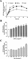
Figure 1
Figure 1
[3H]-Thymidine incorporation induced by cytokines in TSMCs. For time course, after 24 h in serum-free medium, the cells were stimulated with vehicle (basal), 30 ng ml−1 TNF-α or 50 ng ml−1 IL-1β. The cells were labeled with 1 μCi ml−1 [3H]-thymidine for the times indicated in the continuous presence of cytokines (A). For concentration dependence, the cells were stimulated with various concentrations of TNF-α (B) and IL-1β (C). After stimulation for 6 h, cells were labeled with 1 μCi ml−1 [3H]-thymidine for another 18 h in the presence of cytokines. The incorporation of [3H]-thymidine was determined as described in Methods. Data are expressed as the mean±s.e.mean of three separate experiments determined in triplicate. *P<0.05; **P<0.01, as compared with the basal level.
Figure 2
Figure 2
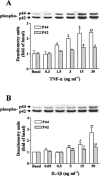
Concentration-dependence of TNF-α- and IL-1β-stimulated p42/p44 MAPK phosphorylation in TSMCs. The cells were grown to confluence, made quiescent by serum-deprivation for 24 h and incubated with various concentrations of TNF-α (A) and IL-1β (B) for 15 min. The cell lysates were subjected to 10% SDS–PAGE and transferred to nitrocellulose membrane. Western blot analysis was performed using an antiserum reactive with an anti-phospho-p42/p44 MAPK polyclonal antibody. Bands were visualized by an ECL method and quantified by a densitometer. Similar results were obtained in three independent experiments. Data are expressed as the mean±s.e.mean of three independent experiments. (Bar graph). *P<0.05; **P<0.01, as compared with the control cells exposed to respective cytokine.
Figure 3
Figure 3
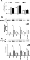
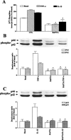
Involvement of G protein and tyrosine kinase in DNA synthesis and MAPK phosphorylation induced by cytokines in TSMCs. The cells were preincubated with pertussis toxin (PTX, 100 ng ml−1, 24 h) or herbimycin A (Herb, 10 μM, 1 h), and then stimulated with vehicle, 30 ng ml−1 TNF-α or 50 ng ml−1 IL-1β. For DNA synthesis, after 6 h incubations, cells were labelled with 1 μCi ml−1 [3H]-thymidine for another 18 h in the continuous presence of TNF-α or IL-1β. The incorporation of [3H]-thymidine was determined as described in Methods. Data are expressed as the mean±s.e.mean of three separate experiments determined in triplicate (A). For MAPK experiment, after treatment with these agents, the cells were stimulated with vehicle, 30 ng ml−1 TNF-α (B) or 50 ng ml−1 IL-1β (C) for 15 min. The cell lysates were subjected to 10% SDS–PAGE and transferred to nitrocellulose membrane. The phosphorylation of p42/p44 MAPK was determined as described in Figure 2. Similar results were obtained in three independent experiments. *P<0.01, as compared with the control cells exposed to respective cytokine.
Figure 4
Figure 4
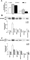
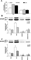
Effects of D609 and U73122 on DNA synthesis and MAPK phosphorylation induced by cytokines in TSMCs. The cells were preincubated with U73122 (10 μM, 1 h), and then stimulated with vehicle or cytokines. For DNA synthesis, after 6 h incubation, cells were labelled with 1 μCi ml−1 [3H]-thymidine for another 18 h in the presence of 30 ng ml−1 TNF-α or 50 ng ml−1 IL-1β. The incorporation of [3H]-thymidine was determined as described in Methods. Data are expressed as the mean±s.e.mean of three separate experiments determined in triplicate (A). For MAPK experiment, after treatment with D609 (30 μM, 1 h) or U73122 (10 μM, 1 h), the cells were stimulated with vehicle, 30 ng ml−1 TNF-α (B) or 50 ng ml−1 IL-1β (C) for 15 min. The phosphorylation of p42/p44 MAPK was determined as described in Figure 2. Similar results were obtained in three independent experiments. *P<0.01, as compared with the control cells exposed to respective cytokine.
Figure 5
Figure 5
Effects of PKC on DNA synthesis and MAPK phosphorylation induced by cytokines in TSMCs. The cells were preincubated with staurosporine (STA, 1 μM, 1 h), and then stimulated with vehicle or cytokines. For DNA synthesis, after 6 h incubation, cells were labelled with 1 μCi ml−1 [3H]-thymidine for another 18 h in the presence of 30 ng ml−1 TNF-α or 50 ng ml−1 IL-1β. The incorporation of [3H]-thymidine was determined as described in Methods. Data are expressed as the mean±s.e.mean of three separate experiments determined in triplicate (A). For MAPK experiment, after treatment with STA, the cells were stimulated with vehicle, 30 ng ml−1 TNF-α (B) or 50 ng ml−1 IL-1β (C) for 15 min. The phosphorylation of p42/p44 MAPK was determined as described in Figure 2. Similar results were obtained in three independent experiments. *P<0.01, as compared with the control cells exposed to respective cytokine.
Figure 6
Figure 6
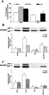
Effect of Ca2+ on DNA synthesis and MAPK phosphorylation induced by cytokines in TSMCs. The cells were preincubated with BAPTA/AM (10 μM) plus EGTA (2 mM) for 1 h, and then stimulated with vehicle or cytokines. For DNA synthesis, after 6 h incubation, cells were labelled with 1 μCi ml−1 [3H]-thymidine for another 18 h in the continuous presence of 30 ng ml−1 TNF-α or 50 ng ml−1 IL-1β. The incorporation of [3H]-thymidine was determined as described in Methods. Data are expressed as the mean±s.e.mean of three separate experiments determined in triplicate (A). For MAPK experiment, after treatment with these agents, the cells were stimulated with vehicle, 30 ng ml−1 TNF-α (B) or 50 ng ml−1 IL-1β (C) for 15 min. The phosphorylation of p42/p44 MAPK was determined as described in Figure 2. Similar results were obtained in three independent experiments. *P<0.01, as compared with the control cells exposed to respective cytokine.
Figure 7
Figure 7
Effect of PD98059 on DNA synthesis and MAPK phosphorylation induced by cytokines in TSMCs. The cells were preincubated with PD98059 (30 μM, 1 h), and then stimulated with vehicle or cytokines. For DNA synthesis, after 6 h incubation, cells were labelled with 1 μCi ml−1 [3H]-thymidine for another 18 h in the presence of 30 ng ml−1 TNF-α or 50 ng ml−1 IL-1β. The incorporation of [3H]-thymidine was determined as described in Methods. Data are expressed as the mean±s.e.mean of three separate experiments determined in triplicate (A). For MAPK experiment, after treatment with PD98059, the cells were stimulated with vehicle, 30 ng ml−1 TNF-α (B) or 50 ng ml−1 IL-1β (C) for 15 min. The phosphorylation of p42/p44 MAPK was determined as described in Figure 2. Similar results were obtained in three independent experiments. *P<0.01, as compared with the control cells exposed to respective cytokine.
Figure 8
Figure 8
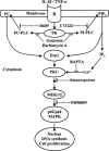
Schematic pathway for TNF-α and IL-1β signaling of cellular proliferation. Each solid line and arrow represents a step in an activating pathway. Each T-shaped and dashed line represents inactivation or inhibition. TNF-α and IL-1β bind to their receptors (R) and activate phosphatidylcholine-phospholipase C (PC-PLC) and phosphatidylinositide-phospholipase C (PI-PLC) through the phosphorylation of tyrosine kinase (TK) to induce protein kinase C (PKC) activation. Activation of PKC leads to sequential phosphorylation of MEK1/2 and p42/p44 MAPK that relays extracellular signalling into nuclei. TNF-α and IL-1β induce activation of the components of downstream p42/p44 MAPK and enhance DNA synthesis and cell proliferation in TSMCs.

Similar articles

Cited by

References

    1. ALESSI D.R., CUENDA A., COHEN P., DUDLEY D.T. , SATIEL A.R. PD098059 is a specific inhibitor of the activation of mitogen-activated protein kinase kinase in vitro and in vivo. J. Biol. Chem. 1995;270:27489–27494. - PubMed
    1. AMRANI Y., PANETTIERI R.A., FROSSARD N. , BRONNER C. Activation of the TNFα-p55 receptor induces myocyte proliferation and modulates agonist-evoked calcium transients in cultured human tracheal smooth muscle cells. Am. J. Respir. Cell Mol. Biol. 1996;15:55–63. - PubMed
    1. ARMOUR C.L., DIMENT L.M. , BLACK J.L. Relationship between smooth muscle volume and contractile response in airway tissue. Isometric versus isotonic measurement. J. Pharmacol. Exp. Ther. 1988;245:687–691. - PubMed
    1. BROIDE D.H., LOTZ M., CUOMO A.J., COBURN D.A., FEDERMAN E.C. , WASSERMAN S.I. Cytokines in symptomatic asthma airways. J. Allergy Clin. Immunol. 1992;89:958–967. - PubMed
    1. CEMBRZYNSKA-NOWAK M., SZKLARZ E., INGLOT A.D. , TEODORCZYK-INJEYAN J.A. Elevated release of tumor necrosis factor-alpha and interferone-gamma by bronchoalveolar leukocytes from patients with bronchial asthma. Am. Rev. Respir. Dis. 1993;147:291–295. - PubMed

Publication types

MeSH terms

Substances