. 2000 Jul 1;20(13):4829-43.
doi: 10.1523/JNEUROSCI.20-13-04829.2000.

Ionic mechanisms underlying repetitive high-frequency burst firing in supragranular cortical neurons

Affiliations

Ionic mechanisms underlying repetitive high-frequency burst firing in supragranular cortical neurons

J C Brumberg et al. J Neurosci. .

Abstract

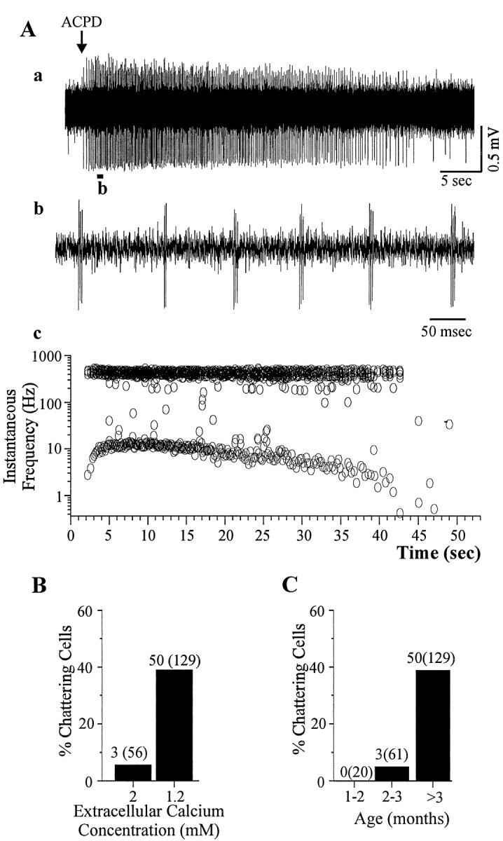
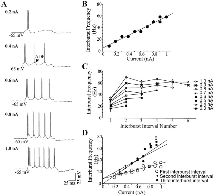
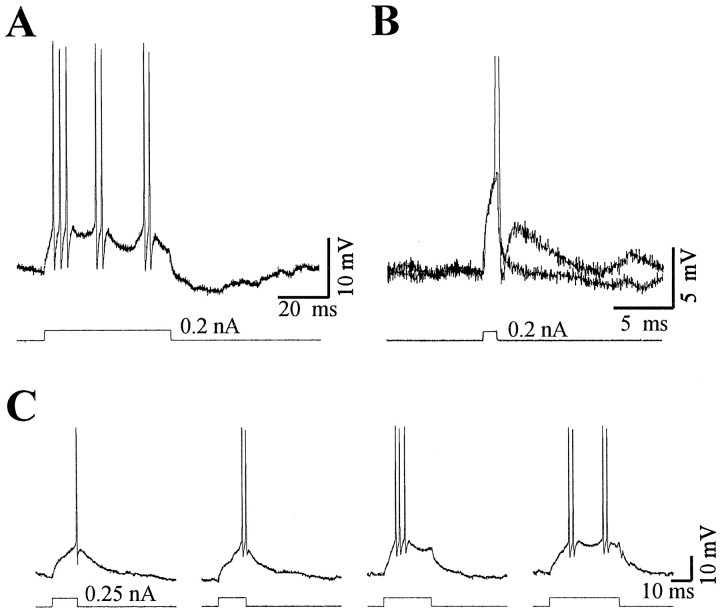
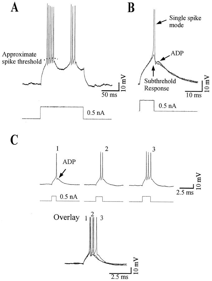
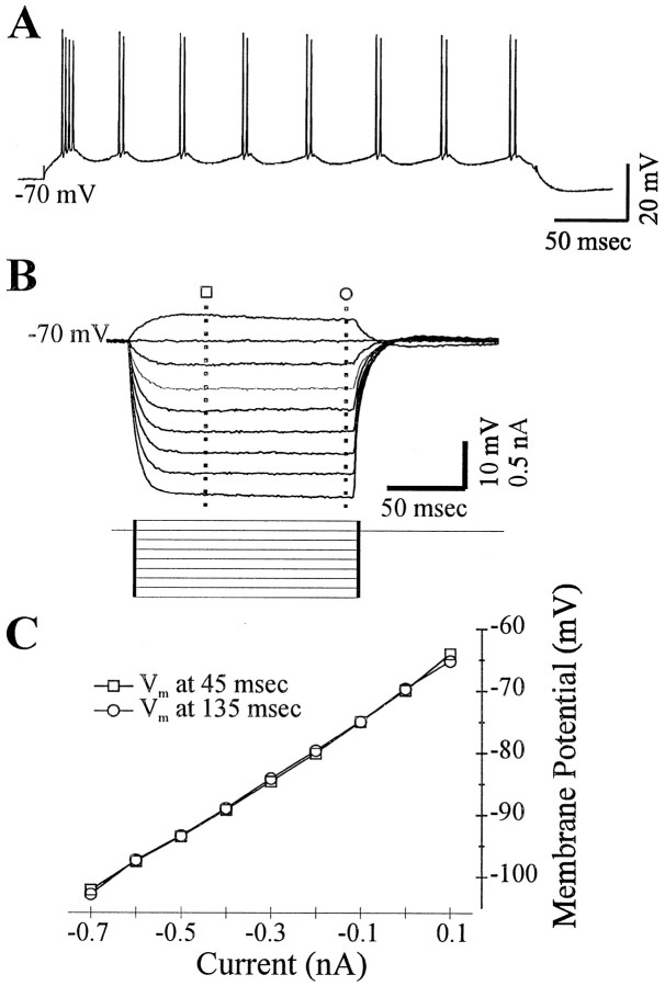
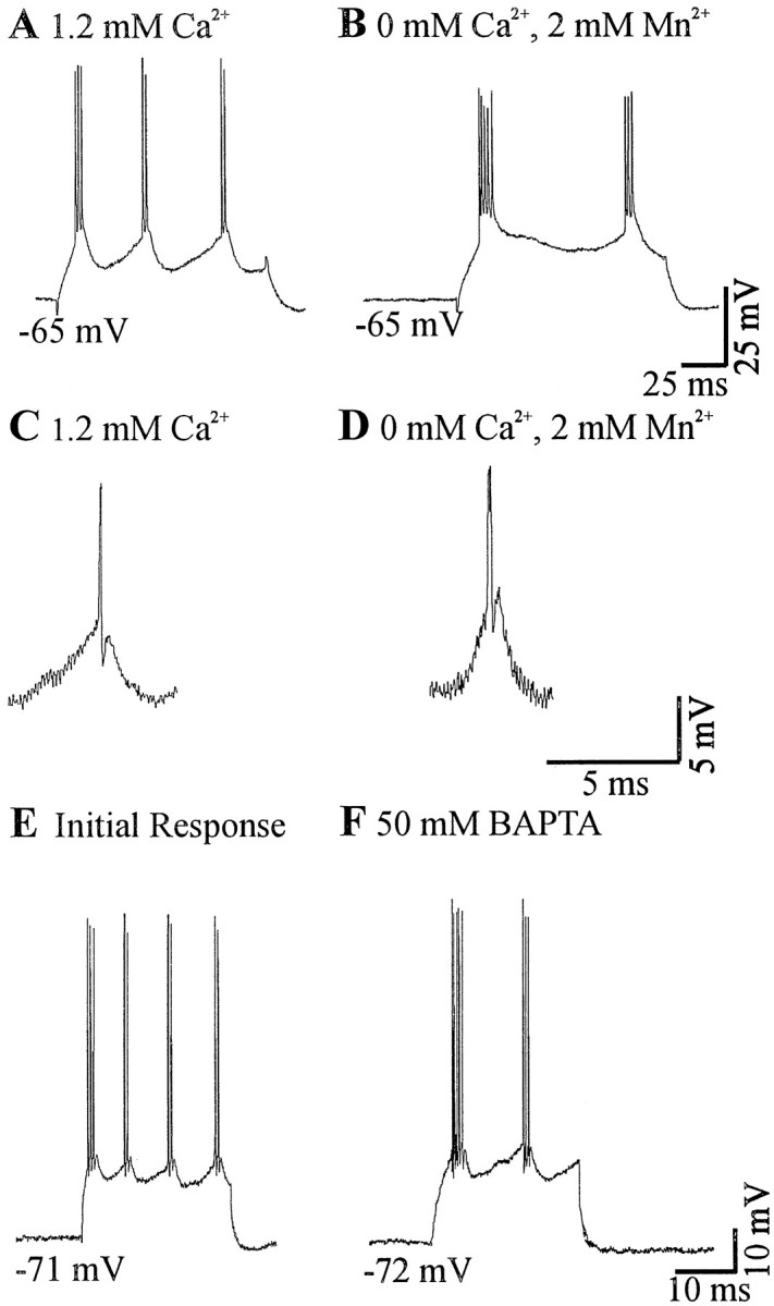
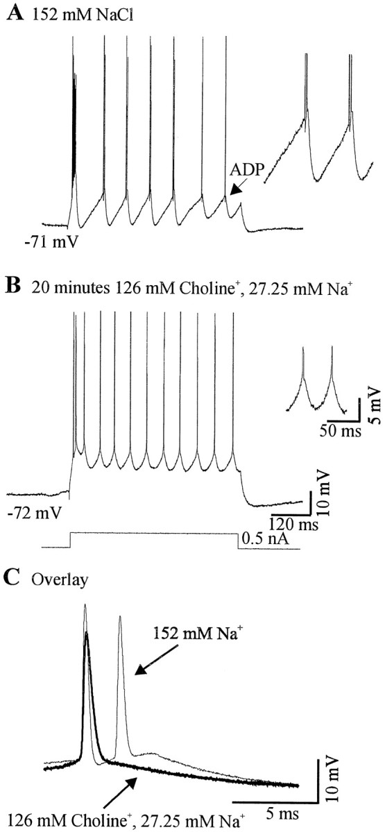
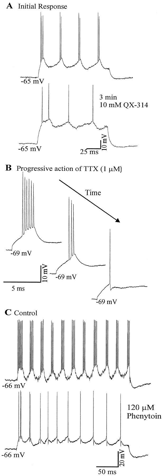
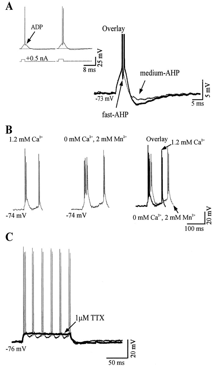
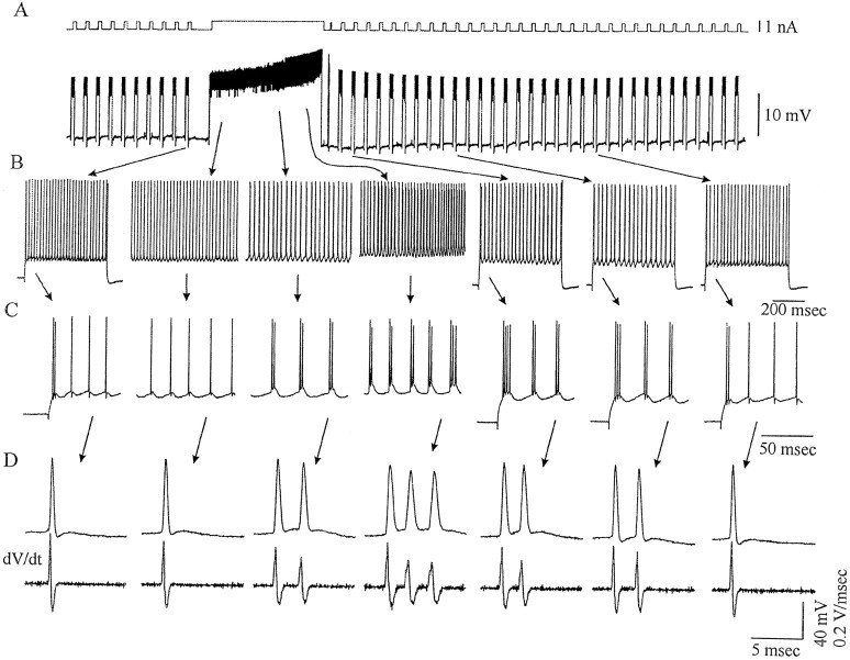
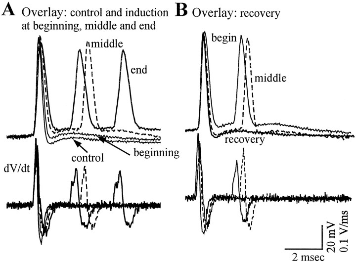
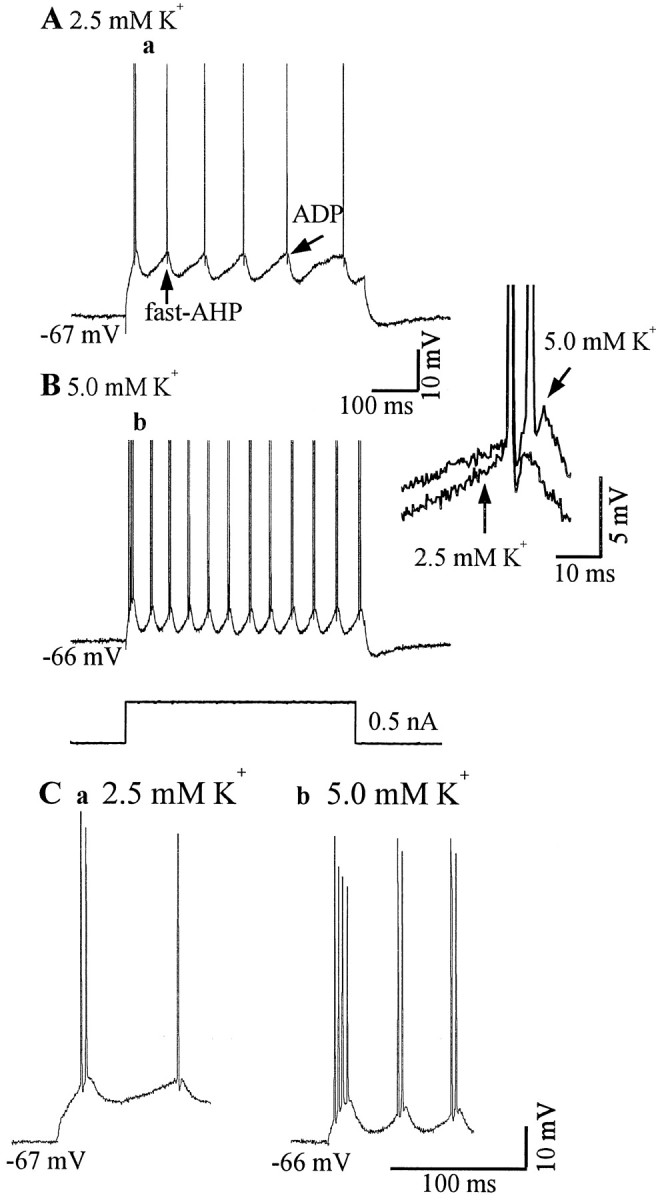
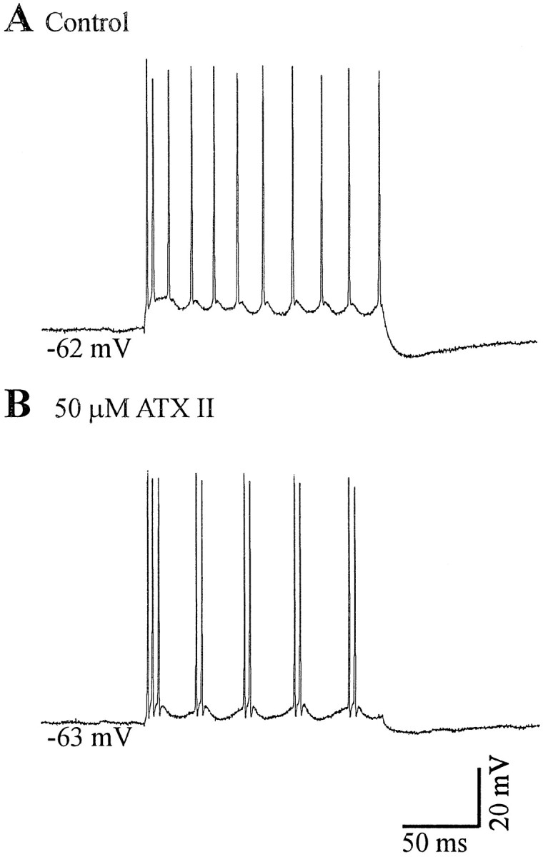
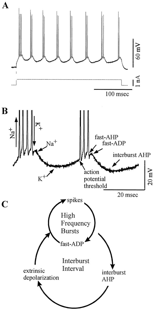
Neocortical neurons in awake, behaving animals can generate high-frequency (>300 Hz) bursts of action potentials, either in single bursts or in a repetitive manner. Intracellular recordings of layer II/III pyramidal neurons were obtained from adult ferret visual cortical slices maintained in vitro to investigate the ionic mechanisms by which a subgroup of these cells generates repetitive, high-frequency burst discharges, a pattern referred to as "chattering." The generation of each but the first action potential in a burst was dependent on the critical interplay between the afterhyperpolarizations (AHPs) and afterdepolarizations (ADPs) that followed each action potential. The spike-afterdepolarization and the generation of action potential bursts were dependent on Na(+), but not Ca(2+), currents. Neither blocking of the transmembrane flow of Ca(2+) nor the intracellular chelation of free Ca(2+) with BAPTA inhibited the generation of intrinsic bursts. In contrast, decreasing the extracellular Na(+) concentration or pharmacologically blocking Na(+) currents with tetrodotoxin, QX-314, or phenytoin inhibited bursting before inhibiting action potential generation. Additionally, a subset of layer II/III pyramidal neurons could be induced to switch from repetitive single spiking to a burst-firing mode by constant depolarizing current injection, by raising extracellular K(+) concentrations, or by potentiation of the persistent Na(+) current with the Na(+) channel toxin ATX II. These results indicate that cortical neurons may dynamically regulate their pattern of action potential generation through control of Na(+) and K(+) currents. The generation of high-frequency burst discharges may strongly influence the response of postsynaptic neurons and the operation of local cortical networks.


Figures

Fig. 1.
Fig. 1.
ACPD (500 μm in pipette) activates chattering cells in a previously quiescent slice (A,a). Expanding the extracellular recording (A,b) reveals high-frequency repetitive bursts. The interburst frequencies are thebottom data points in A,c, and the intraburst frequency of this is >300 Hz (A,c,top traces). The developmental progression of the incidence of chattering is displayed in C. Thebars represent the percentage of bursting neurons observed in the different aged animals or in B how the incidence of chattering was affected by different concentrations of extracellular Ca2+. The numbers above the bars (B, C) represent the number of chattering cells responsive to ACPD and the total number of units recorded.
Fig. 2.
Fig. 2.
Response of a chattering cell to different amplitudes of current injection. Current pulses lasted 120 msec, and the amplitude of each pulse is to the left of each voltage trace (A). Increasing the amplitude of the injected current pulse increases the interburst frequency (B). C plots the interburst frequency (y-axis) as a function of the burst number (x-axis) for another neuron. The interburst frequency is lowest for the first interval and then shortens and reaches a plateau. This effect is independent of the intensity of the injected current (D). Plotted are the interburst frequencies (y-axis) of the first (○), second (+), and third (●) interburst intervals as a function of the strength of the injected current pulse (x-axis). In every case the interburst interval is longest for the first interval, and there is no difference between the second and third intervals. Note the amplitude and time courses of the afterhyperpolarizations after a single burst (A, 0.2 nA) and after repetitive burst firing (A, 1.0 nA).
Fig. 3.
Fig. 3.
Properties of a chattering cell in the cat visual cortex in vivo. A, Intracellular injection of a depolarizing current pulse reveals repetitive burst firing at 71 Hz. B, Intracellular injection of a short depolarizing current pulse results in the activation of a single action potential that exhibits a prominent fast afterhyperpolarization and afterdepolarization. Short depolarizing current pulses did not activate bursts of spikes that outlasted the duration of the current pulse.C, Increasing the duration of the current pulse resulted in the generation of bursts of action potentials and eventually in the generation of repetitive burst firing.
Fig. 4.
Fig. 4.
Properties of chattering cells in ferret visual cortex in vitro. A, Response of a chattering cell to a 120 msec depolarizing current pulse (0.5 nA). Thedotted line indicates the approximate threshold for action potential initiation for each action potential within the burst. Note that the threshold assumes progressively more depolarized levels, and the burst is terminated when the ADP no longer exceeds threshold. Decreasing the duration of the current pulse duration at threshold evokes either single spikes or subthreshold responses (B). Increasing the duration of the current pulse increases the number of action potentials in response, from one to three (C). Overlaying the three traces suggests that the additional action potentials result from the activation of additional spikes by the ADP. Action potentials have been clipped in the overlay. Membrane potential is −73 mV for all three traces.
Fig. 5.
Fig. 5.
Chattering cells exhibit relatively linear membrane properties. A, The response of a supragranular chattering cell in response to a 300 msec 0.4 nA depolarizing current pulse. B, The same cell as in A in response to a family of hyperpolarizing and depolarizing current pulses. C, Plot of the membrane response at 45 and 135 msec (see B) after the onset of the current pulses versus the amount of current injected. There is no evidence for substantial time- or voltage-dependent rectification.
Fig. 6.
Fig. 6.
Transmembrane calcium entry is not essential to burst generation. Neither removal of Ca2+ from the bathing medium (compare A, B) nor chelation of intracellular Ca2+ by BAPTA (compareE, F) inhibited bursting.A and B represent a different cell than pictured in E and F. Note that the interburst interval is lengthened due to both manipulations. Recordings obtained from another neuron reveal that ADPs are present in nominal Ca2+ in the bathing medium (compareC, D). In C andD the action potentials have been clipped to highlight the presence of the ADPs.
Fig. 7.
Fig. 7.
Decreasing the extracellular Na+ concentration inhibits chattering.A, In normal bathing solution, intracellular injection of a depolarizing pulse results in repetitive doublets of action potentials and spike afterdepolarizations. B, Twenty minutes after replacement of NaCl with CholineCl, the repetitive doublets and the initial burst of action potentials are suppressed.C, Overlay of the action potentials before and during wash in low Na+ reveals that the low Na+ solution suppressed the spike ADP.
Fig. 8.
Fig. 8.
A persistent Na+ current underlies chattering. A, Intracellular injection of a depolarizing pulse immediately after obtaining this intracellular recording revealed this cell to be a chattering neuron. After 3 min of recording with an electrode filled with 10 mm QX-314 (dissolved in 2 m KAc), the repetitive burst firing stopped before action potential generation was inhibited. Puffer application of TTX (1 μm) inhibited bursting over time before blocking action potential initiation (B). Bath application of the anti-epileptic phenytoin (120 μm) also inhibited chattering (C) in another neuron (current pulse was +0.5 nA).
Fig. 9.
Fig. 9.
The interburst hyperpolarization is actively generated. A, Intracellular injection of a short depolarizing current pulse in a chattering cell generates either one or two action potentials with an increase in the duration of the pulse. Following the action potentials is a hyperpolarization that lasts for ∼20–30 msec. B, Block of transmembrane Ca2+ currents with low Ca2+ and raised Mn2+ does not block the interburst AHP.C, Block of action potentials with tetrodotoxin reveals how the interburst AHP depends on the generation of a burst of action potentials.
Fig. 10.
Fig. 10.
Induction of chattering in a regular spiking neuron. AD, Same data presented in increasingly fine detail by expanding the time base.A, Induction protocol. The spike discharge pattern was examined by injecting a 500 msec duration depolarizing current pulse once every 2 sec before and after constant depolarization for 16 sec.B, C, The neuron responds to the depolarizing current pulses with a train of single action potentials (except for a doublet at the beginning of the pulse) before tonic current injection. After current injection, the neuron now responds to the current pulse with the generation of repetitive bursts. Action potentials and their derivative reveal the presence of fast spike afterhyperpolarizations and afterdepolarization.
Fig. 11.
Fig. 11.
Overlay of action potentials before, during, and recovery from induced repetitive bursting. A, Overlay of the four traces in Figure 10D before and during induction reveals the action potentials to become broader in duration and a marked decrease in the fast spike afterhyperpolarization. Subsequently, the fast afterdepolarization is able to generate additional action potentials. B, During recovery from chattering induction, the action potential returns to its original duration and the fast afterhyperpolarization returns.
Fig. 12.
Fig. 12.
Raising extracellular K+induces bursting. Raising extracellular K+ from 2.5 mm (a) to 5.0 mm(b) induced bursting to the same depolarizing current pulse (bottom trace, +0.5 nA).Inset shows that in 5.0 mmK+, the ADP reaches threshold for the generation of a second action potential. C, Magnification of the initial responses for 2.5 mm K+(a) and 5.0 mm K+(b) highlighting the transition from single spiking to bursting.
Fig. 13.
Fig. 13.
Potentiation of the persistent Na+ current induces bursting. Local application of ATX II (50 μm in pipette) evoked bursting in response to a 120 msec +0.5 nA current pulse (B).A represents the response before ATX II application.
Fig. 14.
Fig. 14.
Summary of the proposed mechanisms for chattering. Repetitive high-frequency bursting in response to a depolarizing current pulse is typical of chattering cells in vitro (A). Magnification of the bursts highlights the presence of fast AHPs after each action potential. Each burst is terminated by a Na+-dependent ADP that does not reach spike threshold (B) and is followed by the interburst AHP. The high-frequency bursts that typify chattering cells result as an interaction between fast Na+spikes and a fast ADP; the interburst interval is governed by the rate at which the membrane repolarizes (C, interburst AHP) and the amplitude of any extrinsic depolarizing influences.

References

    1. Aghajanian GK, Rasmussen K. Intracellular studies in the facial nucleus illustrating a simple new method for obtaining viable motoneurons in adult rat brain slices. Synapse. 1989;3:331–338. - PubMed
    1. Agmon A, Connors BW. Repetitive burst-firing in the deep layers of mouse somatosensory cortex. Neurosci Lett. 1989;99:137–141. - PubMed
    1. Ahmed B, Anderson JC, Douglas RJ, Martin KA, Whitteridge D. Estimates of the net excitatory currents evoked by visual stimulation of identified neurons in cat visual cortex. Cereb Cortex. 1998;8:462–476. - PubMed
    1. Allen C, Stevens CF. An evaluation of causes for unreliability of synaptic transmission. Proc Natl Acad Sci USA. 1994;91:10380–10383. - PMC - PubMed
    1. Alsen C. Biological significance of peptides from Anemonia sulcata. FASEB J. 1983;42:101–108. - PubMed

Publication types

MeSH terms

LinkOut - more resources