. 2000 Jul 1;20(13):4912-21.
doi: 10.1523/JNEUROSCI.20-13-04912.2000.

Active zones on motor nerve terminals contain alpha 3beta 1 integrin

Affiliations

Active zones on motor nerve terminals contain alpha 3beta 1 integrin

M W Cohen et al. J Neurosci. .

Abstract

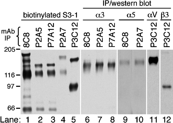
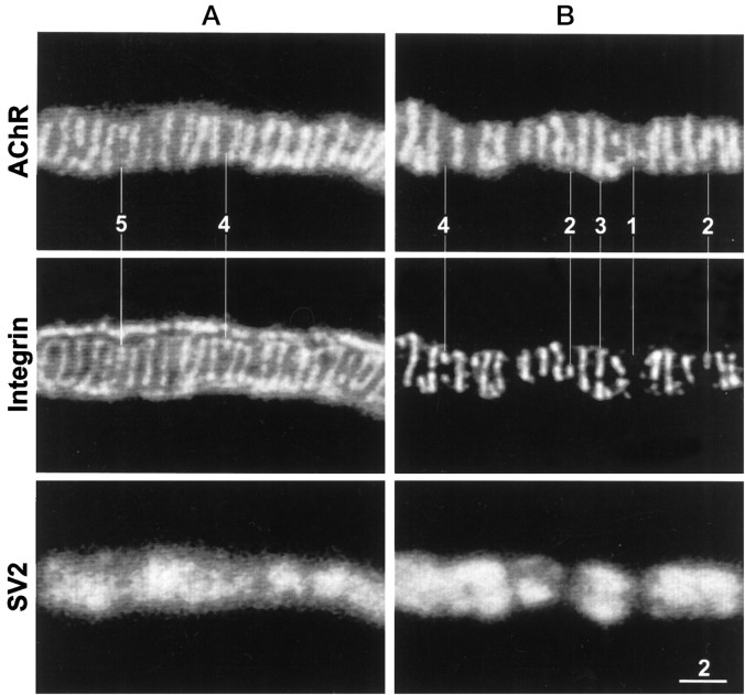
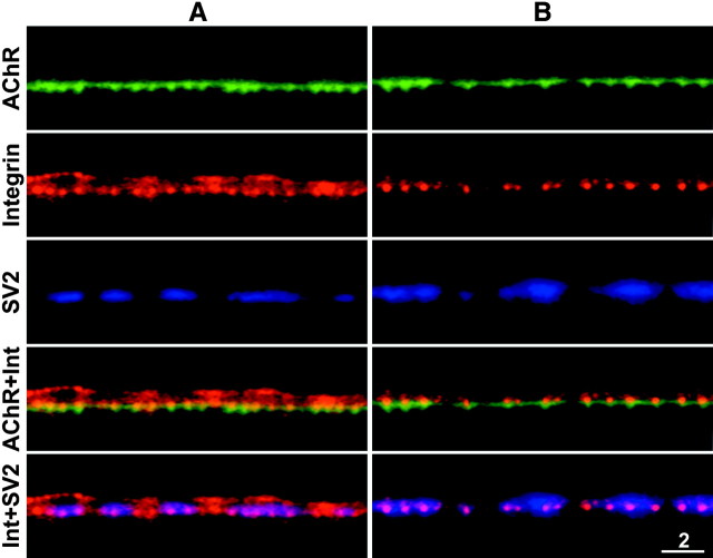
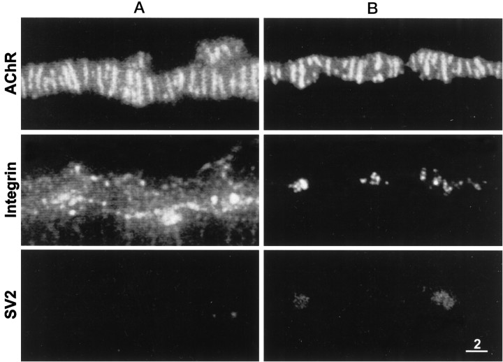
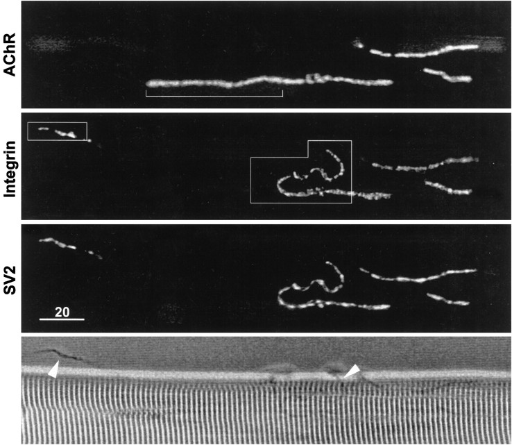
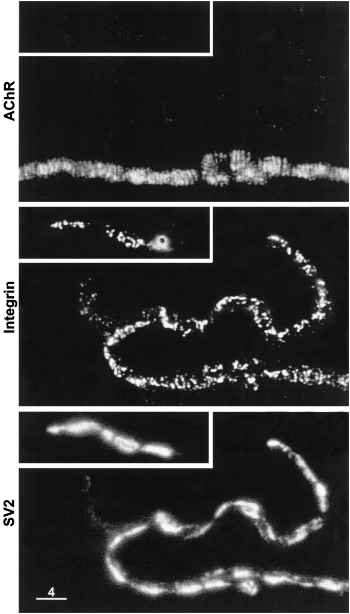
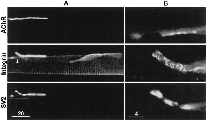
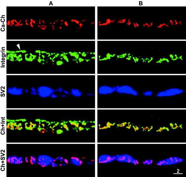
Active zones are the sites along nerve terminals where synaptic vesicles dock and undergo calcium-dependent exocytosis during synaptic transmission. Here we show, by immunofluorescent staining with antibodies generated against Xenopus laevis integrins, that alpha3beta1 integrin is concentrated at the active zones of Xenopus motor nerve terminals. Because integrins can link extracellular matrix molecules to cytoskeletal elements and participate in the formation of signaling complexes, the localization of integrin at active zones suggests that it may play a role in the adhesion of the nerve terminals to the synaptic basal lamina, in the formation and maintenance of active zones, and in some of the events associated with calcium-dependent exocytosis of neurotransmitter. Our findings also indicate that the integrin composition of the terminal Schwann cells differs from that of the motor nerve terminals, and this may account at least in part for differences in their adhesiveness to the synaptic basal lamina.


Figures

Fig. 1.
Fig. 1.
Specificity of mAbs directed againstXenopus integrins. For lanes 1–5,Xenopus S3–1 cells were labeled by cell-surface biotinylation, extracted in IP buffer, and immunoprecipitated using the following mAbs: 8C8 (anti-β1; Gawantka et al., 1992), P2A5 and P7A12 (anti-α3β1), P2A7 (anti-α5β1), and P3C12 (anti-αV). Immunoprecipitated proteins were separated by SDS-PAGE, transferred to nitrocellulose, and probed with streptavidin-HRP. For lanes 6–12, unlabeled S3–1 cell extracts were immunoprecipitated using the mAbs indicated and Western-blotted using HRP-conjugated secondary antibodies. Western blots were probed with subunit-specific polyclonal antibodies directed against α3 (Ab D3FAP; Meng et al., 1997), α5 (Ab 881; Joos et al., 1995), αV (Ab 1930; Chemicon, Temecula, CA), and β3 (Ab VA-28; D. G. Ransom, M. D. Hens, and D. W. DeSimone, unpublished observations). All immunoprecipitations were run on 6.5% SDS-PAGE gels under nonreducing conditions (with the exceptions of lanes 9–10, which were run under reducing conditions). The <66 kDa band observed in the 8C8, P2A5, and P7A12 immunoprecipitates of biotin-labeled cells (lanes 1–3) corresponds to a proteolytic fragment of α3 reported previously (Gawantka et al., 1994; Meng et al., 1997).
Fig. 2.
Fig. 2.
Face views of two neuromuscular junctions (A, B) stained for AChRs (top panels), α3β1 integrin (middle panels), and synaptic vesicle protein SV2 (bottom panels). The integrin in this and other figures was stained with mAb P2A5. Note extensive correspondence as well as subtle differences between the transverse bands of AChR fluorescence and of integrin immunofluorescence. The differences, marked by the numbered lines, include: (1) a faint AChR band without a corresponding integrin band, (2) integrin bands that are shorter than the corresponding AChR bands, (3) an integrin band that is segmented, whereas its corresponding AChR band is not, (4) short integrin bands without corresponding AChR bands, and (5) an integrin band that is longer than its corresponding AChR band. Integrin immunofluorescence that outlines the neuromuscular junction inA but not in B is associated with the outer surface of the terminal Schwann cell. Scale bar, 2 μm.
Fig. 3.
Fig. 3.
Side views of two neuromuscular junctions (A, B) stained for AChRs (top panels), α3β1 integrin (second panels), and SV2 (third panels). The short periodic downward extensions of AChR fluorescence are the sites where the postsynaptic membrane invaginates to form the junctional folds. Merged images indicate that the bright dots of integrin immunofluorescence were aligned with the tops of the junctional folds (fourth panels) and with the synaptic side of the motor nerve terminals (bottom panels). The outer surface of the Schwann cell was revealed by the integrin immunofluorescence in A but not inB. Scale bar, 2 μm.
Fig. 4.
Fig. 4.
Disappearance of synaptic bands of α3β1 integrin after denervation. A, Denervated for 6 d. Transverse bands of AChRs (top panel) are apparent, but corresponding bands of integrin immunofluorescence (middle panel) are not, and SV2 immunofluorescence (bottom panel) is undetectable. The regions of bright integrin immunofluorescence may reflect changes in the Schwann cell in response to degeneration of the motor nerve terminals. B, Denervated for 3 d. There are no transverse integrin bands. Instead there are some isolated sites of integrin stain and some faint SV2 immunofluorescence. These sites are probably portions of degenerating nerve terminals that have not yet been phagocytosed. Scale bar, 2 μm.
Fig. 5.
Fig. 5.
Low-magnification view of a neuromuscular junction treated with collagenase. Comparison of the AChR fluorescence (top panel) and the SV2 immunofluorescence (third panel) reveals that the motor nerve terminal was displaced from a portion of the postsynaptic membrane (top panel, bracket). Displaced nerve terminal branches are also apparent in the phase-contrast image (bottom panel, arrowheads). The α3β1 integrin immunofluorescence (second panel) is codistributed with the nerve terminal branches and is not detectable at the portion of postsynaptic membrane that lacks nerve terminal. Framed portions of the field are shown at higher magnification in Figure 6. Scale bar, 20 μm.
Fig. 6.
Fig. 6.
Higher magnification view of framed portions of Figure 5. Top panels, AChR stain; middle panels, α3β1 integrin stain; bottom panels,SV2 stain. Note that in the region where the nerve terminal was still present at the postsynaptic membrane, the collagenase treatment partially disrupted the integrin bands and altered their orientation such that many of them were no longer aligned with the AChR bands. A similar disorganization of the integrin bands is apparent along the displaced portions of the nerve terminal. Scale bar, 4 μm.
Fig. 7.
Fig. 7.
Synaptic and nonsynaptic staining patterns after treatment with collagenase. A, Low magnification view of AChR stain (top panel), α3β1 integrin stain (middle panel), and SV2 stain (bottom panel). The framed portion of the field (bottom panel) contains a neuromuscular junction and a displaced nerve terminal. Note that integrin stain was associated with the displaced nerve terminal and not with the terminal-free portion of postsynaptic membrane (middle panel, arrowhead). The integrin stain also reveals costameres and a satellite cell.B, Higher magnification of framed area. Bands of integrin stain are apparent on the displaced nerve terminal. The absence of integrin stain on the terminal-free portion of postsynaptic membrane is also apparent. Scale bars: A, 20 μm;B, 4 μm.
Fig. 8.
Fig. 8.
Active zones altered by collagenase treatment contain α3β1 integrin. A,B, Two different examples. Panels from top tobottom, Calcium channels at active zones stained with RωCT; α3β1 integrin stain; SV2 stain; merge of calcium channel and α3β1 integrin images; and merge of calcium channel and SV2 images. Integrin stain is present at all active zones revealed by the calcium channel stain, including those whose orientation and shape was altered by the collagenase treatment. Conversely, calcium channel stain was not detected at sites of integrin immunofluorescence that were associated with the terminal Schwann cell (A, arrowhead). Scale bar, 2 μm.

References

    1. Anderson MJ, Cohen MW. Fluorescent staining of acetylcholine receptors in vertebrate skeletal muscle. J Physiol (Lond) 1974;237:385–400. - PMC - PubMed
    1. Anderson MJ, Shi ZQ, Zackson SL. Proteolytic disruption of laminin-integrin complexes on muscle cells during synapse formation. Mol Cell Biol. 1996;16:4972–4984. - PMC - PubMed
    1. Aplin AE, Howe A, Alahari SK, Juliano RL. Signal transduction and signal modulation by cell adhesion receptors: the role of integrins, cadherins, immunoglobulin-cell adhesion molecules, and selectins [Review]. Pharmacol Rev. 1998;50:197–263. - PubMed
    1. Astrow SH, Qiang H, Ko CP. Perisynaptic Schwann cells at neuromuscular junctions revealed by a novel monoclonal antibody. J Neurocytol. 1998;27:667–681. - PubMed
    1. Bahr B, Staubli U, Xiao P, Chun D, Ji Z, Esteban E, Lynch G. Arg-Gly-Asp-Ser-selective adhesion and the stabilization of long-term potentiation: pharmacological studies and the characterization of a candidate matrix receptor. J Neurosci. 1997;17:1320–1329. - PMC - PubMed

Publication types