. 2000 Jul;20(14):5041-7.
doi: 10.1128/MCB.20.14.5041-5047.2000.

AIB1 is a conduit for kinase-mediated growth factor signaling to the estrogen receptor

Affiliations

AIB1 is a conduit for kinase-mediated growth factor signaling to the estrogen receptor

J Font de Mora et al. Mol Cell Biol. 2000 Jul.

Abstract

Growth factor modulation of estrogen receptor (ER) activity plays an important role in both normal estrogen physiology and the pathogenesis of breast cancer. Growth factors are known to stimulate the ligand-independent activity of ER through the activation of mitogen-activated protein kinase (MAPK) and the direct phosphorylation of ER. We found that the transcriptional activity of AIB1, a ligand-dependent ER coactivator and a gene amplified preferentially in ER-positive breast cancers, is enhanced by MAPK phosphorylation. We demonstrate that AIB1 is a phosphoprotein in vivo and can be phosphorylated in vitro by MAPK. Finally, we observed that MAPK activation of AIB1 stimulates the recruitment of p300 and associated histone acetyltransferase activity. These results suggest that the ability of growth factors to modulate estrogen action may be mediated through MAPK activation of the nuclear receptor coactivator AIB1.


Figures

FIG. 1
FIG. 1
The MAPK pathway promotes ER-signaling. (A) COS cells were transiently cotransfected with the wild-type human ERα and either MKP1 or constitutively activated MEK1 (RΔF). The transfection also included the luciferase reporter under the control of the ERE2 and tk-lacZ as an internal control. One day following transfection, cells were stimulated with 10 nM 17-β-estradiol (E2) (+) or with ethanol alone (−). On the third day, cells were lysed and assayed for the reporter activity. Values represent the ratio between luciferase units and β-galactosidase, relative to the basal activity of either ER or ER(S118A) in the absence of estrogen. (B) ERα containing the S118A mutation was cotransfected into COS cells as described above. Autoradiographs in the insets demonstrate the MAPK activity for each of the transfection conditions, using MBP as a substrate.
FIG. 2
FIG. 2
AIB1 is phosphorylated in vivo. (A) In vitro translated (IVT) AIB1 was labeled with [35S]Met and immunoprecipitated (IP) with either preimmune serum (lane 2) or anti-AIB1 antibodies (lane 3). Subconfluent MCF-7 cells were labeled with [32P]orthophosphate for 6 h and immunoprecipitated with either preimmune serum (lane 4) or anti-AIB1 antibodies (lane 5). Immune complexes were resolved by SDS–8% PAGE. The gel was dried and exposed for autoradiography. The figure is representative of four independent experiments. (B) The band corresponding to AIB1 was excised from the gel and treated with HCl to total hydrolysis, analyzed by TLC together with phosphoamino acid markers, developed with ninhydrin to reveal the mobility of nonradioactive phosphoaminoacids (indicated by dashed circles), and subsequently exposed for autoradiography. (C) Breast cancer cell lines MCF-7, BT-474, and MDA-MB-468 were analyzed by Western blotting (WB) for the expression of AIB1. Each well was loaded with 30 μg of total protein from whole-cell lysates (upper panel). In parallel, cells were labeled with [32P]orthophosphate for 6 h, and 0.8 mg of total protein from each cell line lysate was immunoprecipitated with anti-AIB1 antibodies (lower panel).
FIG. 3
FIG. 3
AIB1 is a substrate of MAPK. Proliferating MCF-7 cells were lysed, and aliquots containing 3 mg of total protein were used for immunoprecipitation (IP) and kinase reactions. Lysates were immunoprecipitated with preimmune serum (lane 1) or with immunopurified anti-AIB1 polyclonal antibodies (lanes 3 to 5). AIB1 immune complexes were split into thirds. One third received no further treatment (lane 3). The other two thirds were treated with protein λ-phosphatase (λPPase) (lane 4), and after a washing, one third was further resuspended in 30 μl of buffer containing 100 μM ATP and treated with 0.1 μg of active Erk2 (Upstate Biotechnology) for 1 hour at 30°C (lane 5). In parallel, the other sample was treated with 0.1 μCi of [32P]ATP and resolved independently. Gels were either Western immunoblotted (WB) with AIB1 antibodies (upper panel) or dried on Whatman paper and exposed for autoradiography (lower panel). Similar results were obtained in three independent experiments.
FIG. 4
FIG. 4
MAPK stimulates AIB1 transcriptional activity. (A) COS cells were transiently transfected with Gal4-DBD fused to full-length or deletion mutants of AIB1, together with a luciferase reporter containing five copies of UAS. tk-lacZ was used as an internal control of the transfection. Transfections were performed in the presence of vector alone, MKP1, or activated MEK1 (RΔF). (B) Structural features of full-length AIB1 and the two deletion mutants. Values represent the ratio between the fold induction observed in the presence of a permanently activated MAPK (RΔF) and that observed in the presence of the phosphatase MKP1, as an indication of sensitivity to regulation by MAPK. Striped boxes indicate the two transactivation domains, AD1 and AD2; LXXLL, NR-interacting domains; Poly Q, stretch of polyglutamines.
FIG. 5
FIG. 5
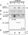
Activation of MAPK recruits p300 to AIB1 complexes. Gal4-DBD alone or fused to AIB1 truncation mutants (amino acids 556 to 1420 or 578 to 1131) was transfected into BOSC cells along with pCIp300. The transfections also included vector alone (−) or vector containing activated MEK1 (RΔF). (A) After 48 h, endogenous Erk2 was immunoprecipitated to determine MAPK activity, using MBP as a substrate. (B) In parallel, 25 μg of whole-cell extract was analyzed for expression of p300 by Western blotting blot (WB). Lysates were further immunoprecipitated with a polyclonal antibody to Gal4-DBD. (C and D) Immune complexes were probed for p300 (C) and Gal4-DBD proteins (D). (E) In parallel, immune complexes were analyzed for HAT activity. Similar results were obtained in five independent experiments.
FIG. 6
FIG. 6
Regulation of steroid receptor transcriptional complexes by MAPK activation. Growth factors such as EGF and IGF-1 stimulate the Ras-MAPK cascade via activation of their respective tyrosine kinase receptors. Activated MAPK may then phosphorylate nuclear targets including ER and AIB1 to modulate gene transcription in response to growth factor and/or steroid stimulation.

References

    1. Anzick S L, Kononen J, Walker R L, Azorsa D O, Tanner M M, Guan X Y, Sauter G, Kallioniemi O P, Trent J M, Meltzer P S. AIB1, a steroid receptor coactivator amplified in breast and ovarian cancer. Science. 1997;277:965–968. - PubMed
    1. Bautista S, Valles H, Walker R L, Anzick S, Zeillinger R, Meltzer P, Theillet C. In breast cancer, amplification of the steroid receptor coactivator gene AIB1 is correlated with estrogen and progesterone receptor positivity. Clin Cancer Res. 1998;4:2925–2929. - PubMed
    1. Bourguet W, Ruff M, Chambon P, Gronemeyer H, Moras D. Crystal structure of the ligand-binding domain of the human nuclear receptor RXR-alpha. Nature. 1995;375:377–382. - PubMed
    1. Brandt B H, Roetger A, Dittmar T, Nikolai G, Seeling M, Merschjann A, Nofer J R, Dehmer-Moller G, Junker R, Assmann G, Zaenker K S. c-erbB-2/EGFR as dominant heterodimerization partners determine a motogenic phenotype in human breast cancer cells. FASEB J. 1999;13:1939–1949. - PubMed
    1. Brownell J E, Allis C D. An activity gel assay detects a single, catalytically active histone acetyltransferase subunit in Tetrahymena macronuclei. Proc Natl Acad Sci USA. 1995;92:6364–6368. - PMC - PubMed

Publication types

MeSH terms

LinkOut - more resources