. 2000 Jul;20(14):5048-63.
doi: 10.1128/MCB.20.14.5048-5063.2000.

A new family of nuclear receptor coregulators that integrate nuclear receptor signaling through CREB-binding protein

Affiliations

A new family of nuclear receptor coregulators that integrate nuclear receptor signaling through CREB-binding protein

M A Mahajan et al. Mol Cell Biol. 2000 Jul.

Abstract

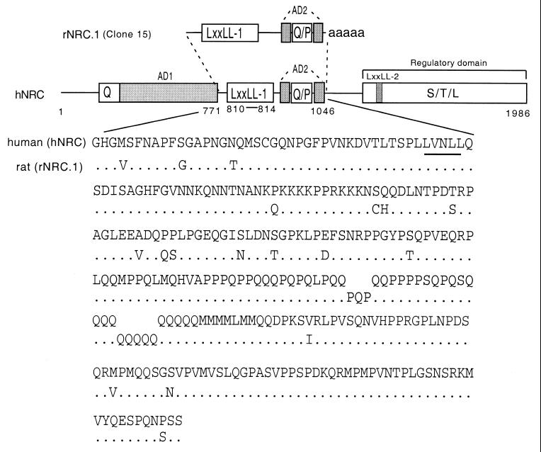
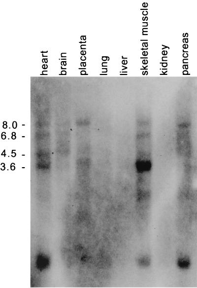
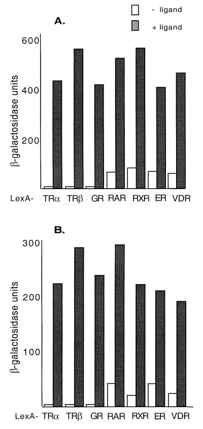
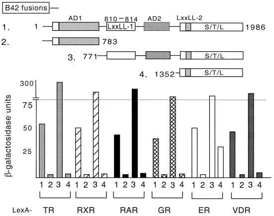
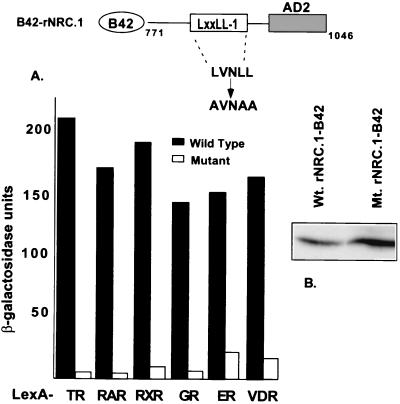
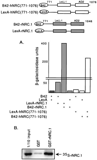
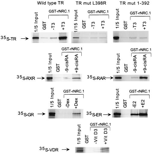
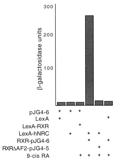
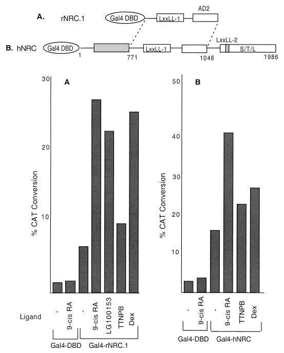
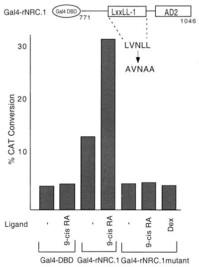
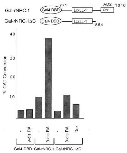
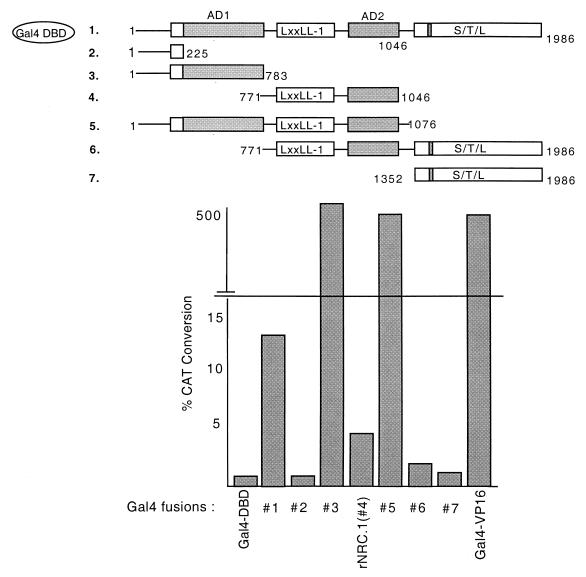
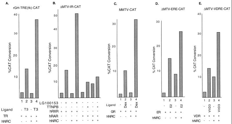
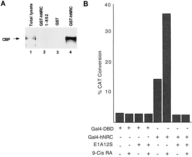
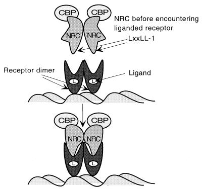
We describe the cloning and characterization of a new family of nuclear receptor coregulators (NRCs) which modulate the function of nuclear hormone receptors in a ligand-dependent manner. NRCs are expressed as alternatively spliced isoforms which may exhibit different intrinsic activities and receptor specificities. The NRCs are organized into several modular structures and contain a single functional LXXLL motif which associates with members of the steroid hormone and thyroid hormone/retinoid receptor subfamilies with high affinity. Human NRC (hNRC) harbors a potent N-terminal activation domain (AD1), which is as active as the herpesvirus VP16 activation domain, and a second activation domain (AD2) which overlaps with the receptor-interacting LXXLL region. The C-terminal region of hNRC appears to function as an inhibitory domain which influences the overall transcriptional activity of the protein. Our results suggest that NRC binds to liganded receptors as a dimer and this association leads to a structural change in NRC resulting in activation. hNRC binds CREB-binding protein (CBP) with high affinity in vivo, suggesting that hNRC may be an important functional component of a CBP complex involved in mediating the transcriptional effects of nuclear hormone receptors.


Figures

FIG. 1
FIG. 1
Structures and comparison of the hNRC and rNRC.1 isoforms. The upper section shows a schematic representation of the functional domains identified in hNRC and rNRC.1, a rat homologue, isolated from the GH4C1 pJG4-5 cDNA library. rNRC.1 is homologous to amino acids 771 to 1046 of hNRC. AD1 and AD2 are two activation domain regions, Q and P represent a glutamine- and proline-rich stretch of amino acids. LXXLL-1 is a nuclear receptor interaction motif. S-T-L is a serine-, threonine-, and leucine-rich sequence in the C-terminal part of hNRC which may be part of regulatory domain. LXXLL-2 is a second LXXLL motif which plays a less important role in nuclear receptor interactions. The lower section shows the amino acid sequence of rNRC.1 and its alignment with the corresponding region in hNRC. LXXLL-1 is underlined. The sequence of KIAA0181 is in GenBank under accession number D80003. We have extended the N terminus of hNRC and have deposited the sequence containing 58 additional amino acids in GenBank (accession number AF245115). The sequence of rNRC.1 has also been deposited in GenBank (accession number AF228043).
FIG. 2
FIG. 2
Northern blot of hNRC mRNAs in different tissues. hNRC mRNAs were detected using an MTN blot (Stratagene) containing RNAs from the various tissues indicated. The various-sized hNRC-related mRNAs were detected by probing the blot with 32P-labeled hNRC cDNA. Numbers on the left are kilobases.
FIG. 3
FIG. 3
Ligand-dependent interaction of rNRC.1 and hNRC with nuclear hormone receptors in yeast. (A) rNRC.1 was expressed as a B42 fusion protein and tested against LexA fusions of cTRα, hTRβ-LBD, hGR-LBD, hRARα-LBD, hRXRα-LBD, hERα, and hVDR-LBD. Both ligand-dependent and ligand-independent interactions were quantified by measuring the activity of β-galactosidase in yeast extracts. Details are given in Materials and Methods, along with the ligands used. (B) Same as panel A except that hNRC was expressed as a B42 fusion and examined for interaction with the various LexA-receptor chimeras.
FIG. 4
FIG. 4
Mapping the region of hNRC necessary for interaction with various receptors. As depicted, various deletion constructs of hNRC were generated as B42 fusions for two-hybrid interaction assays with the LexA-receptor chimeras indicated in Fig. 3.
FIG. 5
FIG. 5
Ligand-dependent interaction of rNRC.1 with nuclear receptors requires LXXLL-1. (A) LexA fusions of various nuclear hormone receptors as indicated were tested in yeast two-hybrid assays against wild-type and mutant rNRC.1 expressed as B42 fusion proteins. The amino acids indicated for rNRC.1 are those which are homologous to amino acids 771 to 1046 of hNRC. LXXLL-1 (LVNLL) was changed to AVNAA. The interaction was quantified by determining the activity of β-galactosidase with and without cognate ligands as described in Materials and Methods. (B) Western blotting using anti-HA antibody indicates the expression of wild-type (Wt.) and mutant (Mt.) proteins as shown.
FIG. 6
FIG. 6
NRCs form homodimers in vivo and in vitro. (A) LexA-rNRC.1 was transformed into yeast along with pJG4-5, which expresses rNRC.1 as a B42 fusion protein (B42-rNRC.1). pJG4-5 (B42) and pEG202 (LexA), pJG4-5 (B42) and LexA-rNRC.1, and B42-rNRC.1 and pEG202 (LexA) were also transformed into yeast and served as controls. Similar studies were carried out with hNRC(771-1076) cloned into LexA and B42 expression vectors. (B) GST-rNRC.1 or GST alone was incubated with 35S-rNRC.1 labeled with l-[35S]methionine in rabbit reticulocyte lysates, and in vitro binding was carried out as described in Materials and Methods. The samples were then electrophoresed in SDS gels, and the amount of 35S-rNRC.1 which bound to GST-rNRC.1 was visualized by fluorography. One-tenth of the input amount of 35S-labeled rNRC.1 used in the incubation with GST-rNRC.1 was electrophoresed in the same gel.
FIG. 7
FIG. 7
Ligand-dependent binding of nuclear receptors with rNRC.1 in vitro. All receptors were labeled with l-[35S]methionine by in vitro transcription-translation using reticulocyte lysates. Bacterially expressed and purified GST-rNRC.1 bound to glutathione-agarose beads was incubated with 35S-labeled receptors with or without the indicated cognate ligands. The samples were then electrophoresed in SDS gels, and the amount of 35S-receptor which bound to GST-rNRC.1 was visualized by fluorography. One-fifth of the amount of 35S-labeled receptor used in the incubation with GST-rNRC.1 was electrophoresed in the same gel. E2, estradiol; VitD3, 1,25-dihydroxy-vitamin D3.
FIG. 8
FIG. 8
Receptor binding induces a conformational change in hNRC. The hRXRα LBD was cloned into pJG4-6. This vector is similar to pJG4-5 but lacks the B42 activation domain. It includes the same HA epitope and same simian virus 40 viral nuclear localization signal as found in pJG4-5, which we introduced. RXR-pJG4-6 expressing the hRXRα LBD was cotransformed into yeast along with LexA-hNRC as indicated. pJG4-6 alone or with pEG202 (LexA), LexA-hRXR-LBD, or LexA-hNRC was transformed into yeast as controls. RXRΔAF2-pJG4-5, contains the hRXRα LBD AF2 mutation expressed as a fusion protein with the B42 activation domain. RXRΔAF2-pJG4-5 was cotransformed with LexA or LexA-hNRC as shown. Yeast cells were incubated with or without 9-cis-RA, and the activity of β-galactosidase was determined.
FIG. 9
FIG. 9
Ligand-dependent interaction of endogenous nuclear receptors with rNRC.1 and hNRC in mammalian cells. (A) HeLa cells were co-transfected with a Gal4-responsive CAT reporter plasmid (pBL-G5-CAT2) and pSG424 vectors which express the Gal4 DBD or Gal4-rNRC.1 with and without the various ligands as indicated. The amino acids indicated for rNRC.1 are those which are homologous to amino acids 771 to 1046 of hNRC. Each sample was analyzed in duplicate, and the experiment was repeated at least three times (see Materials and Methods for details about plasmids and the concentrations of the various ligands). (B) Same as panel A except that a Gal4-hNRC fusion was used instead of Gal4-rNRC.1.
FIG. 10
FIG. 10
LXXLL-1 influences both the ligand-dependent and intrinsic basal activities of rNRC.1. The Gal4 DBD, Gal4-rNRC.1, and the LXXLL-1 Gal4-rNRC.1 mutant were cotransfected with pBL-G5-CAT2 with or without various ligands as indicated. The amino acids indicated for rNRC.1 are those which are homologous to amino acids 771 to 1046 of hNRC. Each sample was analyzed in duplicate, and the experiment was repeated at least two times with similar results.
FIG. 11
FIG. 11
AD2 influences both the ligand-dependent and intrinsic basal activities of rNRC.1. pBL-G5-CAT2 was cotransfected in HeLa cells with the Gal4 DBD and the two Gal4 fusions of rNRC.1 as depicted. The amino acids indicated for the rNRC.1s are those which are homologous to amino acids 771 to 864 and 771 to 1046 of hNRC. The ligands used are as indicated.
FIG. 12
FIG. 12
hNRC harbors a strong activation domain (AD1) and an inhibitory S-T-L region at the C terminus. The indicated Gal4 fusions of hNRC and rNRC.1 were transfected in HeLa cells with pBL-G5-CAT2. The amino acids indicated for the rNRC.1s are those which are homologous to amino acids 771 to 1046 of hNRC. The transfections were analyzed in duplicate, and the experiment was repeated at least three times with similar results. Gal4-VP16 was used as a control for comparison.
FIG. 13
FIG. 13
hNRC functions as a coactivator for wild-type nuclear receptors on native hormone response elements. (A) HeLa cells were transfected with an rGH-TRE-tk-CAT reporter and expression vectors for cTRα and hNRC as indicated. T3 was at 1 μM. (B) Same as panel except that the CAT reporter was ΔMTV-IR-CAT and the receptors expressed were hRXRα and hRARα. The RXR-specific ligand LG100153 and the RAR-specific ligand TTNPB were each used at 200 nM. (C, D, and E) Same as panel except that the corresponding CAT reporters depicted were cotransfected with vectors expressing hGR, hERα, and hVDR and the cells were incubated with and without their cognate ligands at 200 nM. All samples were analyzed in duplicate, and the CAT activity shown is an average of three experiments.
FIG. 14
FIG. 14
hNRC associates with CBP in vivo, and EIA abrogates both the intrinsic activity and ligand-dependent activation function of hNRC. (A) pEBG vectors expressing GST, a GST fusion of hNRC, or GST-hNRC(1-852) were transfected into 293T cells. Extracts were prepared as described in Materials and Methods and incubated with glutathione-agarose. After washing, the proteins bound to the glutathione-agarose beads were resolved by SDS gel electrophoresis and analyzed for CBP by Western blotting using a polyclonal anti-CBP antibody. Lane 1, a sample of the total lysate prior to incubation with glutathione-agarose beads; lane 4, amount of CBP retained by GST-hNRC; lane 3, GST alone, which does not bind CBP; lane 2, hNRC(1-852), which also does not bind CBP. (B) pBL-G5-CAT2 was cotransfected with expression vectors for the Gal4 DBD or for Gal4-hNRC with or without a vector expressing adenovirus 12S E1A. The cells were incubated with or without 500 nM 9-cis-RA. All samples were analyzed in duplicate, and the experiment was repeated two times with similar results.
FIG. 15
FIG. 15
Model for transcriptional activation of nuclear hormone receptors by NRC. The LXXLL-1 motifs of NRC dimers interact with the hydrophobic grooves of ligand-bound receptor homo- or heterodimers. This results in a conformational change in NRC which may expose an activation domain(s), which leads to transcriptional activation. The finding that NRC interacts with CBP in the apparent absence of ligand (Fig. 14A) suggests that a preformed complex of NRC and CBP binds receptors. This model does not exclude the possibility that other factors or metabolic processes influence the association of hNRC with CBP.

References

    1. Anzick S L, Kononen J, Walker R L, Azorsa D O, Tanner M M, Guan X Y, Sauter G, Kallioniemi O P, Trent J M, Meltzer P S. AIB1, a steroid receptor coactivator amplified in breast and ovarian cancer. Science. 1997;277:965–968. - PubMed
    1. Au-Fliegner M, Helmer E, Casanova J, Raaka B M, Samuels H H. The conserved ninth C-terminal heptad in thyroid hormone and retinoic acid receptors mediates diverse responses by affecting heterodimer but not homodimer formation. Mol Cell Biol. 1993;13:5725–5737. - PMC - PubMed
    1. Blanco J C G, Minucci S, Lu J, Yang X J, Walker K K, Chen H, Evans R M, Nakatani V, Ozato K. The histone acetylase PCAF is a nuclear receptor coactivator. Genes Dev. 1998;12:1638–1651. - PMC - PubMed
    1. Carson-Jurica M A, Schrader W T, O'Malley B W. Steroid receptor family: structure and functions. Endocrine Rev. 1990;11:201–218. - PubMed
    1. Chakravarti D, LaMorte V J, Nelson M C, Nakajima T, Schulman I G, Juguilon H, Montminy M, Evans R M. Role of CBP/P300 in nuclear receptor signalling. Nature. 1996;383:99–103. - PubMed

Publication types

MeSH terms

Associated data