. 2000 Jul;20(14):5208-15.
doi: 10.1128/MCB.20.14.5208-5215.2000.

Fatal bilateral chylothorax in mice lacking the integrin alpha9beta1

Affiliations

Fatal bilateral chylothorax in mice lacking the integrin alpha9beta1

X Z Huang et al. Mol Cell Biol. 2000 Jul.

Abstract

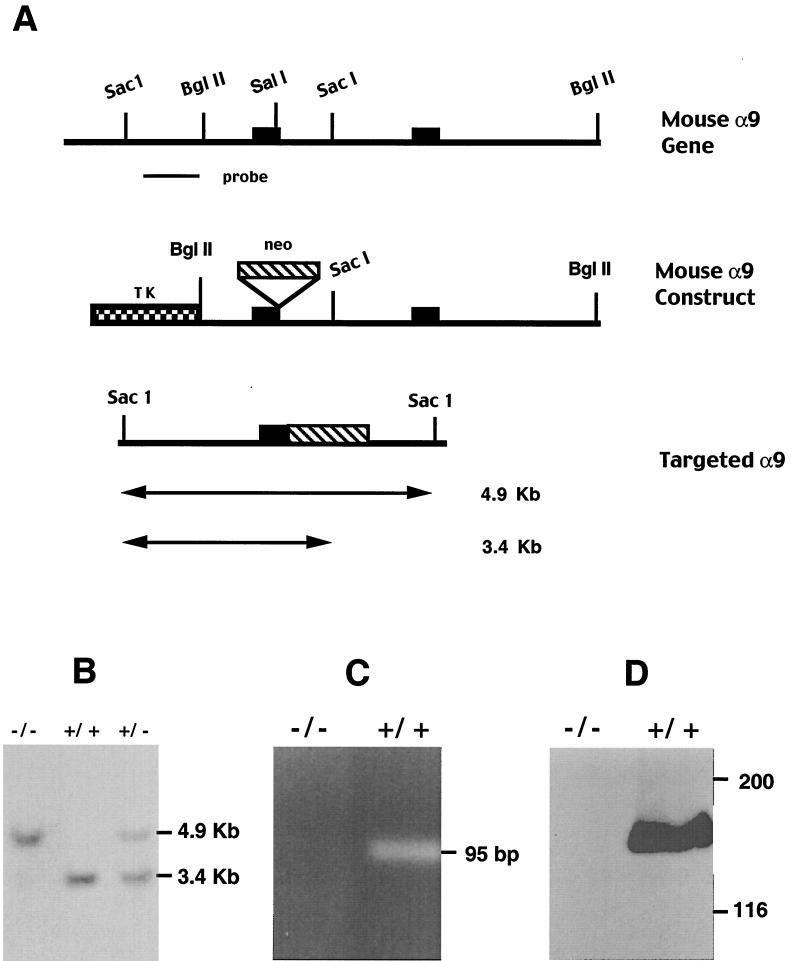
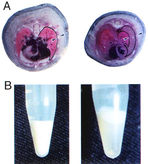
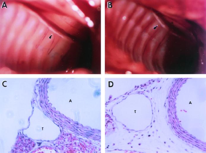
Members of the integrin family of adhesion receptors mediate both cell-cell and cell-matrix interactions and have been shown to play vital roles in embryonic development, wound healing, metastasis, and other biological processes. The integrin alpha9beta1 is a receptor for the extracellular matrix proteins osteopontin and tenacsin C and the cell surface immunoglobulin vascular cell adhesion molecule-1. This receptor is widely expressed in smooth muscle, hepatocytes, and some epithelia. To examine the in vivo function of alpha9beta1, we have generated mice lacking expression of the alpha9 subunit. Mice homozygous for a null mutation in the alpha9 subunit gene appear normal at birth but develop respiratory failure and die between 6 and 12 days of age. The respiratory failure is caused by an accumulation of large volumes of pleural fluid which is rich in triglyceride, cholesterol, and lymphocytes. alpha9(-/-) mice also develop edema and lymphocytic infiltration in the chest wall that appears to originate around lymphatics. alpha9 protein is transiently expressed in the developing thoracic duct at embryonic day 14, but expression is rapidly lost during later stages of development. Our results suggest that the alpha9 integrin is required for the normal development of the lymphatic system, including the thoracic duct, and that alpha9 deficiency could be one cause of congenital chylothorax.


Figures

FIG. 1
FIG. 1
Disruption of the mouse α9 gene by homologous recombination. (A) Structures of mouse α9 wild-type allele, targeting vector, and targeted allele. Two exons are shown as solid boxes. The expected fragment size after SacI digestion is 3.4 kb for the wild-type allele and 4.9 kb for the targeted allele. (B) Southern blot analysis of genomic DNA from mouse tail digested with SacI and hybridized with the external specific probe of mouse α9 indicated in panel A. (C) RT-PCR analysis of mRNA from α9+/+ and α9−/− mice. Total RNA was extracted from mouse liver and transcribed to complementary DNA (cDNA). A 95-bp fragment was amplified from α9+/+ mouse but not from α9−/− mouse using primers specific for wild-type α9 cDNA. (D) Western blotting of cell lysate of mouse liver with a polyclonal antiserum against α9. A band of the appropriate size to be α9 is demonstrated in α9+/+ but not in α9−/− mice. The positions of molecular mass markers (in kilodaltons) are shown to the right.
FIG. 2
FIG. 2
Appearance of pleural effusion in α9−/− mice. (A) Cross section through mouse thorax shows fluid filling an expanded pleural space (demarcated by arrowheads) in an α9−/− mouse compared to the absence of any identifiable space in an α9+/+ littermate. Arrowheads indicate locations of visceral pleura and the external surface of the ribs. (B) Fluid collected from the pleural space of α9−/− mice is milky prior to centrifugation (left) and can be separated into three layers by centrifugation (right): top, white lipid layer; middle, transparent aqueous layer; bottom, cell pellet, typical of chylous effusion.
FIG. 3
FIG. 3
Anatomy and histology of thoracic duct in α9−/− mouse and a wild-type littermate at 8 days of age. Presented are photographs of the opened thorax of α9+/+ (A) and α9−/− (B) mice, showing the presence of a visible thoracic duct in both groups (the white tubular structure along the spine denoted by arrowheads). (C and D) H&E-stained sections of the region including the thoracic duct (T) and the adjacent aorta (A) in α9+/+ (C) and α9−/− mice (D), demonstrating edema and extravascular lymphocytes surrounding the thoracic duct in α9−/− but not in α9+/+ mice.
FIG. 4
FIG. 4
Lymphocyte infiltration in α9 null mice. Presented are H&E-stained cross sections from the chest wall of α9−/− mice, showing mononuclear cell infiltration around lymphatic vessels adjacent to the parietal pleura (A and B), within skeletal muscle and interstitial tissue (C and D), and within the dermis (F) in the α9−/− mice. No such findings were ever observed in these locations in α9+/+ mice. One such example is shown for the dermis (E).
FIG. 5
FIG. 5
Transient α9 expression in the primordial thoracic duct and adjacent aorta of E14 mouse embryo. Frozen sections fixed with acetone were stained with affinity-purified anti-α9 polyclonal antiserum (brown staining). (A) Section of an α9+/+ E19 embryo (original magnification, ×200), demonstrating staining of the aorta but not of the adjacent thoracic duct (Td). (B) Section of an α9+/+ E14 embryo at the same magnification, demonstrating staining of both the aorta and the adjacent tissue that will ultimately form the thoracic duct. Airway smooth muscle (ASM) also demonstrates α9 immunoreactivity. (C) Enlargement of the same section shown in panel B.

References

    1. Bradley A. Teratocarcinomas and embryonic stem cells. In: Robertson E J, editor. Production and analysis of chimaeric mice. Oxford, United Kingdom: IRL Press; 1987. pp. 113–151.
    1. Erle D J, Sheppard D, Breuss J, Rüegg C, Pytela R. Novel integrin α and β subunit cDNAs identified using the polymerase chain reaction. Am J Respir Cell Mol Biol. 1991;5:170–177. - PubMed
    1. Fässler R, Meyer M. Consequences of lack of beta 1 integrin gene expression in mice. Genes Dev. 1995;9:1896–1908. - PubMed
    1. Forsberg E, Hirsch E, Fröhlich L, Meyer M, Ekblom P, Aszodi A, Werner S, Fässler R. Skin wounds and severed nerves heal normally in mice lacking tenascin-C. Proc Natl Acad Sci USA. 1996;93:6594–6599. - PMC - PubMed
    1. Fukamauchi F, Mataga N, Wang Y J, Sato S, Youshiki A, Kusakabe M. Abnormal behavior and neurotransmissions of tenascin gene knockout mouse. Biochem Biophys Res Commun. 1996;221:151–156. - PubMed

Publication types

LinkOut - more resources