Comparative Study
. 2000 Jul;182(14):3955-64.
doi: 10.1128/JB.182.14.3955-3964.2000.

A new two-component regulatory system involved in adhesion, autolysis, and extracellular proteolytic activity of Staphylococcus aureus

Affiliations
Comparative Study

A new two-component regulatory system involved in adhesion, autolysis, and extracellular proteolytic activity of Staphylococcus aureus

B Fournier et al. J Bacteriol. 2000 Jul.

Abstract

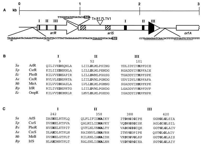
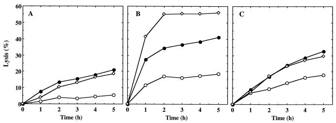
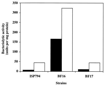
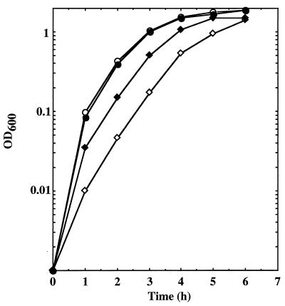
A transposition mutant of Staphylococcus aureus was selected from the parent strain MT23142, a derivative of strain 8325. The site of transposition was near the 5' terminus of the gene arlS. ArlS exhibits strong similarities with histidine protein kinases. Sequence analysis suggested that arlS forms an operon with upstream gene arlR. The predicted product of arlR is a member of the OmpR-PhoB family of response regulators. The arlS mutant formed a biofilm on a polystyrene surface unlike the parent strain and the complemented mutant. Biofilm formation was associated with increased primary adherence to polystyrene, whereas cellular adhesion was only slightly decreased. In addition, the arlS mutant exhibited increased autolysis and altered peptidoglycan hydrolase activity compared to the parental strain and to the complemented mutant. As it has been shown for coagulase-negative staphylococci that some autolysins are able to bind polymer surfaces, these data suggest that the two-component regulatory system ArlS-ArlR may control attachment to polymer surfaces by affecting secreted peptidoglycan hydrolase activity. Finally, the arlS mutant showed a dramatic decrease of extracellular proteolytic activity, including serine protease activity, in comparison to the wild-type strain and the complemented mutant, and cells grown in the presence of phenylmethylsulfonyl fluoride (a serine protease inhibitor) showed an increased autolysin activity. Since the locus arlR-arlS strikingly modifies extracellular proteolytic activity, this locus might also be involved in the virulence of S. aureus.


Figures

FIG. 1
FIG. 1
Schematic map of the arlR-arlS locus. (A) Diagram of the region of the BF15 chromosome including arlR, arlS, and flanking sequences. The site of the Tn917LTV1 insertion in strain BF15 is at position 1339. Arrows, direction of transcription for open reading frames arlR (positions 336 to 995) and arlS (positions 992 to 2347). In arlR, sequences encoding predicted receiver domains are indicated by shaded boxes (positions 363 to 639). In arlS, hatched boxes indicate two predicted hydrophobic membrane-spanning regions (positions 1028 to 1112 and 1442 to 1517) and the black areas represent predicted transmitter domains (positions 1718 to 2252). The −35 and −10 consensus sequences of the putative promoter are underlined, the ribosome-binding sites are double underlined, the start codons are boxed, and the transcription termination signal (positions 2505 to 2534) is indicated by dotted lines. (B) Amino acid sequence alignments of the putative ArlR with other response regulators presenting the strongest similarities with ArlR. Only regions of sequence similarity according to Stock et al. (50) are indicated. Boldface, highly conserved residues. Numbers refer to the amino acids of ArlR. Accession numbers and/or reference numbers for the protein sequences are as follows: CsrR, X98451, 26; PhoB, X04026, 29; CzcR, X98451, 56; MtrA, U01971, 57; IrlR, AF005358, 22; OmpR, 12. (C) Amino acid sequence alignments of the putative ArlS with other protein histidine kinases presenting the strongest similarities with ArlS. Only regions of sequence similarity according to Stock et al. (50) are indicated. Boldface, highly conserved residues. Numbers refer to the amino acids of ArlS. Accession numbers and reference numbers for the protein sequences are as follows: CsrS, AF082668, 26; PhoR, M23549, 47; CzcS, X98451, 56; MtrB, U14909, 57; IrlS, AF005358, 22. Abbreviations of organism names: Sa, S. aureus; Sp, Streptococcus pneumoniae; Ec, E. coli; Ae, Alcaligenes eutrophus; Mt, Mycobacterium tuberculosis; Bp, Burkholderia pseudomallei; Bs, B. subtilis.
FIG. 2
FIG. 2
Quantitative assay of biofilm formation in polystyrene microtiter plates. Lane 1, wild-type strain ISP794; lane 2, mutant BF16; lane 3, complemented mutant BF17.
FIG. 3
FIG. 3
SDS-PAGE analysis of extracellular proteins from different S. aureus strains. Samples were loaded as follows: lane 1, parent strain ISP794; lane 2, strain MT23142; lane 3, mutant BF16. Lane M, protein standard.
FIG. 4
FIG. 4
Autolysis of whole cells of S. aureus ISP794 (○), BF16 (●), and complemented mutant BF17 (◊) by Triton X-100. The results are expressed as lysis percentages as described in Materials and Methods.
FIG. 5
FIG. 5
Lytic activity of S. aureus culture supernatants at different growth phases: OD600, 0.5 (○); OD600, 0.9 (●); overnight (◊). The results are expressed as lysis percentages as described in Materials and Methods. (A) Parent strain ISP794. (B) Mutant BF16. (C) Complemented mutant BF17.
FIG. 6
FIG. 6
Effect of PMSF on the bacteriolytic activity of the culture supernatant. Cells were grown for 18 h in the absence (■) or in the presence (□) of PMSF. The culture supernatants were filtered and tested for bacteriolytic activities. All results shown are the means of at least two independent determinations.
FIG. 7
FIG. 7
Growth curve of the parent strain ISP794 and mutant BF16 in the presence or absence of NaCl. Results are for ISP794 with (●) or without (○) NaCl (50 mM) and for BF16 with (⧫) or without (◊) NaCl (50 mM). OD600 was measured every hour.

References

    1. Archibald A R, Hancock I C, Harwood C R. Cell wall structure, synthesis, and turnover. In: Sonenshein A L, Hoch J A, Losick R, editors. Bacillus subtilis and other gram-positive bacteria. Washington, D.C.: American Society for Microbiology; 1993. pp. 381–410.
    1. Arvidson S, Holme T, Lindholm B. Studies on extracellular proteolytic enzymes from Staphylococcus aureus. I. Purification and characterization of one neutral and one alkaline protease. Biochim Biophys Acta. 1973;302:135–148. - PubMed
    1. Brown N L, Barrett S R, Camakaris J, Lee B T O, Rouch D A. Molecular genetics and transport analysis of the copper-resistance determinant (pco) from Escherichia coli plasmid pRJ10004. Mol Microbiol. 1995;17:1153–1166. - PubMed
    1. Brunskill E W, Bayles K W. Identification and molecular characterization of a putative regulatory locus that affects autolysis in Staphylococcus aureus. J Bacteriol. 1996;178:611–618. - PMC - PubMed
    1. Brunskill E W, Bayles K W. Identification of LytSR-regulated genes from Staphylococcus aureus. J Bacteriol. 1996;178:5810–5812. - PMC - PubMed

Publication types

MeSH terms

Associated data

LinkOut - more resources