

Nonapoptotic neurodegeneration in a transgenic mouse model of Huntington's disease
- PMID: 10869421
- PMCID: PMC16675
- DOI: 10.1073/pnas.110078997
Nonapoptotic neurodegeneration in a transgenic mouse model of Huntington's disease
Abstract
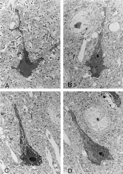
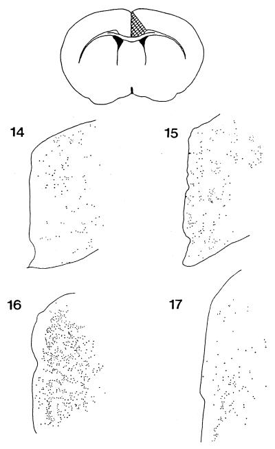
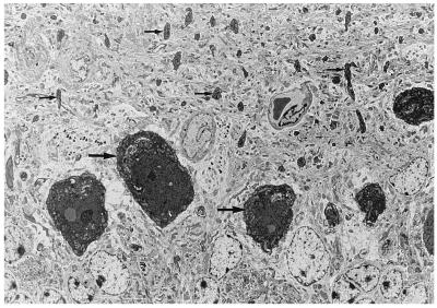
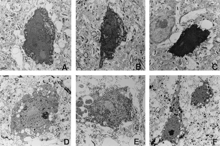
Huntington's disease (HD) is a fatal inherited neurodegenerative disorder characterized by personality changes, motor impairment, and subcortical dementia. HD is one of a number of diseases caused by expression of an expanded polyglutamine repeat. We have developed several lines of mice that are transgenic for exon 1 of the HD gene containing an expanded CAG sequence. These mice exhibit a defined neurological phenotype along with neuronal changes that are pathognomonic for the disease. We have previously observed the appearance of neuronal intranuclear inclusions, but did not find evidence for neurodegeneration. In this study, we report that all lines of these mice develop a late onset neurodegeneration within the anterior cingulate cortex, dorsal striatum, and of the Purkinje neurons of the cerebellum. Dying neurons characteristically exhibit neuronal intranuclear inclusions, condensation of both the cytoplasm and nucleus, and ruffling of the plasma membrane while maintaining ultrastructural preservation of cellular organelles. These cells do not develop blebbing of the nucleus or cytoplasm, apoptotic bodies, or fragmentation of DNA. Neuronal death occurs over a period of weeks not hours. We also find degenerating cells of similar appearance within these same regions in brains of patients who had died with HD. We therefore suggest that the mechanism of neuronal cell death in both HD and a transgenic mouse model of HD is neither by apoptosis nor by necrosis.
Figures
References
-
- Vonsattel J-P G, Meyers R H, Stevens T J, Ferrante R J, Bird E D, Richardson E P. J Neuropathol Exp Neurol. 1985;44:559–577. - PubMed
-
- Vonsattel J P G, DiFiglia M. J Neuropathol Exp Neurol. 1998;57:369–384. - PubMed
-
- Mangiarini L, Sathasivam K, Seller M, Cozens B A, Harper A, Hetherington C, Lawton M, Trottier Y, Lehrach H, Davies S W, et al. Cell. 1996;87:493–506. - PubMed
Publication types
MeSH terms
Substances
Grants and funding
LinkOut - more resources
Full Text Sources
Other Literature Sources
Medical
