. 2000 Jul 5;97(14):7829-34.
doi: 10.1073/pnas.130200197.

Estrogens suppress RANK ligand-induced osteoclast differentiation via a stromal cell independent mechanism involving c-Jun repression

Affiliations

Estrogens suppress RANK ligand-induced osteoclast differentiation via a stromal cell independent mechanism involving c-Jun repression

N K Shevde et al. Proc Natl Acad Sci U S A. .

Abstract

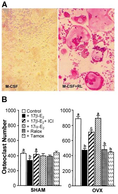
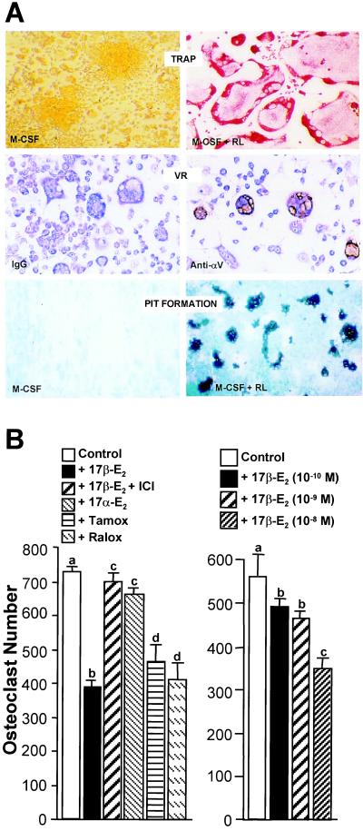
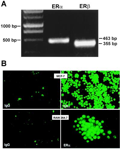
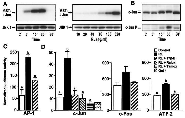
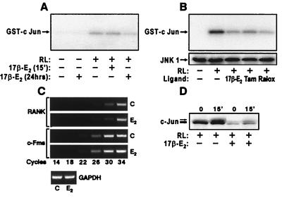
Loss of ovarian function following menopause results in a substantial increase in bone turnover and a critical imbalance between bone formation and resorption. This imbalance leads to a progressive loss of trabecular bone mass and eventually osteoporosis, in part the result of increased osteoclastogenesis. Enhanced formation of functional osteoclasts appears to be the result of increased elaboration by support cells of osteoclastogenic cytokines such as IL-1, tumor necrosis factor, and IL-6, all of which are negatively regulated by estrogens. We show here that estrogen can suppress receptor activator of NF-kappaB ligand (RANKL) and macrophage colony-stimulating factor (M-CSF)-induced differentiation of myelomonocytic precursors into multinucleated tartrate-resistant acid phosphatase-positive osteoclasts through an estrogen receptor-dependent mechanism that does not require mediation by stromal cells. This suppression is dose-dependent, isomer-specific, and reversed by ICI 182780. Furthermore, the bone-sparing analogues tamoxifen and raloxifene mimic estrogen's effects. Estrogen blocks RANKL/M-CSF-induced activator protein-1-dependent transcription, likely through direct regulation of c-Jun activity. This effect is the result of a classical nuclear activity by estrogen receptor to regulate both c-Jun expression and its phosphorylation by c-Jun N-terminal kinase. Our results suggest that estrogen modulates osteoclast formation both by down-regulating the expression of osteoclastogenic cytokines from supportive cells and by directly suppressing RANKL-induced osteoclast differentiation.


Figures

Figure 1
Figure 1
Estrogens suppress M-CSF/RANKL-induced osteoclast formation in primary murine myeloid cells. Nonadherent myeloid progenitors and monocytes were isolated from sham and ovariectomized mice and plated in triplicate at 105 cells/well. (A) Murine monocytes were treated with M-CSF (10 ng/ml) or M-CSF plus RANKL (30 ng/ml) for 10 days, stained for TRAP, and photographed at ×20. (B) Cells from sham (Left) or OVX (Right) mice were treated with M-CSF/RANKL in the presence of vehicle, 17β-estradiol (10−8 M), 17β-estradiol (10−8 M) plus ICI 182780 (10−6 M), 17α-estradiol (10−8 M), 4-hydroxytamoxifen (10−7 M), or raloxifene (10−7 M). Multinucleated (>3 nuclei), TRAP-positive cells were quantitated 10 days later. Numbers represent the mean ± SE, n = 3 (a is significant vs. b and c, and b is significant vs. c at P ≤ 0.05).
Figure 2
Figure 2
Estrogens suppress M-CSF/RANKL-induced osteoclast formation in murine RAW264.7 cells. (A) Properties of osteoclast-like cells generated from RAW264.7 cells. Cells were induced with M-CSF or M-CSF/RANKL for 5 days and then TRAP-stained and photographed at ×20. Cells were incubated with either control IgG or Ab to vitronectin receptor (VR) αV subunit, stained with immunoperoxidase and methylene blue, and then photographed at ×10. Cells were plated on synthetic bone discs and induced with either M-CSF or M-CSF/RANKL for 10 days. Cells were removed and resorption lacunae visualized and photographed under dark-field microscopy at ×10. (B) Cells (2 × 10³/well) were plated in triplicate and multinucleated (>3 nuclei), TRAP-positive osteoclasts quantitated following induction for 5 days with M-CSF/RANKL in the presence of vehicle, 17β-estradiol (10−8 M), 17β-estradiol (10−8 M) plus ICI 182780 (10−6 M), 17α-estradiol (10−8 M), 4-hydroxytamoxifen (10−7 M) or raloxifene (10−7 M) (Left) or increasing concentrations of 17β-estradiol as indicated (Right). Numbers represent the mean ± SE, n = 3 (a is significant vs. b and d; c is significant vs. d at P ≤ 0.05).
Figure 3
Figure 3
Detection of ER in RAW264.7 cells. (A) Expression of ERα and ERβ mRNA transcripts using RT-PCR analysis. (B) Immunocytochemical detection of ERα in human MCF-7 breast cancer cells and in RAW264.7 cells. Cells were fixed and probed with either nonspecific IgG or anti-ERα Ab as indicated. MCF-7 and RAW264.7 cells were photographed at ×20 and ×40, respectively.
Figure 4
Figure 4
Estrogen suppresses RANKL-induced activation of c-Jun and AP-1-mediated transcription in RAW264.7 cells. (A) Cells were treated for the indicated times with RANKL (80 ng/ml) or with the indicated concentrations of RANKL (RL) for 15 min and lysates examined for both JNK1 protein by Western blot and kinase activity (using GST-cJun as substrate). (B) Cells were treated for the indicated times with RANKL (80 ng/ml) and then subjected to Western blot analysis by using antibodies to c-Jun or phospho-c-Jun. Arrows indicate c-Jun and phospho-c-Jun forms. (C) Cells were transfected with a luciferase reporter plasmid (p36) containing three copies of an AP-1 response element. Cells were treated with vehicle or M-CSF and RANKL in the absence or presence of 17β-estradiol (10−8 M). Cells were harvested 24 h later and lysates assessed for luciferase and β-galactosidase activities and protein. Numbers represent the mean ± SE, n = 3 (a is significant vs. b and c; and b is significant vs. c at P ≤ 0.05). The results are representative of three independent experiments. (D) RAW264.7 cells were cotransfected with pFC2-luc and one of the following plasmids: pFA2-cJun (c-Jun), pFA-cFos (c-Fos), pFA-ATF2 (ATF2), or pFC-dbd (Gal4). Following transfection, cells were treated with RANKL in the presence of vehicle, 17β-estradiol (10−8 M), 4-hydroxytamoxifen (10−7 M), or raloxifene (10−7 M). Cells were harvested 24 h later and lysates assessed for luciferase activity, β-galactosidase activity, and protein. Numbers represent the mean ± SE, n = 3 (b is significant vs a and c at P ≤ 0.05). The results are representative of at least three independent experiments.
Figure 5
Figure 5
Estrogen suppresses JNK1 activity but does not alter RANK and c-Fms expression in RAW 264.7 cells. (A) Cells were pretreated with vehicle or 17β-estradiol (10−8 M) (15 min or 24 h) and then stimulated for 5 min with RANKL (80 ng/ml). Cell lysates were examined for JNK1 activity. RANKL-induced JNK1 activity was reduced 0 and 30% following a 15-min and 24-h treatment with estrogen, respectively. (B) Cells were treated for 24 h with vehicle, 17β-estradiol (10−8 M), 4-hydroxytamoxifen (10−7 M), or raloxifene (10−7 M) and then stimulated for 5 min with RANKL (80 ng/ml). Extracts were examined for JNK1 activity and by Western blot analysis. (C) RNA was isolated from cells treated for 24 h with either vehicle or 17β-estradiol (10−8 M) and subjected to RT-PCR by using oligonucleotide primer pairs for murine RANK, c-Fms, and glyceraldehyde-3-phosphate dehydrogenase (GAPDH). The cycle-dependent appearance of specific DNA products was assessed by agarose gel electrophoresis. d, Cells were treated for 24 h with either vehicle or 17β-estradiol (10−8 M), and stimulated for 15 min with RANKL and nuclear extracts subjected to Western blot analysis by using Abs to c-Jun.

References

    1. Manolagas S C, Jilka R L. N Engl J Med. 1995;332:305–311. - PubMed
    1. Pacifici R. J Bone Miner Res. 1996;8:1043–1051. - PubMed
    1. Turner R T, Riggs B L, Spelsberg T C. Endocr Rev. 1994;15:275–300. - PubMed
    1. Roodman G D. Exp Hematol. 1999;27:1229–1241. - PubMed
    1. Jilka R L, Hangoc G, Girosole G, Passeri G, Williams D C, Abrams J S, Boyce B, Broxmeyer H, Manolagas S C. Science. 1992;257:88–91. - PubMed

Publication types

MeSH terms