. 2000 Jul 5;97(14):7835-40.
doi: 10.1073/pnas.140199597.

Regulation of histone deacetylase 4 and 5 and transcriptional activity by 14-3-3-dependent cellular localization

Affiliations

Regulation of histone deacetylase 4 and 5 and transcriptional activity by 14-3-3-dependent cellular localization

C M Grozinger et al. Proc Natl Acad Sci U S A. .

Abstract

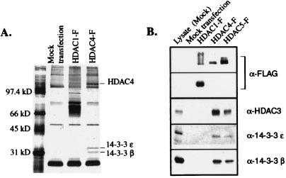
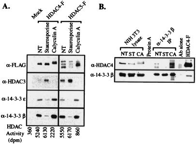
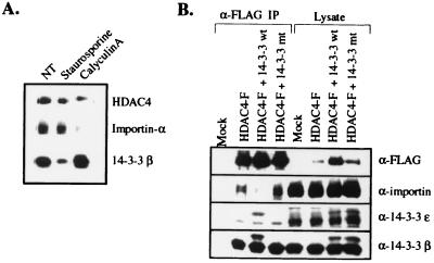
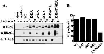
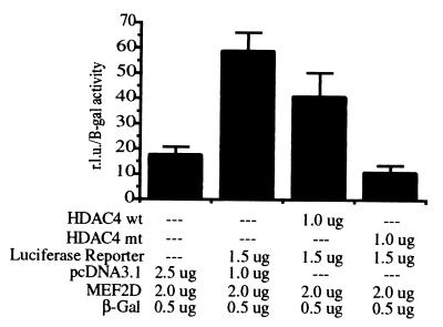
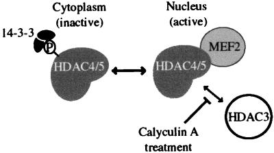
Transcription is controlled in part by the dynamic acetylation and deacetylation of histone proteins. The latter process is mediated by histone deacetylases (HDACs). Previous analysis of the regulation of HDAC activity in transcription has focused primarily on the recruitment of HDAC proteins to specific promoters or chromosomal domains by association with DNA-binding proteins. To characterize the cellular function of the recently identified HDAC4 and HDAC5 proteins, complexes were isolated by immunoprecipitation. Both HDACs were found to interact with14-3-3 proteins at three phosphorylation sites. The association of 14-3-3 with HDAC4 and HDAC5 results in the sequestration of these proteins in the cytoplasm. Loss of this interaction allows HDAC4 and HDAC5 to translocate to the nucleus, interact with HDAC3, and repress gene expression. Regulation of the cellular localization of HDAC4 and HDAC5 by 14-3-3 represents a mechanism for controlling the transcriptional activity of these class II HDAC proteins.


Figures

Figure 1
Figure 1
Association of HDAC4 and HDAC5 with two isoforms of 14-3-3. (A) Recombinant, FLAG-tagged HDAC1 and HDAC4 were transiently expressed in TAg Jurkat cells and were immunoprecipitated by using α-FLAG agarose (Sigma). The immunopurified complexes were separated by SDS/PAGE, and the proteins were visualized by silver stain. (B) The association between HDAC4 and HDAC5 with 14-3-3 was confirmed by Western blot analysis. The recombinant FLAG-tagged proteins were subjected to Western blot analysis using 14-3-3 isoform specific antibodies.
Figure 2
Figure 2
Nuclear-cytoplasmic shuttling of HDAC4 and HDAC5 is correlated to 14-3-3 expression levels. (A) Recombinant HDAC4-EGFP and HDAC5-EGFP were transiently expressed in U2OS cells, and the localization of the protein was observed by fluorescence microscopy. (B) Overexpression of 14-3-3 β causes an increased cytoplasmic localization of HDAC4-EGPF. U2OS cells were transiently transfected with HDAC4-EGFP and either a control plasmid (pcDNA3.1, Invitrogen) or myc-tagged 14-3-3 β. The localization of recombinant HDAC4 and 14-3-3 was analyzed by immunofluorescence.
Figure 3
Figure 3
Phosphorylation-dependent binding of 14-3-3 to HDAC4 and HDAC5. (A) Association of HDAC4 and HDAC5 with 14-3-3 and HDAC3 depends on the phosphorylation state of the proteins. HDAC4-FLAG and HDAC5-FLAG were transiently expressed in TAg Jurkat cells. Forty-eight hours post-transfection, the cells were treated for 1.5 h with staurosporine and calyculin A. The immunopurified complexes were subjected to Western blot analysis and were tested for HDAC activity, as described in Materials and Methods. (B) Association of endogenous HDAC4 with 14-3-3 β in NIH 3T3 cells is phosphorylation-dependent. 14-3-3 β immunoprecipitates from NIH 3T3 cells that had been treated with staurosporine or calyculin A for 1.5 h were analyzed for the presence of endogenous HDAC4 by Western blotting.
Figure 4
Figure 4
Association of 14-3-3 with HDAC4 prevents the binding of importin α. (A) The binding of 14-3-3 to HDAC4 prevents interaction with importin α. Forty-eight hours after transfection with HDAC4-FLAG, TAg Jurkat cells were treated with staurosporine or calyculin A for 1.5 h. HDAC4-FLAG was immunopurified and subjected to Western blot analysis with α-importin α antibodies. (B) Overexpression of 14-3-3 blocks binding of importin α to HDAC4. TAg Jurkat cells were transfected with 1 μg of HDAC4-FLAG and 2 μg each of myc-tagged wild-type or mutant 14-3-3 β and ɛ. pcDNA3.1 (Invitrogen) was used to normalize the amount of DNA transfected. Forty-eight hours post-transfection, HDAC4-FLAG was immunoprecipitated and analyzed for binding to importin α by Western blotting.
Figure 5
Figure 5
Mutation of 14-3-3 binding sites in HDAC4 causes loss of 14-3-3 binding and increased nuclear localization. (A) Single, double, and triple serine to alanine mutations in the three putative 14-3-3 binding sites of HDAC4 were generated. The recombinant proteins were transiently expressed in TAg Jurkat cells, which were treated with calyculin A for 1.5 h before harvesting. The immunopurified complexes were subjected to Western blot analysis. (B) The single and triple 14-3-3 binding site mutants were fused to EGFP and transiently expressed in U2OS cells. The cellular localization was monitored by fluorescence microscopy. These experiments were repeated multiple times with similar results.
Figure 6
Figure 6
Localization-independent loss of HDAC4 and HDAC3 interaction on calyculin A-treatment. Forty-eight hours after transfection with HDAC4-FLAG, TAg Jurkat cells were treated with staurosporine or calyculin A for 1.5 h. HDAC4-FLAG was immunopurified, and the sample was split into thirds. One-third of the immunopurified protein was prepared for Western blot analysis, one-third was incubated for 1 h with untreated TAg Jurkat lysate, and the remaining one-third of the sample was incubated with calyculin A-treated TAg Jurkat lysate for 1 h. These samples were analyzed for 14-3-3 and HDAC3 binding by Western blotting.
Figure 7
Figure 7
Increased nuclear localization of HDAC4 enhances MEF2-dependent transcriptional repression. TAg-Jurkat cells were transfected with MEF2D, a MEF2-luciferase reporter construct, a constitutive β-gal expression construct, and either wild-type HDAC4 or HDAC4 with mutations in all three 14-3-3 binding sites (HDAC4 S246/466/632A). pcDNA3.1 (Invitrogen) was used to normalize the amount of DNA transfected. Thirty-eight hours after transfection, the samples were harvested and divided to perform the subsequent assays in triplicate. The amount of luciferase activity was measured (Promega) and divided by the amount of β-gal activity present to normalize for protein expression levels.
Figure 8
Figure 8
Model for regulation of HDAC4 activity by cellular localization. Binding of phosphorylated HDAC4 or HDAC5 to 14-3-3 sequesters these proteins in the cytoplasm, where they cannot function in repression of transcription. Dephosphorylation allows HDAC4 and HDAC5 to shuttle to the nucleus, where they associate with HDAC3 and the MEF2 transcription factor, and silence transcription of MEF2-dependent genes. Treatment of cells with the phosphatase inhibitor calyculin A also disrupts the interaction of HDAC3 with HDAC4 and HDAC5 by an unknown mechanism, possibly involving additional protein factors.

References

    1. Ayer D E. Trends Cell Biol. 1999;9:193–198. - PubMed
    1. Rundlett S E, Carmen A A, Kobayashi R, Bavykin S, Turner B M, Grunstein M. Proc Natl Acad Sci USA. 1996;93:14503–14508. - PMC - PubMed
    1. Dangond F, Hafler D A, Tong J K, Randall J, Kojima R, Utku N, Gullans S R. Biochem Biophys Res Commun. 1998;242:648–652. - PubMed
    1. Emiliani S, Fischle W, Van Lint C, Al-Abed Y, Verdin E. Proc Natl Acad Sci USA. 1998;95:2795–2800. - PMC - PubMed
    1. Fischle W, Emiliani S, Hendzel M J, Nagase T, Nomura N, Voelter W, Verdin E. J Biol Chem. 1999;274:11713–11720. - PubMed

Publication types

MeSH terms