

Angiographic and clinical characteristics of patients with cerebral arteriovenous malformations associated with hereditary hemorrhagic telangiectasia
- PMID: 10871005
- PMCID: PMC7973909
Angiographic and clinical characteristics of patients with cerebral arteriovenous malformations associated with hereditary hemorrhagic telangiectasia
Erratum in
- AJNR Am J Neuroradiol 2001 Aug;22(7):1446. Manzia JL [corrected to Mandzia JL]
Abstract
Background and purpose: Cerebral arteriovenous malformations (AVMs) are occasionally associated with hereditary hemorrhagic telangiectasia (HHT), which is characterized by the presence of multiple mucocutaneous telangiectasia, epistaxis, and familial inheritance. We analyzed the angiographic and clinical characteristics of patients with cerebral AVMs related to HHT.
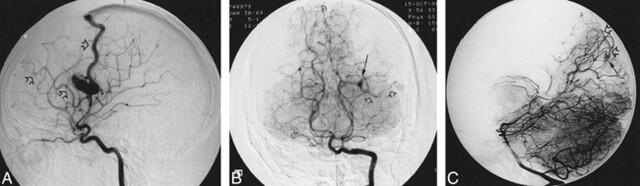
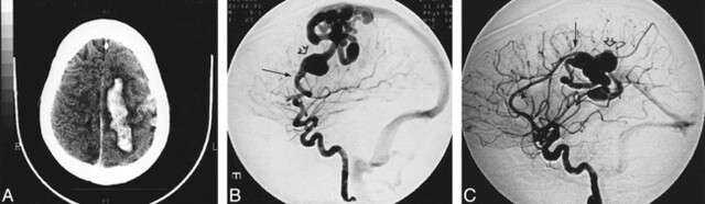
Methods: Among 638 patients with cerebral AVMs, we identified 14 patients with HHT. The AVMs were classified as those with nidi of 1 cm or less (micro AVMs), those with nidi between 1 and 3 cm (small AVMs), and those of the fistulous type (arteriovenous fistulas [AVFs]).
Results: A total of 28 AVMs were found; seven of 14 patients had multiple AVMs. The 28 AVMs were categorized as 12 micro AVMs, eight small AVMs, and eight AVFs. All except one micro AVM were asymptomatic, whereas all small AVMs were symptomatic. Three of eight AVFs were asymptomatic. All 28 AVMs were located on the cortex. All micro AVMs and AVFs had single feeders and single draining veins, whereas the small AVMs had multiple feeders in all lesions and single draining veins in six of eight lesions.
Conclusion: Multiple, cortical, micro AVMs or AVFs harboring single feeding arteries and single draining veins should raise clinical suspicion of HHT-related AVMs.
Figures
References
-
- Guttmacher AE, Marchuk DA, White RIJ. Hereditary hemorrhagic telangiectasia. N Engl J Med 1995;333:918-924 - PubMed
-
- McAllister KA, Grogg KM, Johnson DW, et al. Endoglin, a TGF-B binding protein of endothelial cells, is the gene for hereditary hemorrhagic telangiectasia type I. Nat Genet 1994;8:345-351 - PubMed
-
- Vincent P, Plauchu H, Hazan J, et al. A third locus for hereditary hemorrhagic telangiectasia maps to 12q. Hum Mol Genet 1995;4:945-949 - PubMed
-
- Johnson DW, Berg JN, Gallione CJ, et al. A second locus for hereditary hemorrhagic telangiectasia mapped to chromosome 12. Genome Res 1995;5:21-28 - PubMed
-
- Willinsky RA, Lasjaunias P, Ter Brugge K, Burrows P. Multiple cerebral arteriovenous malformations (AVMs): review of our experience from 203 patients with cerebral vascular lesions. Neuroradiology 1990;32:207-210 - PubMed
MeSH terms
LinkOut - more resources
Full Text Sources