

MR venography of multiple sclerosis
- PMID: 10871010
- PMCID: PMC7973892
MR venography of multiple sclerosis
Abstract
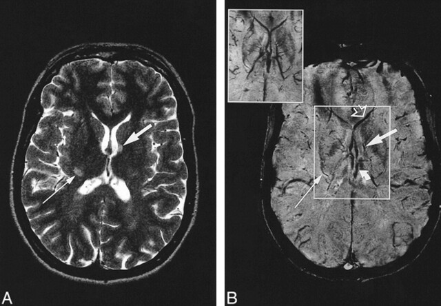
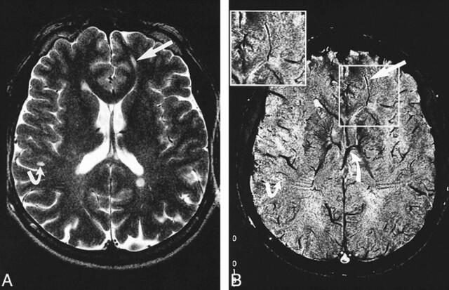
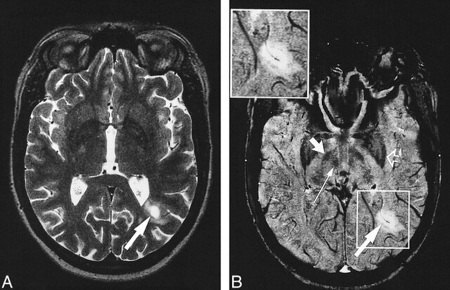
Background and purpose: The distribution of multiple sclerosis (MS) lesions in the brain follows a specific pattern, with most lesions in the periventricular regions and in the deep white matter; histopathologic studies have shown a perivenous distribution. The aim of this study was to illustrate these distribution patterns in vivo using high-resolution MR venography.
Methods: Seventeen MS patients underwent MR imaging at 1.5 T. Venographic studies were obtained with a 3D gradient-echo technique. MS lesions were identified on T2-weighted images, and their shape, orientation, and location were compared with the venous anatomy on the venograms.
Results: The use of contrast material facilitated the visualization of small veins and increased the number of veins seen. A total of 95 MS lesions could be identified on both the T2-weighted series and the venograms; a central vein was visible in all 43 periventricular lesions and in all but one of the 52 focal deep white matter lesions. The typical ovoid shape and orientation of the long axis of the MS lesions correlated well with the course of these veins.
Conclusion: With MR venography, the perivenous distribution of MS lesions in the brain can be visualized in vivo. The venous anatomy defines the typical form and orientation of these lesions.
Figures
References
-
- Miller DH, Grossman RI, Reingold SC, McFarland HF. The role of magnetic resonance techniques in understanding and managing multiple sclerosis. Brain 1998;121:3-24 - PubMed
-
- Fog T. The topography of plaques in multiple sclerosis. Acta Neurol Scand 1965;15:1-161 - PubMed
-
- Raine C. The neuropathology of multiple sclerosis. In: Raine CS, McFarland HF, Tourtelotte WW, eds. Multiple Sclerosis: Clinical and Pathogenetic Basis. London: Chapman and Hall 1997 151-172
-
- Reichenbach JR, Venkatesan R, Schillinger DJ, Kido DK, Haacke EM. Small vessels in the human brain: MR venography with deoxyhemoglobin as an intrinsic contrast agent. Radiology 1997;204:272-277 - PubMed
Publication types
MeSH terms
Substances
LinkOut - more resources
Full Text Sources
Other Literature Sources
Medical