Review
. 2000 Jun-Jul;21(6):1099-109.

Concepts of myelin and myelination in neuroradiology

Affiliations
Review

Concepts of myelin and myelination in neuroradiology

A J Barkovich. AJNR Am J Neuroradiol. 2000 Jun-Jul.

Abstract

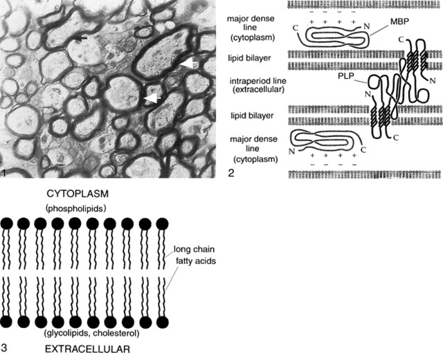
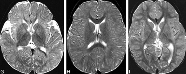
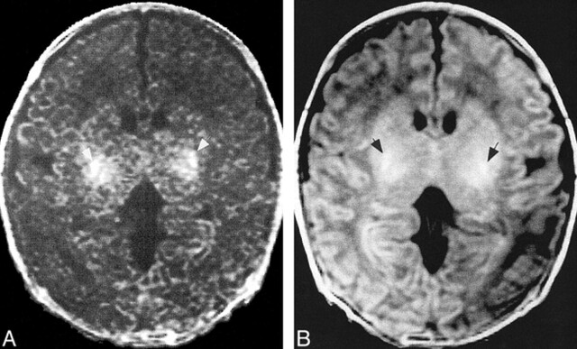
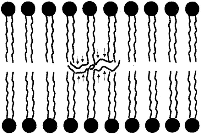
Until the advent of MR imaging, knowledge of the structure of myelin and the process of myelination were of little importance to the neuroradiologist. Other than some mild changes in the attenuation of white matter, myelination resulted in no significant alterations of CT (1) or sonographic studies. MR studies, on the other hand, have been increasingly used for pediatric brain imaging. MR imaging's greater sensitivity to small changes in the water content of brain tissue, to changes in the binding of free water (revealed by magnetization transfer), and to the extent and anisotropy of water diffusion (revealed by diffusion imaging) has cast new light on this very complex and important molecule. Assessing myelination has become a key component of evaluating the child with delayed development. Moreover, better understanding of the nature of myelin and the effect of its different components on MR imaging parameters may help us to understand and diagnose inborn errors of metabolism better. In this review, I discuss what is known regarding the function and structure of CNS myelin and the effects of the various components of myelin on the signal imparted to the MR image. Summary Abnormalities of myelin can cause a wide variety of disorders of the nervous system. MR imaging is a powerful tool for the study of myelin and its disorders. However, only by understanding the physiologic properties and structure of myelin can we use MR imaging to its fullest capacity for studying patients with myelin disorders. In this review, I have discussed the structure of myelin as it relates to MR imaging of normal myelination and to neurologic disorders resulting from abnormalities of myelin. Thinking of myelin and its disorders in this manner will be critical to using MR imaging techniques optimally to diagnose and study these disorders further.


Figures

<sc>fig</sc> 1.
fig 1.
Electron micrograph of myelinated axons in the optic nerve of a 3-week-old rat. Myelin wraps around the axon in multiple spirals to speed transmission of action potentials. Arrows point to the thickest spirals, which are around the largest axons.fig 2. Schematic of the structure of myelin. Myelin is composed of multiple layers having a protein-lipid-protein-lipid-protein structure. The lipid layers are in the cell membrane, composed of a bimolecular layer of hydrocarbon chains, cholesterol, phospholipids, and glycolipids. A highly electron-dense line, the major period line, contains MBP, an intracellular protein attached to the inner surface of the cell membrane and situated mainly in the cytoplasm. A less electron-dense protein line, the intraperiod line, represents PLP in the outer portion of the cell membrane and in the extracellular space. PLP interacts homophilically with similar PLP chains from the surface of the myelin membrane in the next loop of the spiral. In addition, the lipophilic amino acid tryptophan (not shown) is present on the outer surface edge of the PLP and may interact with galactocerebrosides in the outer lipid membrane of the adjacent myelin spiral. MBP forms dimers (chemical bonds with other MBPs) within the cytoplasm of the myelin sheath. MBP is thought to stabilize the myelin spiral at the major dense line by interacting with negatively charged lipids at the cytoplasmic surface of the lipid membrane.fig 3. Close-up schematic of myelin lipid bilayer. The lipid layer of myelin is composed of cholesterol, phospholipid, and glycolipid in an approximately 4:3:2 ratio for adult CNS myelin. It appears that most of the glycolipid (in the form of galactocerebroside and sulfatide) and cholesterol are in the outer layer of the membrane, exposed to the extracellular space. In contrast, the phospholipids, of which ethanolamine-containing plasmalogen is the most abundant type, are hydrophobic and are located exclusively in the inner (cytoplasm) side of the membrane. The area between the outer and inner membrane layers is composed primarily of hydrocarbon chains (long chain fatty acids)
<sc>fig</sc> 4.
fig 4.
MR changes of myelination at the level of the basal ganglia. A and B, Spin-echo (600/11 [TR/TE]) image (A) of a neonate shows that myelination (white matter with short T1 [hyperintensity on T1-weighted images] and short T2 [hypointensity on T2-weighted images]) is limited to the posterior limb of the internal capsule at this level. Spin-echo (3000/120) image (B) of the same patient shown in panel A. C and D, Spin-echo (600/11) image (C) of a 5-month-old patient shows hyperintensity in the entire internal capsule, optic radiations, and splenium of the corpus callosum. Spin-echo (3000/120) image (D) of the same patient shown in panel C shows hypointensity limited to the posterior limb of the internal capsule and a portion of the optic radiations. E and F, Spin-echo (600/11) image (E) of an 8-month-old patient shows hyperintensity in all white matter except the immediate subcortical regions. Spin-echo (3000/120) image (F) of the same patient shown in panel E shows hypointensity in the entire corpus callosum, the entire posterior limb of the internal capsule, and part of the anterior limb of the internal capsule. G, Spin-echo (2500/80) image of a 12-month-old patient shows hypointensity in the entire internal capsule, in the subcortical white matter of the motor cortex, and in the subcortical white matter of the visual cortex. H, Spin-echo (2500/80) image of an 18-month-old patient shows hypointensity in most of the deep white matter but lack of maturity of subcortical white matter. I, Image of a 24-month-old patient shows that essentially all white matter is hypointense.
<sc>fig</sc> 5.
fig 5.
Images of a neonate. A, Magnetization transfer image shows that magnetization transfer (white arrows) in the neonate develops in almost precisely the same locations as does the T1 shortening. B, T1-weighted image shows T1 shortening (black arrows) attributable to myelination.
<sc>fig</sc> 6.
fig 6.
Schematic of postulated membrane destabilization by very long chain fatty acids in ALD. As a result of the inability to import very long chain fatty acids into the peroxisome, they are not broken down into long chain fatty acids. When the very long chain fatty acids (arrows) are incorporated into the bilaminar membrane of myelin, the thermodynamic stability of the membrane is lessened, theoretically making myelin easier to break down.
<sc>fig</sc> 7.
fig 7.
ALD in an 8-year-old patient. Axial-view spin-echo (2500/30) image shows T2 prolongation in the callosal splenium and forceps major, regions where high concentrations of ALD protein are expressed.fig 8. MLD in a 2-year-old patient. Axial-view spin-echo (2500/80) image shows T2 prolongation, representing loss of myelin, in the central hemispheric white matter. The subcortical white matter is relatively spared. The radial stripes extending through the hemispheres are characteristic of the MR imaging appearance in this disorder.fig 9. Pelizaeus-Merzbacher disease in a 5-year-old patient. Axial-view spin-echo (600/15) image shows a paucity of myelin, with hyperintensity only in the internal capsules and optic radiations (compare with fig 4).fig 10. MS. Parasagittal fast spin-echo (3500/102) image shows differing degrees of hyperintensity in the large tumefactive plaque in the posterior frontal lobe, possibly representing different degrees of myelin and axonal destruction.

References

    1. Brant-Zawadzki M, Enzmann DR. Using computed tomography of the brain to correlate low white matter attenuation with early gestational age in neonates. Radiology 1981;139:105-108 - PubMed
    1. Morell P, Quarles RH, Norton WT. Myelin formation, structure, and biochemistry. In: Siegel GJ, ed. Basic Neurochemistry: Molecular, Cellular, and Medical Aspects. 5th ed. New York: Raven Press 1994 117-143
    1. Brady ST, Witt AS, Kirkpatrick LL, et al. Formation of compact myelin is required for maturation of the axonal cytoskeleton. J Neurosci 1999;19:7278-7288 - PMC - PubMed
    1. Kirchner DA, Blaurock AE. Organization, phylogenetic variations and dynamic transitions of myelin. In: Martenson RE, ed. Myelin: Biology and Chemistry. Boca Raton: CRC 1991 413-448
    1. Kirschner DA, Ganser AL. Myelin labeled with mercuric chloride: asymmetric localization of phosphatidylethanolamine plasmalogen. J Mol Biol 1982;157:635-658 - PubMed

MeSH terms

LinkOut - more resources