. 2000 Jun-Jul;21(6):1110-5.

Cranial MR imaging of osteopetrosis

Affiliations

Cranial MR imaging of osteopetrosis

J K Curé et al. AJNR Am J Neuroradiol. 2000 Jun-Jul.

Abstract

Background and purpose: The purpose of this study was to describe the cranial MR imaging manifestations of osteopetrosis. These features have not previously been reported in the literature.

Methods: Cranial MR studies, obtained with a uniform imaging protocol, were reviewed in 47 patients with osteopetrosis. Thirty-four patients had autosomal recessive (malignant) osteopetrosis (AROP), seven had intermediate osteopetrosis (IOP), and six had either type I or type II autosomal dominant osteopetrosis (ADOP I or II). The prevalence of abnormalities was tabulated and compared with the specific osteopetrosis variants.

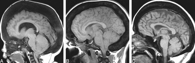
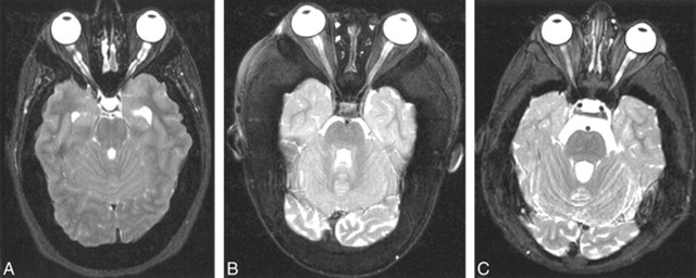
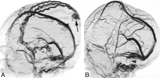
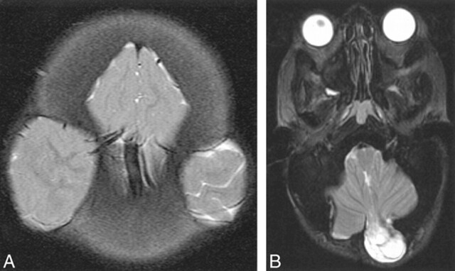
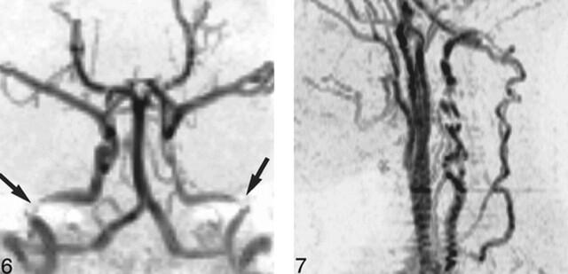
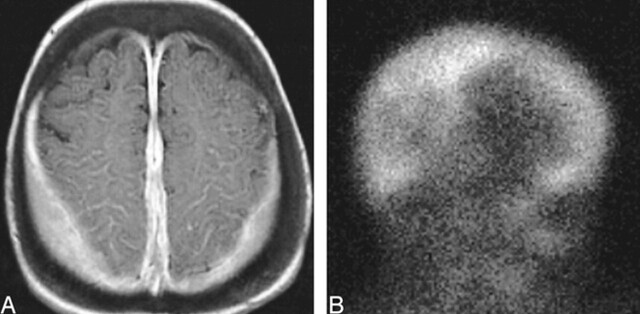
Results: All patients with osteopetrosis had thickening and sclerosis of the calvaria. Ventriculomegaly, tonsillar herniation, proptosis, and dural venous sinus stenosis were observed in the majority of patients with AROP and ADOP I. Optic nerve sheath dilatation occurred in many of the patients with AROP and in all patients with ADOP I. Acquired cephaloceles were also observed only in these two groups. Optic nerve atrophy and optic canal stenosis were observed in a majority of patients with AROP, IOP, and ADOP II. Middle ear fluid was prevalent in AROP and IOP, present in over half the patients in each group. Features seen most prevalently, or exclusively, in AROP included stenosis of the internal carotid and vertebral arteries and extramedullary hematopoiesis.

Conclusion: The cranial MR imaging features of osteopetrosis are both shared and unique among the various subtypes of the disease. The specific cranial and intracranial manifestations reflect the predominant calvarial or skull base patterns of bone thickening. The unique features seen in patients with AROP probably reflect the early age of onset and the greater severity of this form of the disease.


Figures

<sc>fig</sc> 1.
fig 1.
Calvarial thickening. A, AROP. Sagittal T1-weighted MR image shows thickening of the calvaria and facial bones. Note hypointensity of skull and cervical vertebra, and cerebellar tonsillar ectopia. B, ADOP I. Sagittal T1-weighted image shows calvarial thickening and hypointensity, as well as tonsillar ectopia. C, ADOP II. Sagittal T1-weighted image shows only a moderately thickened calvaria; however, the skull base is more significantly thickened than in the example of ADOP I (B). Compare the small sellar volume with that in ADOP I (B).
<sc>fig</sc> 2.
fig 2.
Optic nerve/sheath manifestations. A, Prominent optic nerve sheath dilatation, ADOP I. Axial T2-weighted FSE image shows scalloped inner table. The optic canals are not stenotic. B, AROP. Axial T2-weighted FSE image shows optic canal stenosis and optic nerve atrophy. C, IOP. Axial T2-weighted FSE image shows optic canal stenosis and optic nerve atrophy, with more severe atrophy evident on the right in this case.
<sc>fig</sc> 3.
fig 3.
Dural venous sinus stenosis at 2D-TOF MR venography in two patients with AROP. A, In one patient, lateral projection shows occlusion or high-grade stenosis of both sigmoid sinuses/jugular bulbs. Note prominent scalp collateral venous flow and sinus pericranii (arrow). B, In another patient (same child as in fig 4A), lateral projection shows bilateral sigmoid sinus stenosis or occlusion (arrowhead). Note superior displacement of superior sagittal sinus by protruding brain.
<sc>fig</sc> 4.
fig 4.
Cephaloceles, nonsurgical. A, AROP. Sagittal T1-weighted image shows brain protruding upward into the anterior fontanel as a result of limited intracranial volume. The superior sagittal sinus is superiorly displaced (See fig 3B). Note tonsillar ectopia. B, AROP. Axial T2-weighted FSE image shows bilateral meningoceles at the craniocervical junction (arrows). C, ADOP I. Sagittal T1-weighted image shows brain protruding downward through the foramen ovale (arrow). Note scalloping of the floor of the anterior cranial fossa.
<sc>fig</sc> 5.
fig 5.
Acquired cephaloceles, surgical. A, AROP. Axial T2-weighted FSE image. Bilateral decompressive craniectomies have resulted in a “Mickey Mouse”-like herniation of brain into and through the calvaria. B, AROP. Axial T2-weighted FSE image. A posterior fossa decompressive craniectomy was complicated by a cephalocele. Cerebellar tissue, CSF, and meninges protrude through the surgical defect.
<sc>fig</sc> 6.
fig 6.
AROP. 3D-TOF MR angiogram, anteroposterior projection, shows bilateral petrous ICA stenosis (arrows), with more diffuse stenosis also evident in the left ICA. fig 7. AROP. 2D-TOF MR angiogram, left lateral projection, shows segmental vertebral artery stenoses with collateral flow in the ascending cervical arteries, anastomosing with the vertebral arteries above the level of the most cranial transverse foramen.
<sc>fig</sc> 8.
fig 8.
Extramedullary hematopoiesis, AROP. A, Axial contrast-enhanced T1-weighted image shows homogeneously enhancing extraaxial tissue along the parietal convexities and falx cerebri. B, Right lateral projection of 99mTc-sulfur colloid study shows uptake in these deposits of extramedullary hematopoiesis.

References

    1. Whyte MP. Sclerosing bone dysplasias. In: Favus MJ, ed. Primer on the Metabolic Bone Diseases and Disorders of Mineral Metabolism. 2nd ed. New York: Raven; 1993 327-330
    1. Makin GJV, Coates RK, Pelz D, et al. Major cerebral arterial and venous disease in osteopetrosis. Stroke 1986;17:106-110 - PubMed
    1. Wilms G, Casaer P, Alliet P, et al. Cerebrovascular occlusive complications in osteopetrosis major. Neuroradiology 1990;32:511-513 - PubMed
    1. Curé JK, Key LL, Shankar L, Gross AJ. Petrous carotid canal stenosis in malignant osteopetrosis: CT documentation with MR angiographic correlation. Radiology 1996;199:415-421 - PubMed
    1. Kahler SG, Burns JA, Ayslworth AS. A mild autosomal recessive form of osteopetrosis. Am J Med Gen 1984;17:451-464 - PubMed

MeSH terms