. 2000 Jun 26;149(7):1419-32.
doi: 10.1083/jcb.149.7.1419.

Coupling of Gab1 to c-Met, Grb2, and Shp2 mediates biological responses

Affiliations

Coupling of Gab1 to c-Met, Grb2, and Shp2 mediates biological responses

U Schaeper et al. J Cell Biol. .

Abstract

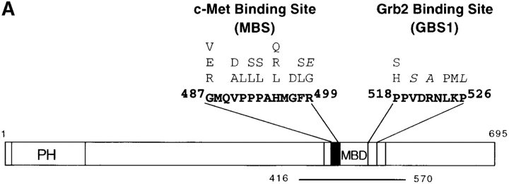
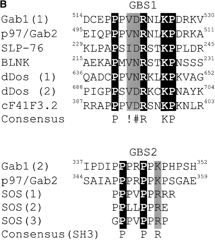
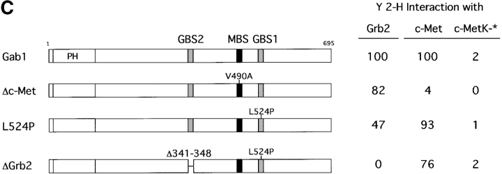
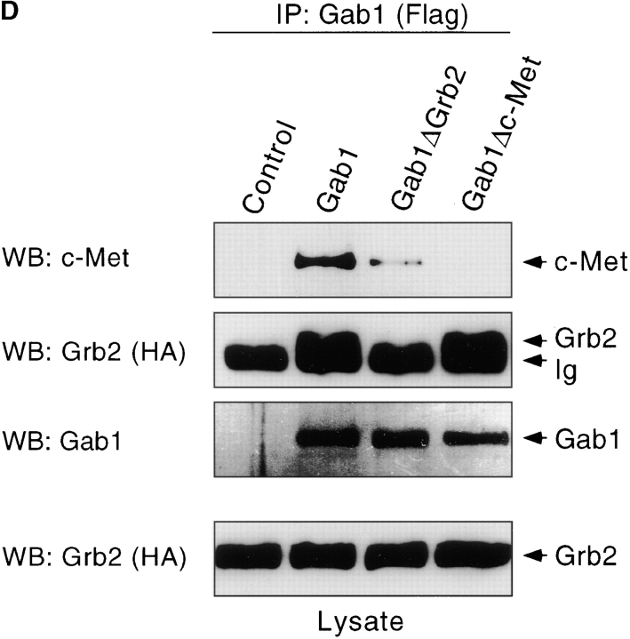
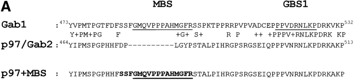
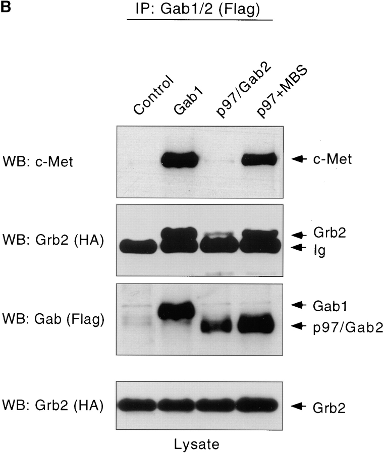
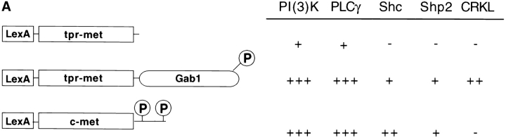
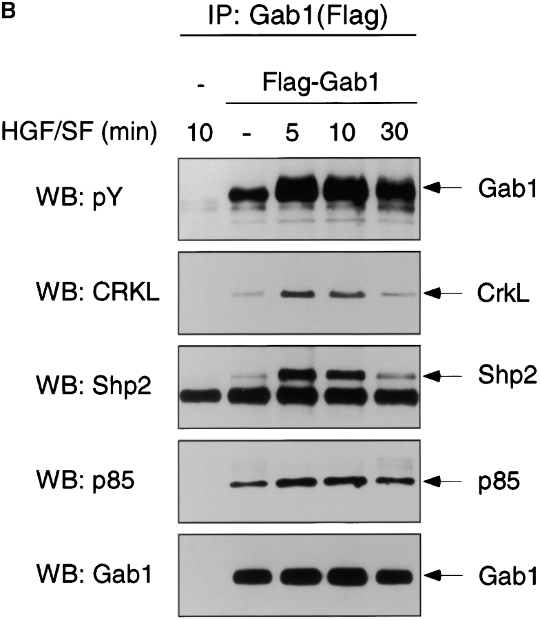
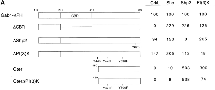
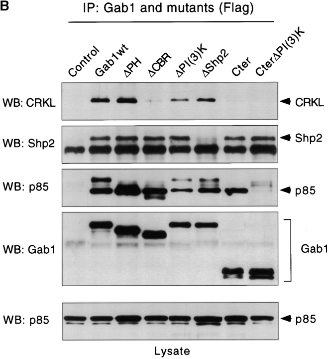
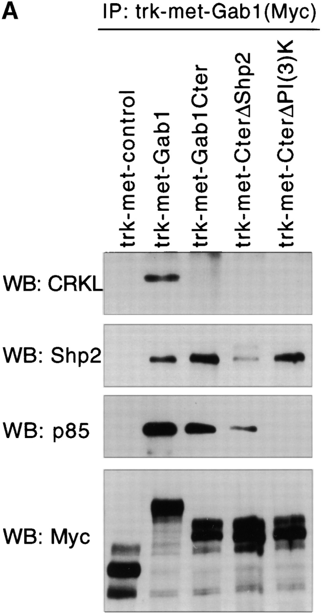
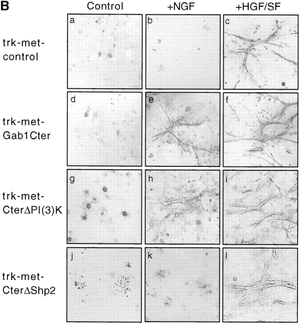
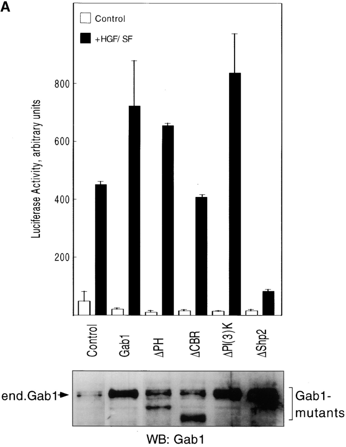
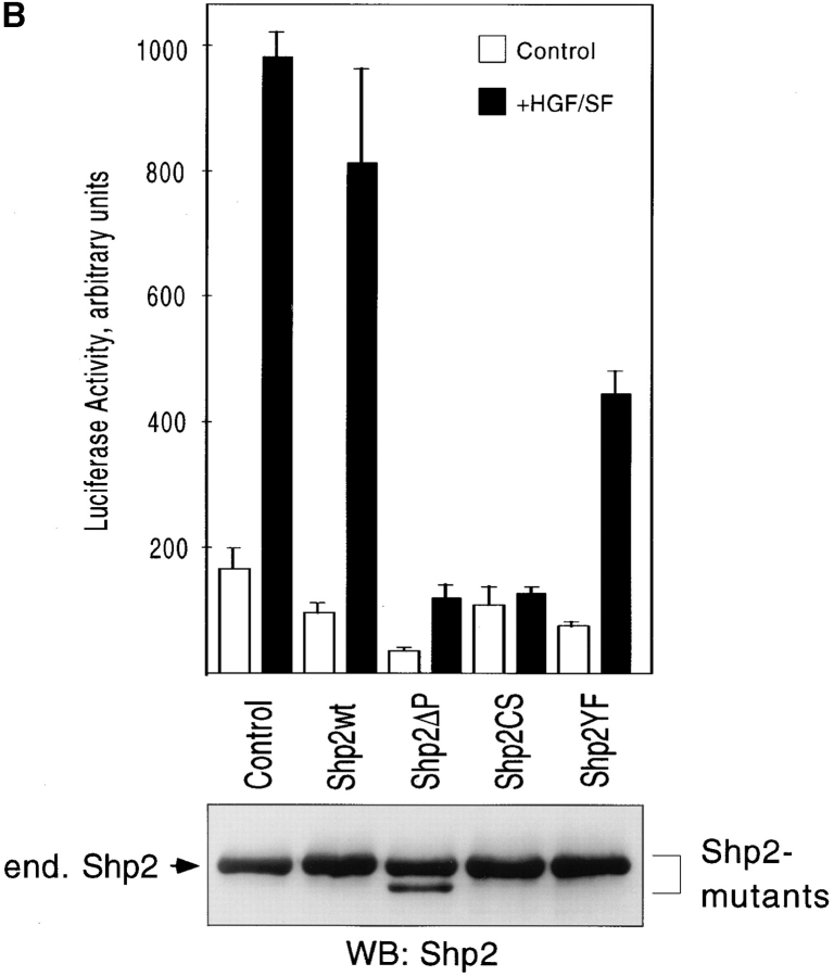
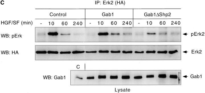
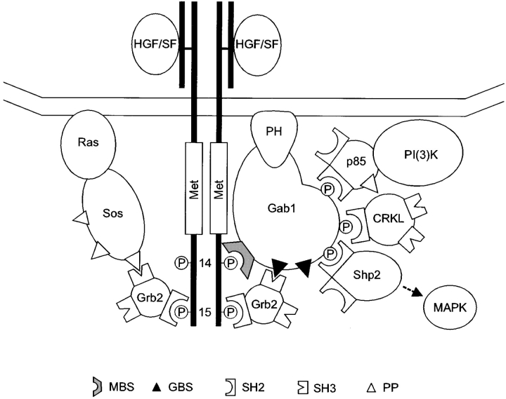
Gab1 is a substrate of the receptor tyrosine kinase c-Met and involved in c-Met-specific branching morphogenesis. It associates directly with c-Met via the c-Met-binding domain, which is not related to known phosphotyrosine-binding domains. In addition, Gab1 is engaged in a constitutive complex with the adaptor protein Grb2. We have now mapped the c-Met and Grb2 interaction sites using reverse yeast two-hybrid technology. The c-Met-binding site is localized to a 13-amino acid region unique to Gab1. Insertion of this site into the Gab1-related protein p97/Gab2 was sufficient to confer c-Met-binding activity. Association with Grb2 was mapped to two sites: a classical SH3-binding site (PXXP) and a novel Grb2 SH3 consensus-binding motif (PX(V/I)(D/N)RXXKP). To detect phosphorylation-dependent interactions of Gab1 with downstream substrates, we developed a modified yeast two-hybrid assay and identified PI(3)K, Shc, Shp2, and CRKL as interaction partners of Gab1. In a trk-met-Gab1-specific branching morphogenesis assay, association of Gab1 with Shp2, but not PI(3)K, CRKL, or Shc was essential to induce a biological response in MDCK cells. Overexpression of a Gab1 mutant deficient in Shp2 interaction could also block HGF/SF-induced activation of the MAPK pathway, suggesting that Shp2 is critical for c-Met/Gab1-specific signaling.


Figures

Figure 1
Figure 1
Identification of c-Met and Grb2-binding sites of Gab1 by yeast two-hybrid analysis. (A) Reverse two-hybrid screens with Gab1 fragments encoding residues 416–570 of Gab1 (see bar). Gab1 cDNA was mutated by PCR and selected against interactions with c-Met or Grb2. Displayed are single point mutations, that interfered with the association of one binding partner (<10% interaction), but not the other (at least 80% interaction remaining). The mutations displayed in italics were generated by site-directed mutagenesis. Mutations interfering with c-Met association are localized in the MBS, residues 487–499, marked in black, mutations interfering with Grb2 association are localized in the GBS1, residues 518–526, marked in grey. (B) The GBS1 of Gab1 constitutes a novel Grb2-binding site. Similar sequences are found in the Grb2-binding adaptor proteins p97/Gab2, SLP-76 and BLNK. Drosophila DOS contains two of these putative Grb2-binding sites. F41F3.2 encodes a hypothetical protein from C. elegans (Wilson et al. 1994). In the alignment, ! can be V or I, and # can be D or N. A second Grb2-binding site (GBS2) is present in Gab1 and p97/Gab2 sequence. Shown below is an alignment of the GBS2 with Grb2-binding peptides derived from Sos. The PXXPXR consensus binding site is found in various SH3 domain binding peptides (Feng et al. 1994). (C) Yeast two-hybrid interactions of full-size Gab1 prey vectors with c-Met, the kinase-inactive c-Met mutant, c-MetK-, or Grb2 as bait. Interactions of mutant Gab1 proteins are displayed as a percentage of wild-type Gab1-c-Met or Gab1-Grb2 association. *Associations with c-MetK- are displayed as percentage of Gab1-c-Met interaction. The proline-rich sequence PPPRPPKP (GBS2) is deleted by the Δ341-348 mutation. (D) Association of Gab1 mutants with c-Met and Grb2 in tissue culture. 293 cells were transfected with HA-tagged Grb2 expression vector plus pBat-Flag (Control) or expression vectors encoding Flag-tagged Gab1 proteins as indicated. After stimulation with HGF/SF, proteins were precipitated with anti-Flag agarose. Proteins were separated by SDS-PAGE and probed with antibodies as indicated.
Figure 1
Figure 1
Identification of c-Met and Grb2-binding sites of Gab1 by yeast two-hybrid analysis. (A) Reverse two-hybrid screens with Gab1 fragments encoding residues 416–570 of Gab1 (see bar). Gab1 cDNA was mutated by PCR and selected against interactions with c-Met or Grb2. Displayed are single point mutations, that interfered with the association of one binding partner (<10% interaction), but not the other (at least 80% interaction remaining). The mutations displayed in italics were generated by site-directed mutagenesis. Mutations interfering with c-Met association are localized in the MBS, residues 487–499, marked in black, mutations interfering with Grb2 association are localized in the GBS1, residues 518–526, marked in grey. (B) The GBS1 of Gab1 constitutes a novel Grb2-binding site. Similar sequences are found in the Grb2-binding adaptor proteins p97/Gab2, SLP-76 and BLNK. Drosophila DOS contains two of these putative Grb2-binding sites. F41F3.2 encodes a hypothetical protein from C. elegans (Wilson et al. 1994). In the alignment, ! can be V or I, and # can be D or N. A second Grb2-binding site (GBS2) is present in Gab1 and p97/Gab2 sequence. Shown below is an alignment of the GBS2 with Grb2-binding peptides derived from Sos. The PXXPXR consensus binding site is found in various SH3 domain binding peptides (Feng et al. 1994). (C) Yeast two-hybrid interactions of full-size Gab1 prey vectors with c-Met, the kinase-inactive c-Met mutant, c-MetK-, or Grb2 as bait. Interactions of mutant Gab1 proteins are displayed as a percentage of wild-type Gab1-c-Met or Gab1-Grb2 association. *Associations with c-MetK- are displayed as percentage of Gab1-c-Met interaction. The proline-rich sequence PPPRPPKP (GBS2) is deleted by the Δ341-348 mutation. (D) Association of Gab1 mutants with c-Met and Grb2 in tissue culture. 293 cells were transfected with HA-tagged Grb2 expression vector plus pBat-Flag (Control) or expression vectors encoding Flag-tagged Gab1 proteins as indicated. After stimulation with HGF/SF, proteins were precipitated with anti-Flag agarose. Proteins were separated by SDS-PAGE and probed with antibodies as indicated.
Figure 2
Figure 2
Insertion of the c-Met–binding site confers c-Met association to p97/Gab2. (A) Alignment of the central regions of Gab1 and p97/Gab2. Underlined are the sequences that define the c-Met–binding site (MBS) or the Grb2-binding site (GBS1) of Gab1. Shown below is the insertion of MBS coding sequences into p97/Gab2 cDNA. Gab1-derived sequences of the p97+MBS hybrid are displayed in bold. (B) p97/Gab2+MBS associates with c-Met in cultured cells. 293 cells were transfected with HA-Grb2 and Flag-tagged Gab1 expression vectors and were stimulated with HGF/SF. After immunoprecipitation with anti-Flag agarose, proteins were detected by Western blotting with anti–c-Met, anti-HA and anti-Flag antibodies as indicated.
Figure 2
Figure 2
Insertion of the c-Met–binding site confers c-Met association to p97/Gab2. (A) Alignment of the central regions of Gab1 and p97/Gab2. Underlined are the sequences that define the c-Met–binding site (MBS) or the Grb2-binding site (GBS1) of Gab1. Shown below is the insertion of MBS coding sequences into p97/Gab2 cDNA. Gab1-derived sequences of the p97+MBS hybrid are displayed in bold. (B) p97/Gab2+MBS associates with c-Met in cultured cells. 293 cells were transfected with HA-Grb2 and Flag-tagged Gab1 expression vectors and were stimulated with HGF/SF. After immunoprecipitation with anti-Flag agarose, proteins were detected by Western blotting with anti–c-Met, anti-HA and anti-Flag antibodies as indicated.
Figure 3
Figure 3
Phosphorylation-dependent interaction of Gab1 with substrates. (A) Modified yeast two-hybrid assay. Interaction of Gab1 with substrates is induced by fusion with LexA-tpr-met kinase. Tpr-met bait vectors lack the multiple docking site of Met, and efficient interaction with substrates is dependent on Gab1 cDNA. Prey vectors encode the SH2 domain of p85α PI(3)K (SH2-2), CRKL, PLC-γ, or full-size cDNAs of Shc and Shp2. The CRKL cDNA clone was identified in a screen for Gab1 interacting cDNAs. It does not associate with c-Met. +++ > 100 , ++ > 50, + > 25 β-galactosidase units. (B) HGF/SF enhances Gab1 phosphorylation and association with signaling molecules. 293 cells were transfected with expression vectors encoding Flag-tagged Gab1 and p85α PI(3)K. After serum starvation for 1 h, cells were stimulated with 50 U/ml HGF/SF for the time period indicated. Proteins were precipitated with anti-Flag beads and detected by Western blotting.
Figure 3
Figure 3
Phosphorylation-dependent interaction of Gab1 with substrates. (A) Modified yeast two-hybrid assay. Interaction of Gab1 with substrates is induced by fusion with LexA-tpr-met kinase. Tpr-met bait vectors lack the multiple docking site of Met, and efficient interaction with substrates is dependent on Gab1 cDNA. Prey vectors encode the SH2 domain of p85α PI(3)K (SH2-2), CRKL, PLC-γ, or full-size cDNAs of Shc and Shp2. The CRKL cDNA clone was identified in a screen for Gab1 interacting cDNAs. It does not associate with c-Met. +++ > 100 , ++ > 50, + > 25 β-galactosidase units. (B) HGF/SF enhances Gab1 phosphorylation and association with signaling molecules. 293 cells were transfected with expression vectors encoding Flag-tagged Gab1 and p85α PI(3)K. After serum starvation for 1 h, cells were stimulated with 50 U/ml HGF/SF for the time period indicated. Proteins were precipitated with anti-Flag beads and detected by Western blotting.
Figure 4
Figure 4
Mapping the substrate-binding sites of Gab1. (A) Gab1 mutants were expressed as LexA-tpr-met fusion proteins and tested for yeast two-hybrid interactions with substrates as in Fig. 3 A. CBR represents the CRKL-binding region of Gab1. Point mutations were introduced into Gab1ΔPH or Gab1Cter sequence as indicated. Interactions were quantified by β-galactosidase liquid assays and compared with the interaction of Gab1ΔPH (set as 100). (B) Gab1 proteins and p85 PI(3)K were expressed in 293 cells and following stimulation with HGF/SF, examined for interactions with downstream interaction partners. The Gab1 mutants are the same as in Fig. 4 A, except that the ΔCBR, ΔPI(3)K, and ΔShp2 mutations were introduced into full-size Gab1 cDNA. Gab1 proteins were precipitated with anti-Flag beads and immune complexes detected by Western blotting.
Figure 4
Figure 4
Mapping the substrate-binding sites of Gab1. (A) Gab1 mutants were expressed as LexA-tpr-met fusion proteins and tested for yeast two-hybrid interactions with substrates as in Fig. 3 A. CBR represents the CRKL-binding region of Gab1. Point mutations were introduced into Gab1ΔPH or Gab1Cter sequence as indicated. Interactions were quantified by β-galactosidase liquid assays and compared with the interaction of Gab1ΔPH (set as 100). (B) Gab1 proteins and p85 PI(3)K were expressed in 293 cells and following stimulation with HGF/SF, examined for interactions with downstream interaction partners. The Gab1 mutants are the same as in Fig. 4 A, except that the ΔCBR, ΔPI(3)K, and ΔShp2 mutations were introduced into full-size Gab1 cDNA. Gab1 proteins were precipitated with anti-Flag beads and immune complexes detected by Western blotting.
Figure 5
Figure 5
Trk-met-Gab1 hybrids interact with downstream substrates and induce branching morphogenesis of MDCK cells. (A) Association of trk-met-Gab1 fusion proteins with signaling proteins in 293 cells. After stimulation with NGF, trk-met-Gab1 proteins were precipitated using myc epitope-tag. Coprecipitating proteins were detected by immuno-blotting. Trk-met-control encodes trk-met lacking the multiple docking site of Met. Gab1 proteins were fused COOH-terminally of trk-met. Gab1Cter encodes residues 450–695 of wild-type Gab1, and CterΔPI(3)K and CterΔShp2 express the COOH-terminal coding sequences of the Gab1 mutants ΔPI(3)K and ΔShp2 (as in Fig. 4 A). (B) Association of Gab1 with Shp2 is essential for c-Met–dependent branching morphogenesis activity. MDCK cells were stably transfected with the trk-met-control and trk-met-Gab1Cter constructs as in A. Cell clones were seeded in collagen gel matrix and maintained in 10% FCS (a, d, g, and j) or stimulated with NGF (b, e, h, and k) or HGF/SF (c, f, i, and l) for up to 7 d to induce tubulogenesis. Trk-met-GabCter, and trk-met-CterΔPI3K respond to NGF treatment (e and h), but not trk-met-CterΔShp2 (k) in which the Shp2-binding site of Gab1 is mutated.
Figure 5
Figure 5
Trk-met-Gab1 hybrids interact with downstream substrates and induce branching morphogenesis of MDCK cells. (A) Association of trk-met-Gab1 fusion proteins with signaling proteins in 293 cells. After stimulation with NGF, trk-met-Gab1 proteins were precipitated using myc epitope-tag. Coprecipitating proteins were detected by immuno-blotting. Trk-met-control encodes trk-met lacking the multiple docking site of Met. Gab1 proteins were fused COOH-terminally of trk-met. Gab1Cter encodes residues 450–695 of wild-type Gab1, and CterΔPI(3)K and CterΔShp2 express the COOH-terminal coding sequences of the Gab1 mutants ΔPI(3)K and ΔShp2 (as in Fig. 4 A). (B) Association of Gab1 with Shp2 is essential for c-Met–dependent branching morphogenesis activity. MDCK cells were stably transfected with the trk-met-control and trk-met-Gab1Cter constructs as in A. Cell clones were seeded in collagen gel matrix and maintained in 10% FCS (a, d, g, and j) or stimulated with NGF (b, e, h, and k) or HGF/SF (c, f, i, and l) for up to 7 d to induce tubulogenesis. Trk-met-GabCter, and trk-met-CterΔPI3K respond to NGF treatment (e and h), but not trk-met-CterΔShp2 (k) in which the Shp2-binding site of Gab1 is mutated.
Figure 6
Figure 6
Association of Gab1 with Shp2 is required for activation of the MAPK pathway. (A) Elk1-dependent reporter gene expression is blocked by Gab1ΔShp2 mutant. 293 cells were transiently transfected with 0.05 μg Gal4-Elk, 0.5 μg Gal4-luciferase and 0.5 μg SV40 LacZ reporter plasmids plus 1 μg pBat-Flag (Control) or Gab1 expression vectors. Gab1 mutants are the same as in Fig. 4 B. Cells were treated with HGF/SF for 5 h and luciferase activity assessed from normalized cell extract. Aliquots of the cell lysates were examined for Gab1 protein expression by Western blotting using Gab1 anti-serum (shown below). (B) Catalytic inactive Shp2 mutants block HGF/SF-induced Elk1 activation. Luciferase reporter assays were carried out as in A. Cells were cotransfected with empty vector (Control) or Shp2 expression vectors as indicated. Shp2ΔP carries a 31–amino acid internal deletion, and Shp2CS a cysteine to serine point mutation at amino acid 453 in the phosphatase domain. In Shp2YF, tyrosine 542 and 580 are mutated to phenylalanine (Bennett et al. 1996). Shown below, Shp2 protein expression of cell lysates. (C) Gab1ΔShp2 attenuates HGF/SF-stimulated phosphorylation of Erk2. 293 cells transfected with HA-tagged Erk2 and either pBat-Flag (Control) or Gab1 expression vectors. After serum starvation, cells were stimulated with HGF/SF as indicated. HA-Erk2 was precipitated and phosphorylation detected by immunoblotting with anti-active MAPK antiserum (pErk). Below: Gab1 protein expression was examined from the total cell lysates (lane C represents control lysate from unstimulated cells).
Figure 6
Figure 6
Association of Gab1 with Shp2 is required for activation of the MAPK pathway. (A) Elk1-dependent reporter gene expression is blocked by Gab1ΔShp2 mutant. 293 cells were transiently transfected with 0.05 μg Gal4-Elk, 0.5 μg Gal4-luciferase and 0.5 μg SV40 LacZ reporter plasmids plus 1 μg pBat-Flag (Control) or Gab1 expression vectors. Gab1 mutants are the same as in Fig. 4 B. Cells were treated with HGF/SF for 5 h and luciferase activity assessed from normalized cell extract. Aliquots of the cell lysates were examined for Gab1 protein expression by Western blotting using Gab1 anti-serum (shown below). (B) Catalytic inactive Shp2 mutants block HGF/SF-induced Elk1 activation. Luciferase reporter assays were carried out as in A. Cells were cotransfected with empty vector (Control) or Shp2 expression vectors as indicated. Shp2ΔP carries a 31–amino acid internal deletion, and Shp2CS a cysteine to serine point mutation at amino acid 453 in the phosphatase domain. In Shp2YF, tyrosine 542 and 580 are mutated to phenylalanine (Bennett et al. 1996). Shown below, Shp2 protein expression of cell lysates. (C) Gab1ΔShp2 attenuates HGF/SF-stimulated phosphorylation of Erk2. 293 cells transfected with HA-tagged Erk2 and either pBat-Flag (Control) or Gab1 expression vectors. After serum starvation, cells were stimulated with HGF/SF as indicated. HA-Erk2 was precipitated and phosphorylation detected by immunoblotting with anti-active MAPK antiserum (pErk). Below: Gab1 protein expression was examined from the total cell lysates (lane C represents control lysate from unstimulated cells).
Figure 7
Figure 7
Scheme of Gab1 associations. Gab1 binds to the bipartide docking site (Y14, Y1349 and Y15, Y1356) of activated c-Met receptor. Direct association of Gab1 to c-Met requires the c-Met–binding site (MBS) of Gab1, while the interaction with Grb2 is constitutive and mediated via the Grb2-binding sites (GBS). The MBS is essential for the cellular association of Gab1 with c-Met, but the Grb2-binding sites also contribute to the strength of the interaction. Grb2 binds Y15 of c-Met via SH2 domain and Gab1 via SH3 domains, thereby stabilizing Gab1-c-Met associations in vivo. Upon phosphorylation, Gab1 associates with PI(3)K, CRKL and Shp2. Recruitment of Shp2 is essential for MAPK activation and c-Met/Gab1–dependent morphogenesis. (PP) indicates poly proline SH3 interaction site.

Similar articles

Cited by

References

    1. Altschul S.F., Gish W., Miller W., Myers E.W., Lipman D.J. Basic local alignment search tool. J. Mol. Biol. 1990;215:403–410. - PubMed
    1. Astier A., Manie S.N., Law S.F., Canty T., Haghayghi N., Druker B.J., Salgia R., Golemis E.A., Freedman A.S. Association of the Cas-like molecule HEF1 with CrkL following integrin and antigen receptor signaling in human B-cellspotential relevance to neoplastic lymphohematopoietic cells. Leuk. Lymphoma. 1997;28:65–72. - PubMed
    1. Ausubel, F.M., R. Brent, R. Kingston, D. Moore, J. Seidmann, J. Smith, and K. Struhls. 1992. Curr. Prot. Mol. Biol.
    1. Bardelli A., Longati P., Gramaglia D., Stella M.C., Comoglio P.M. Gab1 coupling to the HGF/Met receptor multifunctional docking site requires binding of Grb2 and correlates with the transforming potential. Oncogene. 1997;15:3103–3111. - PubMed
    1. Bennett A.M., Tang T.L., Sugimoto S., Walsh C.T., Neel B.G. Protein-tyrosine-phosphatase SHPTP2 couples platelet-derived growth factor receptor beta to Ras. Proc. Natl. Acad. Sci. USA. 1994;91:7335–7339. - PMC - PubMed

Publication types

MeSH terms

Substances