. 2000 Jun 26;149(7):1443-54.
doi: 10.1083/jcb.149.7.1443.

Shared and unique roles of CAP23 and GAP43 in actin regulation, neurite outgrowth, and anatomical plasticity

Affiliations

Shared and unique roles of CAP23 and GAP43 in actin regulation, neurite outgrowth, and anatomical plasticity

D Frey et al. J Cell Biol. .

Abstract

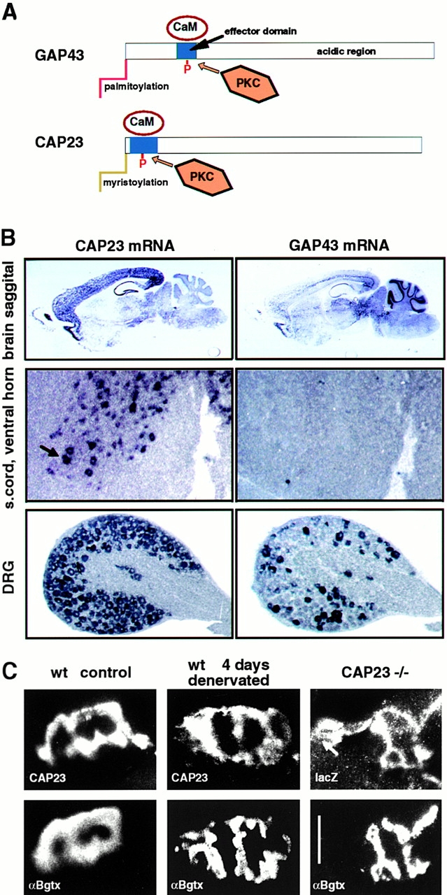
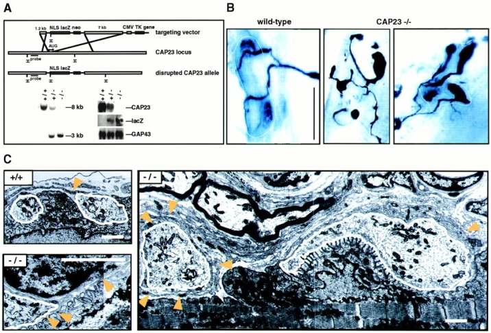
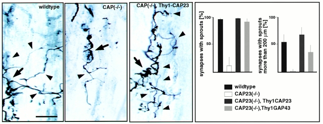
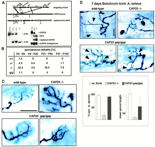
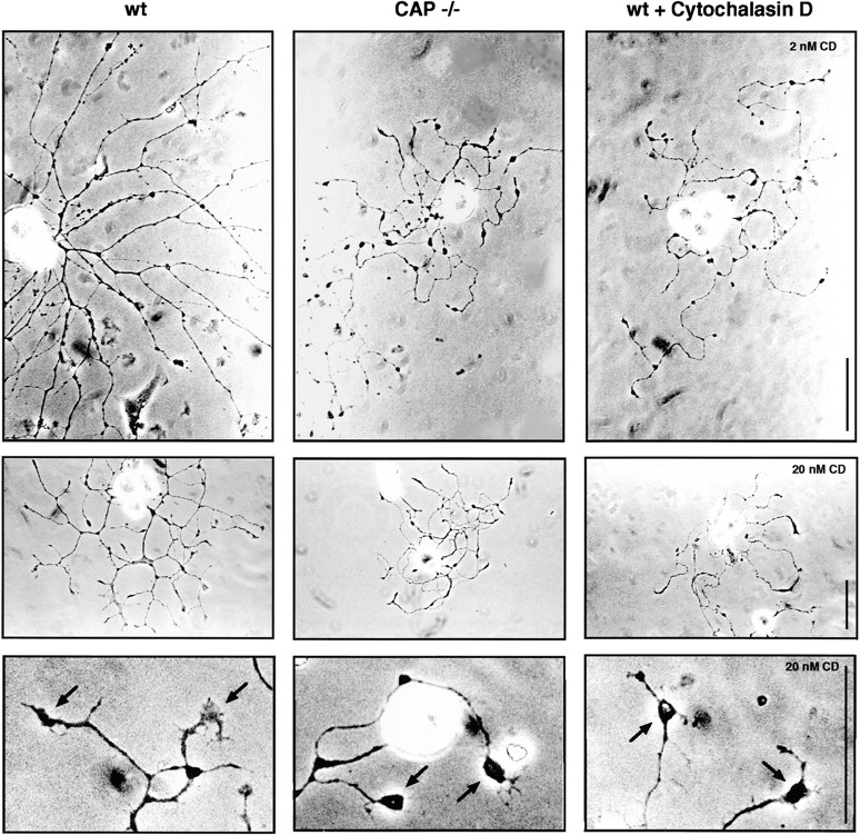
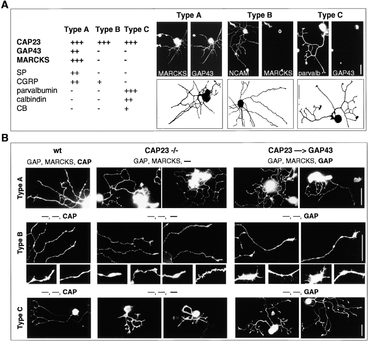
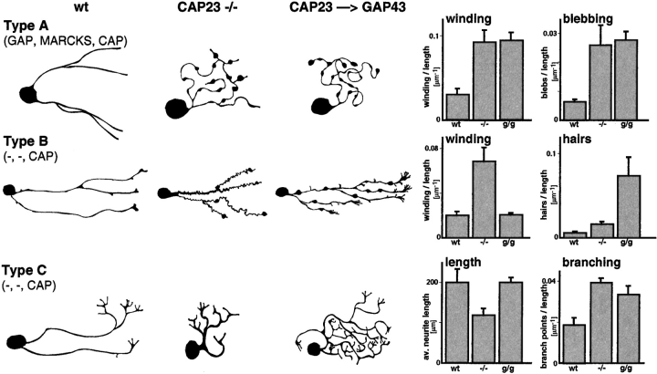
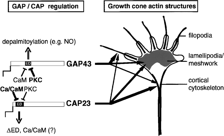
CAP23 is a major cortical cytoskeleton-associated and calmodulin binding protein that is widely and abundantly expressed during development, maintained in selected brain structures in the adult, and reinduced during nerve regeneration. Overexpression of CAP23 in adult neurons of transgenic mice promotes nerve sprouting, but the role of this protein in process outgrowth was not clear. Here, we show that CAP23 is functionally related to GAP43, and plays a critical role to regulate nerve sprouting and the actin cytoskeleton. Knockout mice lacking CAP23 exhibited a pronounced and complex phenotype, including a defect to produce stimulus-induced nerve sprouting at the adult neuromuscular junction. This sprouting deficit was rescued by transgenic overexpression of either CAP23 or GAP43 in adult motoneurons. Knockin mice expressing GAP43 instead of CAP23 were essentially normal, indicating that, although these proteins do not share homologous sequences, GAP43 can functionally substitute for CAP23 in vivo. Cultured sensory neurons lacking CAP23 exhibited striking alterations in neurite outgrowth that were phenocopied by low doses of cytochalasin D. A detailed analysis of such cultures revealed common and unique functions of CAP23 and GAP43 on the actin cytoskeleton and neurite outgrowth. The results provide compelling experimental evidence for the notion that CAP23 and GAP43 are functionally related intrinsic determinants of anatomical plasticity, and suggest that these proteins function by locally promoting subplasmalemmal actin cytoskeleton accumulation.


Figures

Figure 1
Figure 1
Expression patterns of mouse CAP23 and GAP43 in the adult. (A) Schematic representation of the structures of GAP43 and CAP23. The NH2-terminal with the acylation domains is to the left; (blue box) basic ED with PKC phosphorylation sites; and (white rectangles) predominantly acidic and highly hydrophilic regions with similar amino acid compositions. The relative dimensions of the proteins and the EDs are in scale. (B) In situ hybridization. Note the differential expression of CAP43 and GAP43 in adult brain regions, expression of CAP23 in large ventral horn neurons (arrow), and expression of GAP43 in DRG neuron subgroups. (C) CAP23 immunoreactivity at the adult nmj. Double labeling for postsynaptic acetylcholine receptors (bottom, αBgtx) and CAP23 or reporter gene lacZ (top). Note the persistence of CAP23 signal at denervated (i.e., nerve-free) nmj, and synaptic, plus extrasynaptic (arrow) accumulation of reporter gene signal in homozygous CAP23−/− mice. Bar, 50 μm.
Figure 2
Figure 2
nmj phenotype of CAP23−/− mice. (A) Gene disruption strategy (top), and genomic Southern (left) and Northern (right; adult brain mRNA) blots of wild-type, CAP23+/−, and CAP23−/− mice. In the mutant mice, GAP43 expression was not noticeably affected (immunocytochemistry and in situ hybridization data not shown). (B) Altered arrangement of the presynaptic nerve at the nmj of CAP23-deficient mice. Combined silver esterase stains of fast-type medial gastrocnemius nmjs (2 mo); each photograph shows one nmj (black, nerve; blue, esterase reaction product). The dotted lines mark the synaptic regions. Note the highly irregular shapes of the nerve terminal branches in the mutants, with swellings and bloblike endings. More than 90% of the nmjs exhibited obvious structural abnormalities in CAP23−/− mice. (C) Ultrastructural appearance of adult medial gastrocnemius nmjs in wild-type and CAP23−/− mice. Muscle is down and nerve is up. The nerve terminal branches are outlined in white. Arrowheads point to tSC processes. Note the hypertrophy of tSC processes, and nerve blob (left end of the photograph, white outline) surrounded by tSC processes in the photograph on the right. The photograph on the lower left shows postsynaptic junctional folds unopposed by a nerve profile (local denervation), and tSC (nucleus on the upper part of the photograph) processes insinuating into the synaptic cleft on the left. Bars: (B) 50 μm; (C) 1.6 μm.
Figure 3
Figure 3
Critical role of motoneuron CAP23 for nerve sprouting at the adult nmj. Defective ultraterminal nerve sprouting at the soleus nmj of CAP23−/− mice, and rescue of nerve sprouting by reintroduction of CAP23 in motoneurons (CAP23−/− × Thy1CAP23 mice). (left) Combined silver esterase stains of soleus nmjs (arrowheads) 30 d after BotA injection. One nmj is shown in each photograph (arrows, broader nerve regions); some of the sprouts are indicated by arrowheads. (right) Quantitative analysis of nerve sprouting at the soleus, 30 d after BotA injection. The graphs refer to the fraction of synapses with sprouts (top), and to the fraction of synapses with a total length of sprouts >200 μm. The values are averages from three independent experiments (n = 50). Bar, 50 μm.
Figure 4
Figure 4
GAP43 can functionally substitute for CAP23. (A) Generation of CAP23gap43/gap43 knockin mice. (top) Targeting construct for gene replacement, (bottom) genomic Southern blot (left), and Northern blot of adult brain mRNA. The signs (+) and (−) refer to a wild-type or mutated CAP23 allele. (B) Enhanced mortality of CAP23+/− and CAP23−/− mice, and its rescue by GAP43. (g/g) CAP23gap43/gap43 background. (C) Correction by GAP43 of an nmj morphology phenotype because of the absence of CAP23. Combined silver esterase stains of fast-type medial gastrocnemius nmjs at 2 mo. Note the irregular diameter and blobs at nerve terminal branches of CAP23−/− mice. In the presence of GAP43, instead of CAP23, nerve terminal branches at the nmj were slightly broader than in a wild-type background, but regular in size, and without blobs. (D) Rescue of BotA-induced nerve sprouting at the soleus nmj of CAP23gap43/gap43 mice. Combined silver esterase stains and quantitative analysis of sprouting of soleus nmj's, 7 d after BotA injection; sprouts are indicated by arrows. Sprouts in the presence of GAP43, instead of CAP23, were shorter and more branched. Bar, 50 μm.
Figure 5
Figure 5
Alterations in neurite outgrowth because of the absence of CAP23 can be phenocopied by cytochalasin D, which interferes with actin filament polymerization. Representative phase-contrast photographs of 15-h DRG cultures on laminin, in the presence of NGF. Note the thin, meandering neurites, varicosities, and bulbous growth cones (arrows, bottom row) in the absence of CAP23 or in the presence of cytochalasin D. Bars: 50 μm.
Figure 6
Figure 6
Common and unique roles of CAP23 and GAP43 in neurite outgrowth. (A) Operational definition of three subtypes of DRG neurons, based on the expression of marker antigens, and morphology. The camera lucida drawings show one representative neuron per subtype; the double labeling immunocytochemistry photographs show one further example per subtype. (B) Neurite outgrowth patterns of DRG neuron subtypes from wild-type (wt), CAP23−/−, and CAP23gap43/gap43 (CAP23→GAP43) mice (15-h cultures on 40 μg/ml laminin). The expression (or absence of expression, minus sign) of GAP43, MARCKS, and CAP23 is indicated on top of the photographs. Labeling antigens: MARCKS (type A), NCAM (type B), and parvalbumin (type C). Two details each of type B neuron photographs are also shown. Bars: 50 μm.
Figure 7
Figure 7
Common and unique roles of CAP23 and GAP43 in neurite outgrowth. Schematic representation of characteristic features (drawings on the left), and quantitative analysis of 15-h DRG cultures as shown in Fig. 6 B. See Materials and Methods for details. n = 15.
Figure 8
Figure 8
Proposed model of common and unique regulation and roles of CAP23 and GAP43 in growth cone actin dynamics. (left) CAP23 and GAP43 are regulated differently at the level of ED masking (relative roles of calmodulin [CaM] and PKC), and retention at the plasmalemma (ED masking versus depalmitoylation). These differences may result in differential impacts to the dynamics and accumulation of different types of actin structures at the growth cone (right).

Similar articles

Cited by

References

    1. Aderem A. The MARCKS family of protein kinase-C substrates. Biochem. Soc. Trans. 1995;23:587–591. - PubMed
    1. Aigner A., Caroni P. Absence of persistent spreading, branching, and adhesion in GAP-43-depleted growth cones. J. Cell Biol. 1995;128:647–660. - PMC - PubMed
    1. Aigner, L., S. Arber, J.P. Kapfhammer, T. 0, C. Schneider, F. Botteri, H.-R. Brenner, and P. Caroni. 1995. Overexpression of the neural growth-associated protein GAP-43 induces nerve sprouting in the adult nervous system of transgenic mice. Cell. 83:269–278. - PubMed
    1. Antonini A., Fagiolini M., Stryker M.P. Anatomical correlates of functional plasticity in mouse visual cortex. J. Neurosci. 1999;19:4388–4406. - PMC - PubMed
    1. Baumeister A., Arber S., Caroni P. Accumulation of muscle ankyrin repeat protein transcript reveals local activation of primary myotube end compartments during muscle morphogenesis. J. Cell Biol. 1997;139:1231–1242. - PMC - PubMed

MeSH terms