. 2000 Jun 15;28(12):2315-23.
doi: 10.1093/nar/28.12.2315.

Dbp10p, a putative RNA helicase from Saccharomyces cerevisiae, is required for ribosome biogenesis

Affiliations

Dbp10p, a putative RNA helicase from Saccharomyces cerevisiae, is required for ribosome biogenesis

F Burger et al. Nucleic Acids Res. .

Abstract

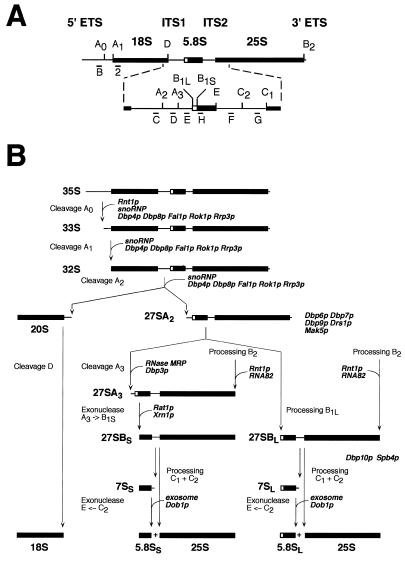
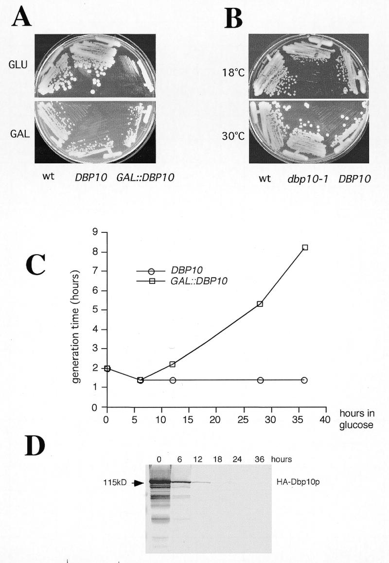
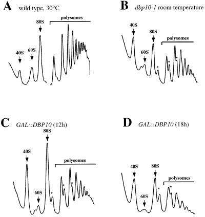
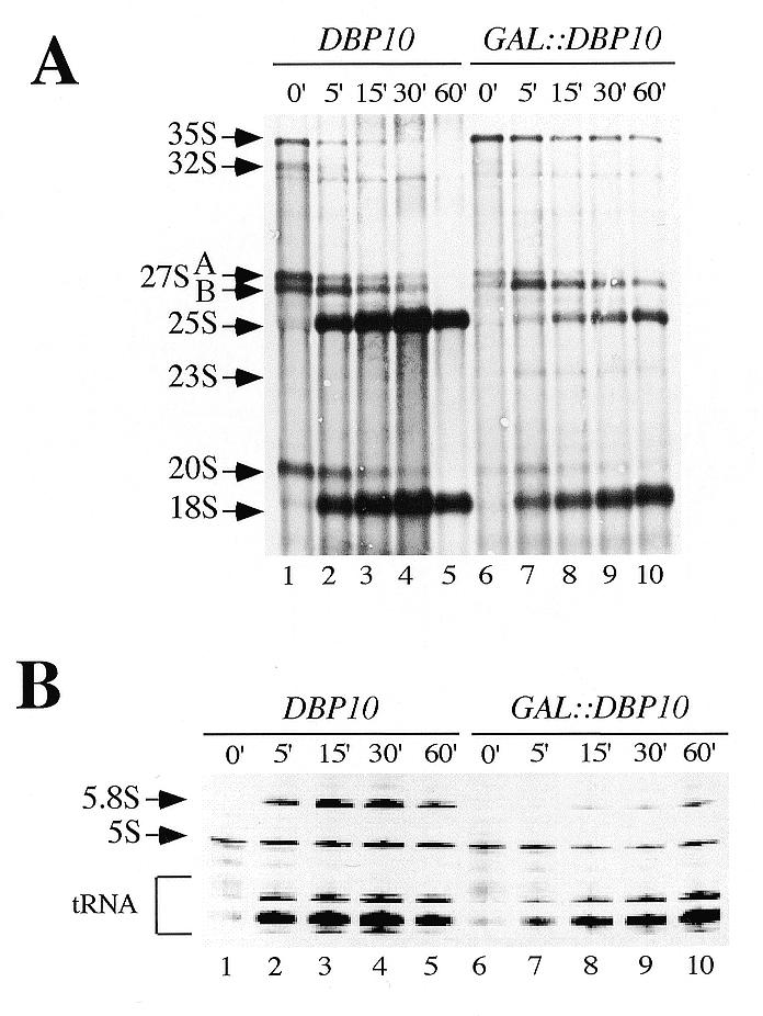
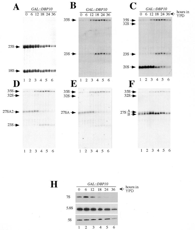
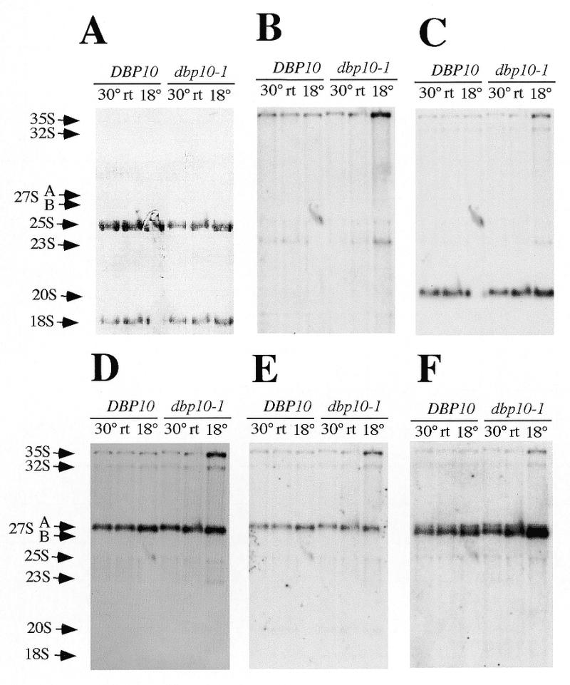
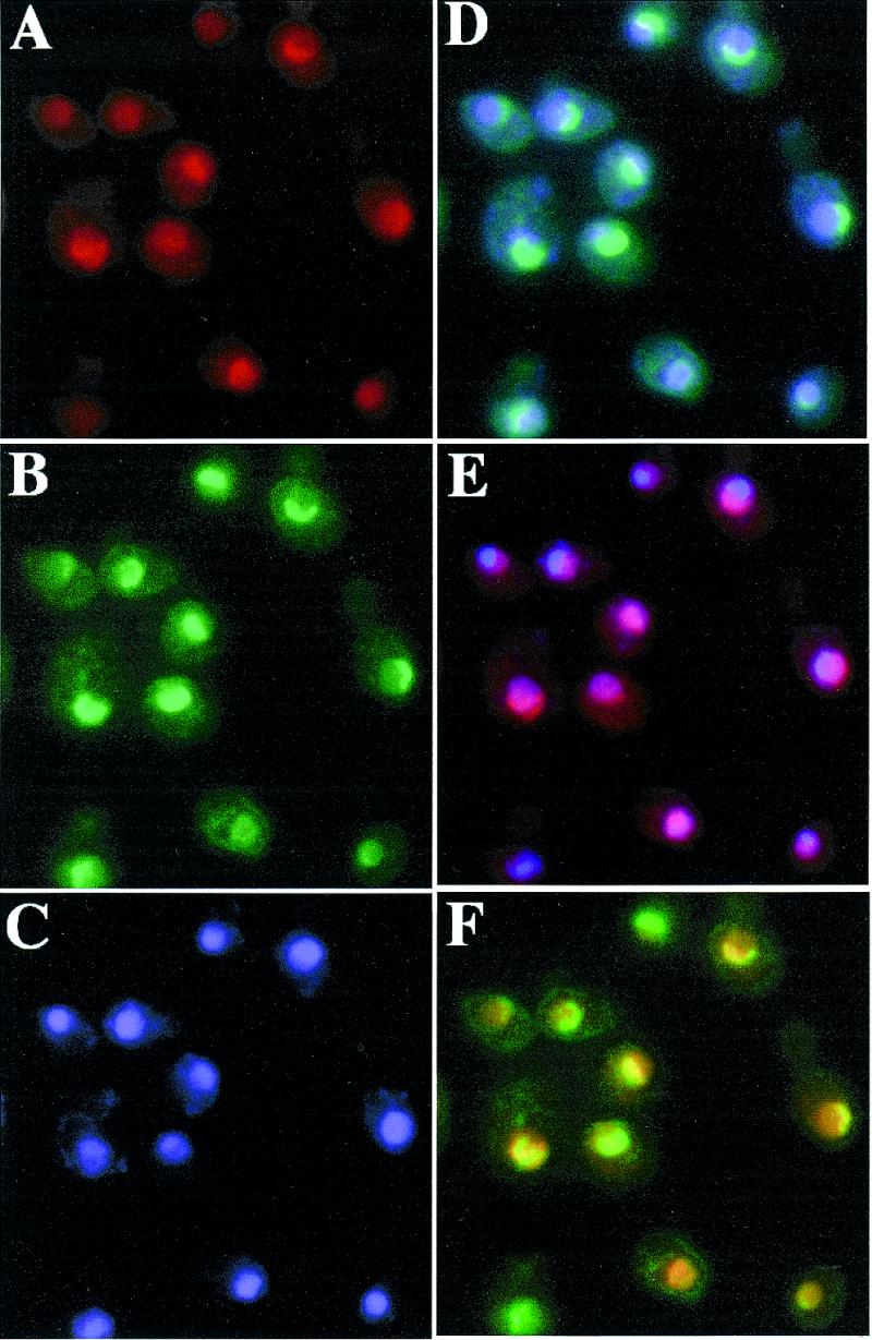
Ribosome biogenesis requires, in addition to rRNA molecules and ribosomal proteins, a multitude of trans-acting factors. Recently it has become clear that in the yeast Saccharomyces cerevisiae many RNA helicases of the DEAD-box and related families are involved in ribosome biogenesis. Here we show that the previously uncharacterised open reading frame YDL031w (renamed DBP10 for DEAD-box protein 10) encodes an essential putative RNA helicase that is required for accurate ribosome biogenesis. Genetic depletion of Dbp10p results in a deficit in 60S ribosomal subunits and an accumulation of half-mer polysomes. Furthermore, pulse-chase analyses of pre-rRNA processing reveal a strong delay in the maturation of 27SB pre-rRNA intermediates into 25S rRNA and 7S pre-rRNA. Northern blot analyses indicate that this delay leads to higher steady-state levels of 27SB species and reduced steady-state levels of 7S pre-rRNA and 25S/5.8S mature rRNAs, thus explaining the final deficit in 60S subunit and the formation of half-mer polysomes. Consistent with a direct role in ribosome biogenesis, Dbp10p was found to be located predominantly in the nucleolus.


Figures

Figure 1
Figure 1
Pre-rRNA processing in S.cerevisiae. (A) Structure and processing sites of the 35S pre-rRNA. This precursor contains the sequences for the mature 18S, 5.8S and 25S rRNAs, which are separated by the two internal transcribed spacers ITS1 and ITS2 and flanked by two external transcribed spacers 5′ ETS and 3′ ETS. The location of various probes (labelled from B to H) used in this study are indicated. Bars represent mature rRNA species and lines the transcribed spacers. (B) Pre-rRNA processing pathway. The 35S pre-rRNA is cleaved at site A0 by the endonuclease Rnt1p, generating the 33S pre-rRNA. This molecule is subsequently processed at sites A1 and A2, resulting in the separation of the pre-rRNAs destined for the small and large ribosomal subunits. It is proposed that the early pre-rRNA cleavages A0–A2 require a large snoRNP complex, which may be assisted by the putative ATP-dependent RNA helicases Dbp4p, Dbp8p, Fal1p, Rok1p and Rrp3p. Final maturation of the 20S precursor takes place in the cytoplasm, where endonucleolytic cleavage at site D yields the mature 18S rRNA. The 27SA2 precursor is processed by two alternative pathways that both lead to the formation of mature 5.8S and 25S rRNAs. In the major pathway, the 27SA2 precursor is cleaved at site A3 by the RNase MRP complex. The putative ATP-dependent RNA helicase Dpb3p assists in this processing step. The 27SA3 precursor is exonucleolytically digested 5′→3′ up to site B1S to yield the 27SBS precursor, a reaction requiring the exonucleases Xrn1p and Rat1p. A minor pathway processes the 27SA2 molecule at site B1L, producing the 27SBL pre-rRNA. While processing at site B1 is completed, the 3′ end of mature 25S rRNA is generated by processing at site B2. The subsequent processing at ITS2 appears to be identical for both 27SB species. Cleavage at sites C1 and C2 releases the mature 25S rRNA and the 7S pre-rRNA. The latter undergoes exosome-dependent 3′ to 5′ exonuclease digestion to the 3′ end of the mature 5.8S rRNA. It has been proposed that Dob1p/Mtr4p, a putative ATP-dependent RNA helicase, assists the exosome activity. The data presented in this study suggest that Dbp10p is required for a late step in the assembly of 60S ribosomal subunits, a process that may also involve six other putative ATP-dependent RNA helicases: Dbp6p, Dbp7p, Dbp9p, Spb4p, Drs1p and Mak5p. The pre-rRNA processing phenotype of the Drs1p-depleted and Mak5p-depleted strains has not been described. Data concerning Dbp8p and Dbp9p are unpublished results from our laboratory.
Figure 2
Figure 2
Mutations in DBP10 or depletion of Dbp10p results in growth inhibition. (A) Growth of YBF1-1A (wt), YBF1-1B [YCplac33-DBP10] (DBP10) and YBF1-1B [pAS24-DBP10] (GAL::DBP10) on YPD (glucose) or YPGal (galactose) plates. The plates were incubated at 30°C for 3 and 2 days, respectively. (B) Growth of YBF1-1A (wt), YBF1-1B [YCplac33-DBP10] (DBP10) and YBF1-1B [YCplac33-dbp10-1] (dbp10-1) strains on YPD plates at 16 and 30°C. The plates were incubated for 6 and 2 days, respectively. (C) Growth curves of YBF1-1B [YCplac33-DBP10] (DBP10, circles) and YBF1-1B [pAS24-DBP10] (GAL::DBP10, squares) strains at 30°C after shifting logarithmic cultures from YPGal to YPD for up to 36 h. Data are given as doubling times at different times in YPD medium. (D) Depletion of Dbp10p. The GAL::DBP10 strain was grown in YPGal and shifted to YPD for up to 36 h and cell extracts were prepared from samples harvested at the indicated times and assayed by western blotting. Equal amounts of proteins were loaded in each lane (~70 µg) as judged by red ponceau staining of the blot (data not shown). Pre-stained markers (Bio-Rad) were used as standards for molecular mass estimation. Monoclonal mouse anti-HA 16B12 antibodies followed by alkaline phosphatase coupled goat anti-rabbit IgG were used to detect HA-Dbp10p. The HA-Dbp10p signal is indicated by an arrow.
Figure 3
Figure 3
The dbp10-1 mutation and the depletion of Dbp10p result in a deficit in free 60S ribosomal subunits and in the accumulation of half-mer polysomes. The wild-type strain YFB1-1A (A) and the dbp10-1 mutant strain YFB1-1B [YCplac33-dbp10-1] (B) were grown at 30°C and room temperature, respectively. The GAL::DBP10 strain YFB1-1B [pAS24-DBP10] was grown at 30°C in YPGal and shifted to YPD for 12 h (C) and 18 h (D). Cells were harvested at an OD600 of 0.8, and cell extracts were resolved in 7–50% (w/v) sucrose gradients. The A254 was measured continuously. Sedimentation is from left to right. The peaks of free 40S and 60S ribosomal subunits, 80S free couples/monosomes and polysomes are indicated. Half-mers are labelled with asterisks.
Figure 4
Figure 4
Dbp10p depletion leads to reduced synthesis of the mature 25S and 5.8S rRNAs. Strains YBF1-1A [YCplac33] (DBP10) and YBF1-1B [pAS24-DBP10, YCplac33] (GAL::DBP10) were grown at 30°C in Sgal–Ura and shifted to SD–Ura for 18 h. Cells were pulse-labelled with [5,6-3H]uracil for 2 min, and then chased with an excess of cold uracil for 5, 15, 30 and 60 min. Total RNA was extracted and separated on 1.2% agarose–6% formaldehyde (A) or 7% polyacrylamide–50% urea gels (B), transferred to nylon membrane and visualised by fluorography. Approximately 25 000 c.p.m. were loaded in each lane. The positions of the different pre-rRNAs and mature rRNAs and tRNAs are indicated.
Figure 5
Figure 5
Depletion of Dbp10p affects the steady-state levels of pre-rRNA and mature rRNA species. Strain YBF1-1B [pAS24-DBP10] (GAL::DBP10) was grown in YPGal and shifted for up to 36 h in YPD. Cells were harvested at the indicated times and total RNA was extracted. Equal amounts (5 µg) of total RNA were resolved on 1.2% agarose–6% formaldehyde and transferred to a nylon membrane, which was stained with methylene blue (A), and then consecutively hybridised with the different probes indicated in Figure 1A [(BF) probes B–F respectively]. Equal amounts (2.5 µg) of the same RNA samples were also resolved on 7% polyacrylamide–50% urea gel, transferred to a nylon membrane and hybridised consecutively with probe E, 5.8S and 5S (H). The positions of the different pre-rRNAs and mature rRNAs are indicated.
Figure 6
Figure 6
Steady-state levels of pre-rRNAs and mature rRNAs are altered in the dbp10-1mutant strain. Strains YBF1-1A (DBP10) and YBF1-1B [YCplac33-dbp10-1] (dbp10-1) were grown in YPD at 30°C, room temperature (rt) and 18°C as indicated on the top of the lanes. When the cultures reached the exponential phase, cells were harvested and total RNA was extracted. Equal amounts (5 µg) of total RNA were resolved on 1.2% agarose–6% formaldehyde and transferred to a nylon membrane, which was stained with methylene blue (A), and then consecutively hybridised with the different probes indicated in Figure 1A [(BF) probes B–F respectively]. The positions of the different pre-rRNAs and mature rRNAs are indicated. The absence of 18S rRNA and 20S signals in the lane corresponding to DBP10 strain grown at 18°C is due to a technical artefact during migration of the gel.
Figure 7
Figure 7
HA-Dbp10p localises to the nucleolus. Indirect immunofluorescence was performed with cells expressing HA-Dbp10p from the cognate promoter (strain YBF1-1B [YCplac111-HA-DBP10]) (A). HA-Dbp10p was detected by the monoclonal mouse anti-HA 16B12 antibody, followed by decoration with a goat anti-mouse rhodamine-conjugated antibody. (B) Nop1p was detected by polyclonal rabbit anti-Nop1p antibodies, followed by decoration with a goat anti-rabbit fluorescein-conjugated antibody. (C) Chromatin DNA was stained using DAPI. Pseudo-colours were assigned to the digitised micrographs (A–C) and images were merged. The overlapping distributions are revealed in: (D) cyan for Nop1p and chromatin DNA colocalisation; (E) magenta for HA-Dbp10p and chromatin DNA colocalisation; and (F) yellow for Nop1p and HA-Dbp10p colocalisation.

References

    1. Warner J.R. (1999) Trends Biol. Sci., 24, 437–440. - PubMed
    1. Li B., Nierras,C.R. and Warner,J.R. (1999) Mol. Cell. Biol., 19, 5393–5404. - PMC - PubMed
    1. Mager W.H., Planta,R.J., Ballesta,J.-P.G., Lee,J.C., Mizuta,K., Suzuki,K., Warner,J.R. and Woolford,J. (1997) Nucleic Acids Res., 25, 4872–4875. - PMC - PubMed
    1. Mélèse T. and Xue,Z. (1995) Curr. Opin. Cell Biol., 7, 319–324. - PubMed
    1. Scheer U. and Hock,R. (1999) Curr. Opin. Cell Biol., 11, 385–390. - PubMed

Publication types

MeSH terms