. 2000 Jun 15;28(12):2324-32.
doi: 10.1093/nar/28.12.2324.

Characterisation of the catalytically active form of RecG helicase

Affiliations

Characterisation of the catalytically active form of RecG helicase

P McGlynn et al. Nucleic Acids Res. .

Abstract

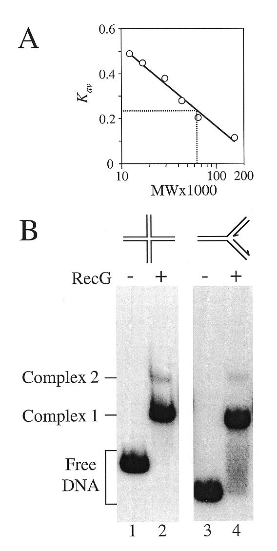
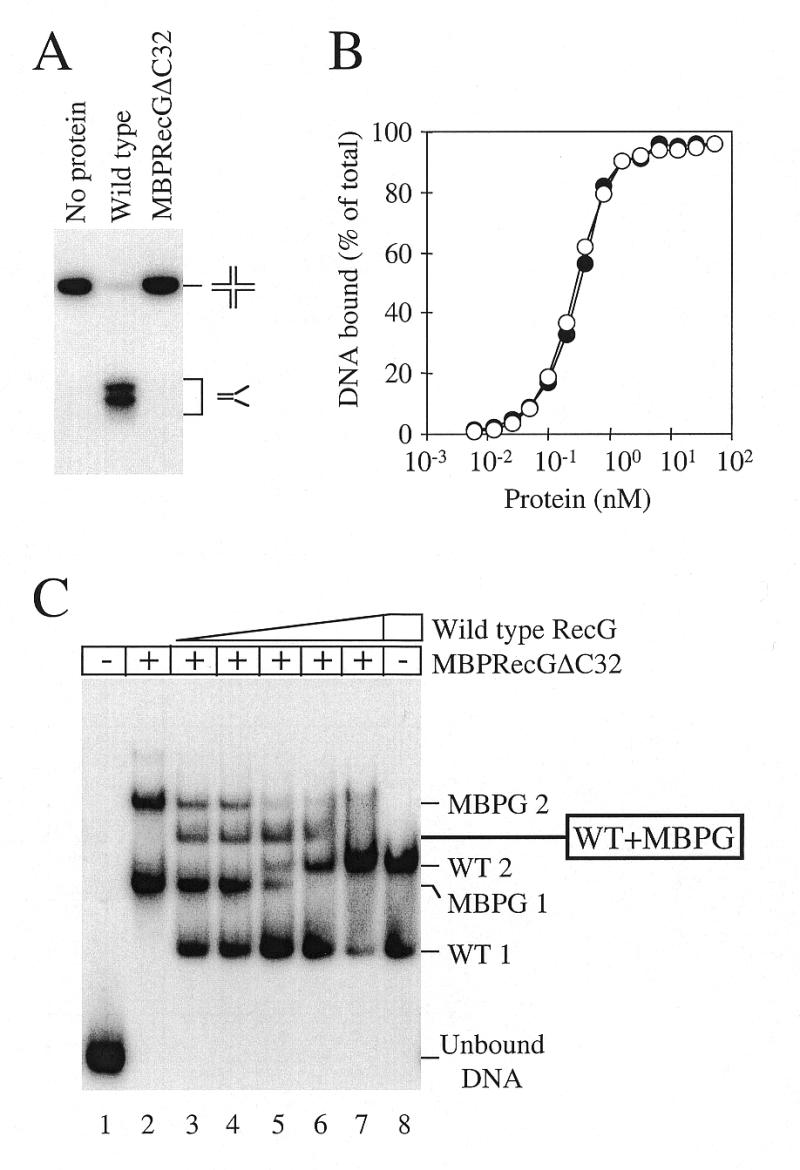
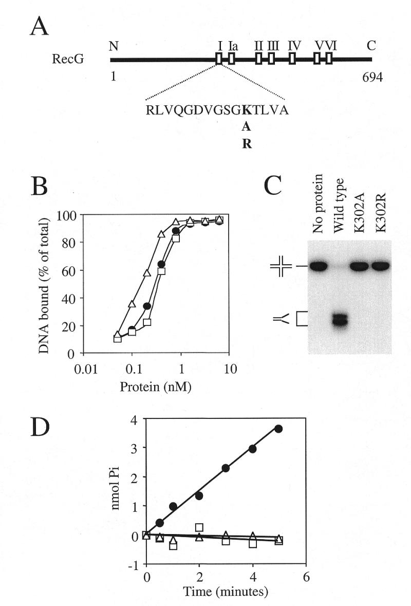
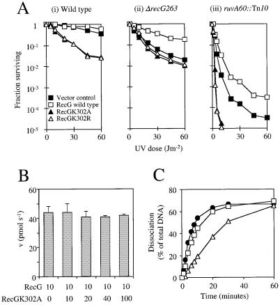
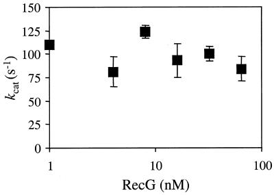
Replication of DNA is fraught with difficulty and chromosomes contain many lesions which may block movement of the replicative machinery. However, several mechanisms to overcome such problems are beginning to emerge from studies with Escherichia coli. An important enzyme in one or more of these mechanisms is the RecG helicase, which may target stalled replication forks to generate a four-stranded (Holliday) junction, thus facilitating repair and/or bypass of the original lesion. To begin to understand how RecG might catalyse regression of fork structures, we have analysed what the catalytically active form of the enzyme may be. We have found that RecG exists as a monomer in solution as measured by gel filtration but when bound to junction DNA the enzyme forms two distinct protein-DNA complexes that contain one and two protein molecules. However, mutant inhibition studies failed to provide any evidence that RecG acts as a multimer in vitro. Additionally, there was no evidence for cooperativity in the junction DNA-stimulated hydrolysis of ATP. These data suggest that RecG functions as a monomer to unwind junction DNA, which supports an 'inchworm' rather than an 'active rolling' mechanism of DNA unwinding. The observed in vivo inhibition of wild-type RecG by mutant forms of the enzyme was attributed to occlusion of the DNA target and correlates with the very low abundance of replication forks within an E.COLI: cell, even during rapid growth.


Figures

Figure 1
Figure 1
Native molecular weight of RecG and binding stoichiometry to junction DNA. (A) Gel filtration of RecG. The Kav value of RecG was compared to that of protein standards of known size to give an estimated molecular weight of 62 000. (B) Binding reactions containing 50 nM wild-type RecG and 0.2 nM junction 1 (lanes 1 and 2) or junction 2 (lanes 3 and 4) were analysed on band shift gels. The arrows on the fork DNA structure indicate the 3′-termini of the DNA strands. The reactions were electrophoresed on the same gel but for clarity the intervening lanes have been deleted.
Figure 2
Figure 2
DNA binding and unwinding properties of a MBP–RecGΔC32 fusion. (A) Helicase activity of 100 nM wild-type RecG and 100 nM MBPRecGΔC32 on 0.2 nM junction 3 DNA. Intact junction DNA and flayed duplex products are marked. (B) Comparison of the DNA binding affinities of wild-type RecG (closed circles) and MBPRecGΔC32 (open circles) as measured by band shift assays with 0.2 nM Holliday junction DNA (junction 3). (C) Binding of Holliday junction DNA (junction 3) by a mix of wild-type RecG and MBPRecGΔC32. An aliquot of 50 nM MBPRecGΔC32 fusion polypeptide was bound to 0.2 nM DNA (lanes 2–7), forming two protein–DNA complexes designated MBPG 1 and MBPG 2. Aliquots of 5, 10, 20, 40 and 80 nM wild-type RecG were also added to lanes 3–7, respectively, and 80 nM to lane 8, forming the two protein–DNA complexes marked WT 1 and WT 2. In the presence of both wild-type and MBPRecGΔC32 a novel protein–DNA complex was detected whose mobility suggested it contained one molecule each of wild-type RecG and MBPRecGΔC32, as indicated. Both proteins were omitted from lane 1.
Figure 3
Figure 3
In vitro properties of RecG mutant proteins deficient in ATP hydrolysis. (A) Schematic diagram of the RecG polypeptide with the locations of the seven motifs conserved across a wide range of DNA and RNA helicases. The numbers refer to amino acid residues and the N- and C-termini are also indicated. The amino acid sequence of motif I is shown together with the lysine residue within this sequence which was mutated to alanine (RecGK302A) and arginine (RecGK302R). (B) Comparison of the DNA binding affinities as measured in band shift assays of wild-type RecG (circles), K302A (squares) and K302R (triangles) with 0.2 nM Holliday junction DNA (junction 3). (C) Helicase activity of 100 nM wild-type RecG, K302A and K302R on 0.2 nM Holliday junction DNA (junction 3). The positions of the junction DNA and flayed duplex products after gel electrophoresis are indicated. (D) Rate of ATP hydrolysis by 10 nM wild-type RecG (circles), K302A (squares) and K302R (triangles) in the presence of 100 nM Holliday junction DNA (junction 3). ATP hydrolysis was measured as the release of inorganic phosphate in a 10 µl sample.
Figure 4
Figure 4
In vivo and in vitro effects of ATPase-deficient RecG proteins on wild-type activity. (A) The effects of plasmids encoding wild-type RecG, RecGK302A and RecGK302R on the survival of UV-irradiated E.coli strains. A plasmid vector control (pT7-7) and plasmids encoding wild-type RecG (pGS772), K302A (pAM239) and K302R (pAM240) were transformed into (i) AB1157 wild-type (recG+ruv+), (ii) N3793 ΔrecG263 and (iii) N2057 ruvA60::Tn10 strains and the effect on cell survival upon exposure to UV light determined. (B) Effect of RecGK302A on wild-type RecG ATPase activity. Rates of ATP hydrolysis by 10 nM wild-type enzyme were measured in the presence of increasing concentrations of RecGK302A (indicated in nM) with 250 nM Holliday junction DNA (junction 3). ATP hydrolysis was measured as the amount of inorganic phosphate released per second. Error bars represent standard deviations from the mean. (C) Wild-type RecG helicase activity in the presence of RecGK302A. The rate of dissociation of 0.2 nM Holliday junction DNA (junction 3) by 0.01 nM wild-type RecG was monitored in the absence (circles) or presence of 0.1 (squares) or 1 nM (triangles) RecGK302A.
Figure 5
Figure 5
Hydrolysis of ATP by RecG does not display cooperativity. kcat was measured over a range of RecG concentrations, as indicated, in the presence of 250 nM Holliday junction DNA (junction 3). Error bars represent standard deviations from the mean.

References

    1. Sandler S.J. and Marians,K.J. (2000) J. Bacteriol., 182, 9–13. - PMC - PubMed
    1. Krasilnikova M.M., Samadashwily,G.M., Krasilnikov,A.S. and Mirkin,S.M. (1998) EMBO J., 17, 5095–5102. - PMC - PubMed
    1. McGlynn P. and Lloyd,R.G. (2000) Cell, 101, 35–45. - PubMed
    1. Friedberg E.C., Walker,G.C. and Siede,W. (1995) In Friedberg,E.C., Walker,G.C. and Siede,W. (eds), DNA Repair and Mutagenesis. ASM Press, Washington, DC.
    1. Tang M., Bruck,I., Eritja,R., Turner,J., Frank,E.G., Woodgate,R., O’Donnell,M. and Goodman,M.F. (1998) Proc. Natl Acad. Sci. USA, 95, 9755–9760. - PMC - PubMed

Publication types

MeSH terms