. 2000 Jun 15;28(12):2353-62.
doi: 10.1093/nar/28.12.2353.

Bacterial cryptochrome and photolyase: characterization of two photolyase-like genes of Synechocystis sp. PCC6803

Affiliations

Bacterial cryptochrome and photolyase: characterization of two photolyase-like genes of Synechocystis sp. PCC6803

K Hitomi et al. Nucleic Acids Res. .

Abstract

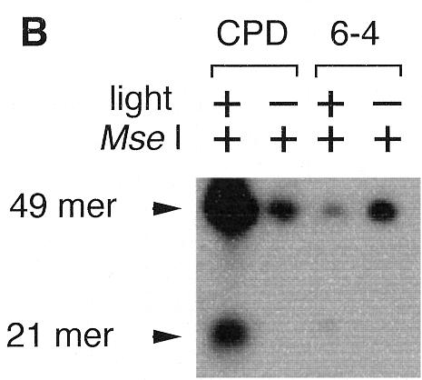
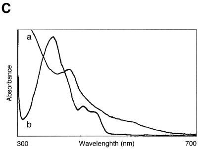
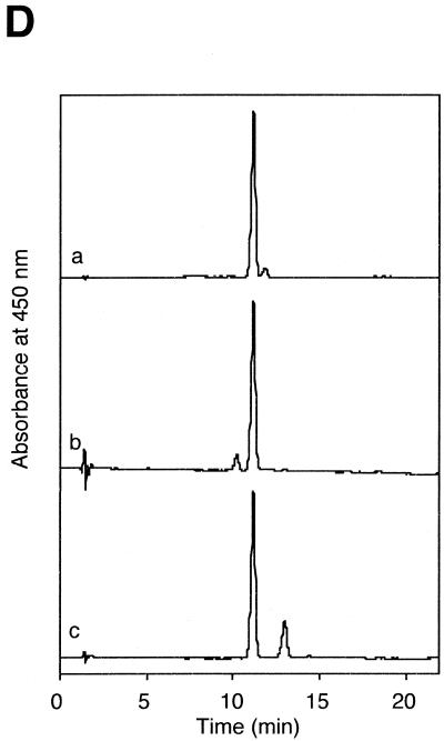
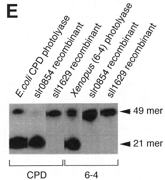
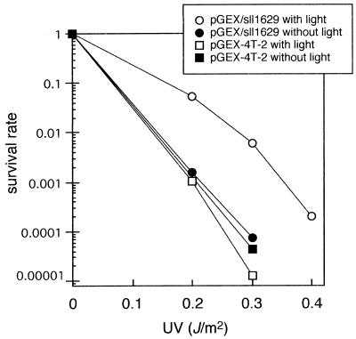
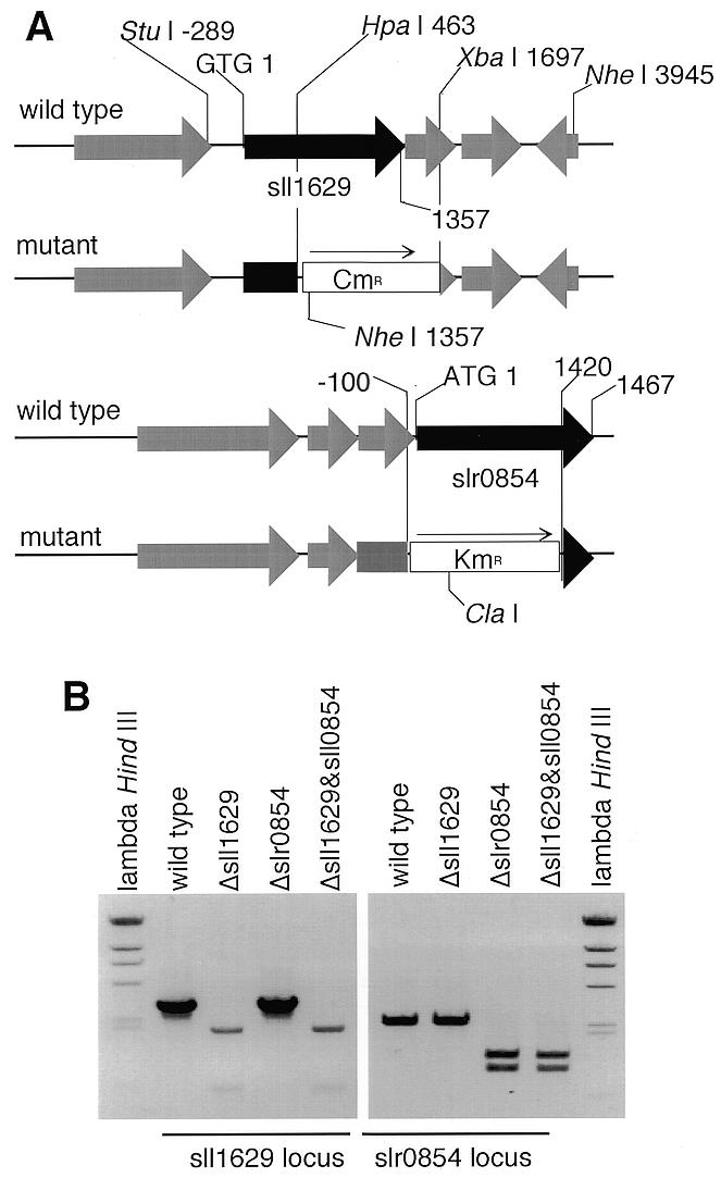
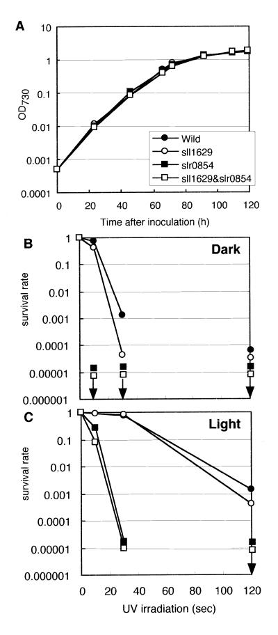
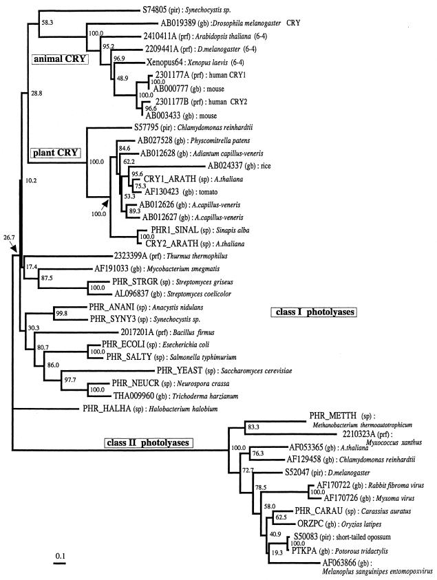
Photolyase is a DNA repair enzyme that reverses UV-induced photoproducts in DNA in a light-dependent manner. Recently, photolyase homologs were identified in higher eukaryotes. These homologs, termed crypto-chromes, function as blue light photoreceptors or regulators of circadian rhythm. In contrast, most bacteria have only a single photolyase or photolyase-like gene. Unlike other microbes, the chromosome of the cyanobacterium SYNECHOCYSTIS: sp. PCC6803 contains two ORFs (slr0854 and sll1629) with high similarities to photolyases. We have characterized both genes. The slr0854 gene product exhibited specific, light-dependent repair activity for a cyclo-butane pyrimidine dimer (CPD), whereas the sll1629 gene product lacks measurable affinity for DNA in vitro. Disruption of either slr0854 or sll1629 had little or no effect on the growth rate of the cyanobacterium. A mutant lacking the slr0854 gene showed severe UV sensitivity, in contrast to a mutant lacking sll1629. Phylogenetic analysis showed that sll1629 is more closely related to the cryptochromes than photolyases. We conclude that sll1629 is a bacterial cryptochrome. To our knowledge, this is the first description of a bacterial cryptochrome.


Figures

Figure 1
Figure 1
Photoreversal activity of Synechocystis sp. PCC6803. (A) Oligonucleotide sequence containing a photoproduct. (B) Photoenzymatic repair of the CPD and (6–4) photoproducts in the 49mer DNA by a Synechocystis sp. PCC6803 cell extract.
Figure 1
Figure 1
Photoreversal activity of Synechocystis sp. PCC6803. (A) Oligonucleotide sequence containing a photoproduct. (B) Photoenzymatic repair of the CPD and (6–4) photoproducts in the 49mer DNA by a Synechocystis sp. PCC6803 cell extract.
Figure 2
Figure 2
Characterization of the recombinant slr0854 and sll1629 gene products. (A) SDS–polyacrylamide gel (4–20% gradient). Lane 1, molecular weight markers; lane 2, an extract of E.coli pGEX/slr0854; lane 3, semi-purified slr0854 fusion protein from a glutathione–Sepharose column. a, GST-fused slr0854 recombinant. The asterisk shows an unknown protein. (B) SDS–polyacrylamide gel (4–20% gradient). Lane 1, molecular weight markers; lane 2, an E.coli pGEX/sll1629 extract; lane 3, the eluate from a glutathione–Sepharose column; lane 4, cleavage of GST by thrombin; lane 5, purified sll1629 protein after purification on a heparin–Sepharose column. b, GST-fused sll1629 recombinant; c, sll1629 recombinant; d, GST. The asterisk shows an unknown protein. (C) Absorption spectra of the slr0854 fusion protein (a) and the sll1629 protein (b). (D) Analysis of the prosthetic groups of the slr0854 and sll1629 fusion proteins by HPLC. a, standard FAD; b, the slr0854 protein; c, the sll1629 protein. (E) Photoenzymatic repair of photoproducts in a 49mer DNA.
Figure 2
Figure 2
Characterization of the recombinant slr0854 and sll1629 gene products. (A) SDS–polyacrylamide gel (4–20% gradient). Lane 1, molecular weight markers; lane 2, an extract of E.coli pGEX/slr0854; lane 3, semi-purified slr0854 fusion protein from a glutathione–Sepharose column. a, GST-fused slr0854 recombinant. The asterisk shows an unknown protein. (B) SDS–polyacrylamide gel (4–20% gradient). Lane 1, molecular weight markers; lane 2, an E.coli pGEX/sll1629 extract; lane 3, the eluate from a glutathione–Sepharose column; lane 4, cleavage of GST by thrombin; lane 5, purified sll1629 protein after purification on a heparin–Sepharose column. b, GST-fused sll1629 recombinant; c, sll1629 recombinant; d, GST. The asterisk shows an unknown protein. (C) Absorption spectra of the slr0854 fusion protein (a) and the sll1629 protein (b). (D) Analysis of the prosthetic groups of the slr0854 and sll1629 fusion proteins by HPLC. a, standard FAD; b, the slr0854 protein; c, the sll1629 protein. (E) Photoenzymatic repair of photoproducts in a 49mer DNA.
Figure 3
Figure 3
Photoreactivation of UV-induced damage in the repair-defective E.coli SY2 (uvrA, recA, phr –) strain with pGEX-4T-2 (squares) or pGEX/sll1629 (circles). After UV irradiation the E.coli cells were either kept in the dark (closed symbols) or illuminated with white light (open symbols).
Figure 4
Figure 4
Disruption of the sll1629 and slr0854 genes. (A) The sll1629 and slr0854 loci in wild-type and mutant genomic DNA of the Synechocystis sp. PCC6803 strain. CmR and KmR stand for the chloramphenicol and kanamycin resistance genes, respectively. (B) Agarose gel analysis of PCR products. The region amplified by PCR to check for disruption of sll1629 was –520 to 2430, digested with NheI. To confirm the slr0854 disruption, the region from –505 to 1900 was amplified by PCR and digested with ClaI.
Figure 5
Figure 5
(A) Growth rate of Synechocystis sp. PCC6803 wild-type and mutants with disrupted slr0829 and sll1629 genes in liquid culture under white light. Cell density was measured as optical density at 730 nm. (B and C) Viability of each strain after UV irradiation. After UV irradiation (14.6 W/m2), samples were incubated in the dark for 60 min and then cultured under red (B) or white light (C). Arrowheads indicate that viability is less than 0.0001 and cannot be measured at the specified irradiation time.
Figure 6
Figure 6
An unrooted phylogenetic tree of the photolyase/cryptochrome family. The tree was constructed according to the amino acid sequence alignment of 44 members of the family. The number associated with each node indicates the bootstrap probability of the cluster at the node. Instead of the citations of sequence data, the ID codes and the corresponding databases for the sequence data are shown as the leaves of the tree. The abbreviated names for the databases are shown in the figure, where pir, prf, sp and gb indicate PIR, PRF, SwissProt and GenBank, respectively. The animal CRY cluster comprises not only cryptochromes but also (6–4) photolyases. To distinguish them in the cluster, (6–4) or CRY is attached to the names of the species only when the data have been identified as (6–4) photolyase or cryptochrome. The scale bar under the tree indicates the branch length, corresponding to 0.1 amino acid substitutions per site.

References

    1. Friedberg E.C., Walker,G.C. and Siede,W. (1995) DNA Repair and Mutagenesis, American Society for Microbiology, Washington, DC.
    1. Sancar A. (1994) Biochemistry, 33, 2–9. - PubMed
    1. Todo T., Takemori,H., Ryo,H., Ihara,M., Matsunaga,T., Nikaido,O., Sato,K. and Nomura,T. (1993) Nature, 361, 371–374. - PubMed
    1. Kato T. Jr, Todo,T., Ayaki,H., Ishizaki,K., Morita,T., Mitra,S. and Ikenaga,M. (1994) Nucleic Acids Res., 22, 4119–4124. - PMC - PubMed
    1. Yasui A., Eker,A.P., Yasuhira,S., Yajima,H., Kobayashi,T., Takao,M. and Oikawa,A. (1994) EMBO J., 13, 6143–6151. - PMC - PubMed

Publication types

MeSH terms